

Search Count: 27
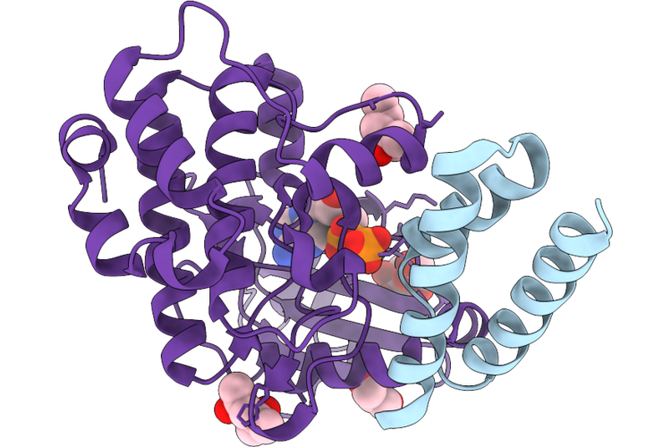 |
Crystal Structure Of Aurora-A Bound To Dbl5
Organism: Synthetic construct, Homo sapiens
Method: X-RAY DIFFRACTION Resolution:1.79 Å Release Date: 2026-07-22 Classification: TRANSFERASE Ligands: CL, ADP, GOL, MPD |
Organism: Synthetic construct, Homo sapiens
Method: X-RAY DIFFRACTION
Release Date: 2026-07-22
Ligands: CL, ADP, GOL, MPD
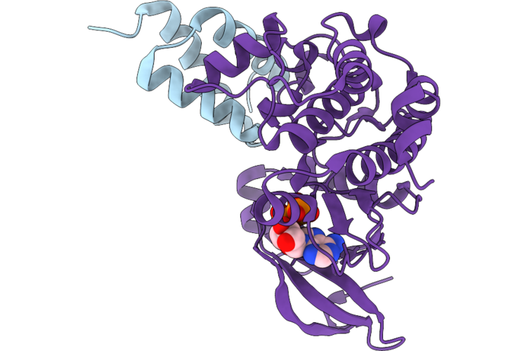 |
Crystal Structure Of Aurora-A Bound To Dbl1
Organism: Synthetic construct, Homo sapiens
Method: X-RAY DIFFRACTION Resolution:2.36 Å Release Date: 2026-07-22 Classification: TRANSFERASE Ligands: ADP, MG |
Organism: Synthetic construct, Homo sapiens
Method: X-RAY DIFFRACTION
Release Date: 2026-07-22
Ligands: ADP, MG
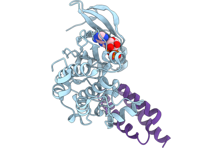 |
Crystal Structure Of Aurora-A Bound To Dbl3
Organism: Homo sapiens, Synthetic construct
Method: X-RAY DIFFRACTION Resolution:2.07 Å Release Date: 2026-07-22 Classification: TRANSFERASE Ligands: ADP |
Organism: Homo sapiens, Synthetic construct
Method: X-RAY DIFFRACTION
Release Date: 2026-07-22
Ligands: ADP
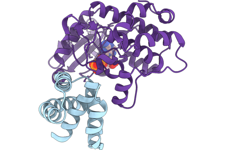 |
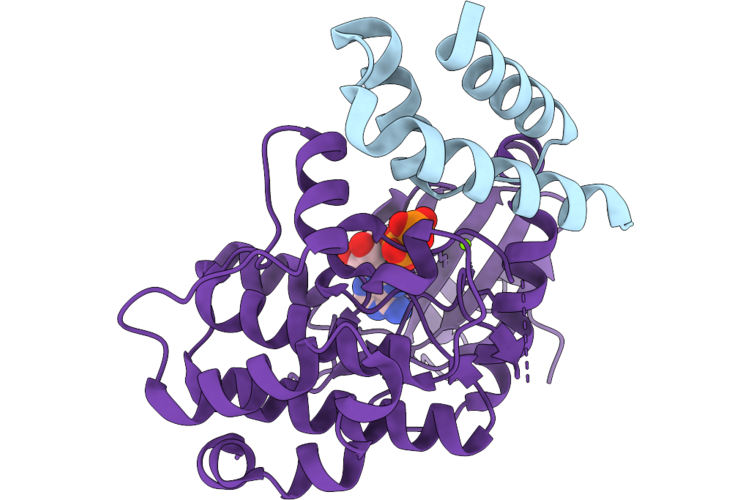
Crystal Structure Of Aurora-A Bound To Dbs1
Organism: Synthetic construct, Homo sapiens
Method: X-RAY DIFFRACTION Resolution:2.00 Å Release Date: 2026-07-22 Classification: TRANSFERASE Ligands: ADP |
Organism: Synthetic construct, Homo sapiens
Method: X-RAY DIFFRACTION
Release Date: 2026-07-22
Ligands: ADP
 |
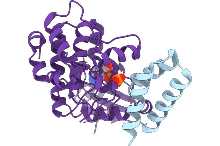
Crystal Structure Of Aurora-A Bound To Dbl6
Organism: Synthetic construct, Homo sapiens
Method: X-RAY DIFFRACTION Resolution:2.36 Å Release Date: 2026-07-22 Classification: TRANSFERASE Ligands: ADP, MG |
Organism: Synthetic construct, Homo sapiens
Method: X-RAY DIFFRACTION
Release Date: 2026-07-22
Ligands: ADP, MG
 |
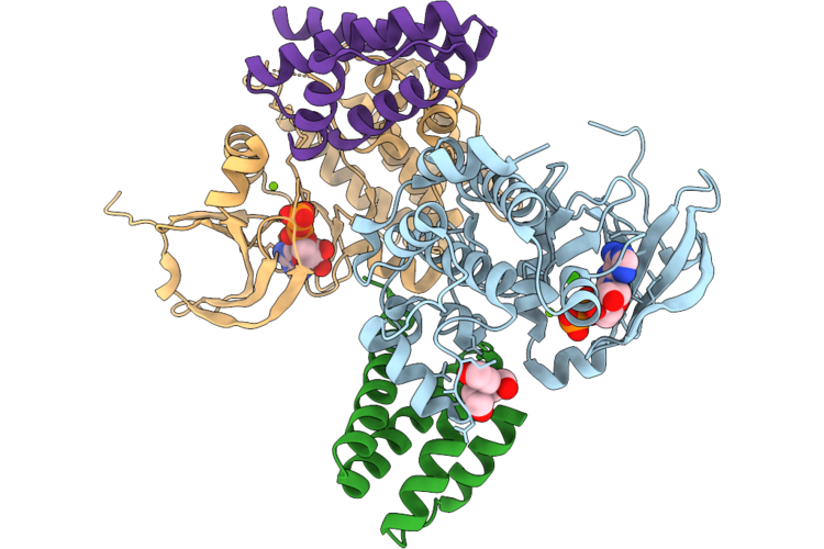
Crystal Structure Of Aurora-A Bound To Dbl8
Organism: Synthetic construct, Homo sapiens
Method: X-RAY DIFFRACTION Resolution:2.37 Å Release Date: 2026-07-22 Classification: TRANSFERASE Ligands: ADP |
Organism: Synthetic construct, Homo sapiens
Method: X-RAY DIFFRACTION
Release Date: 2026-07-22
Ligands: ADP
 |
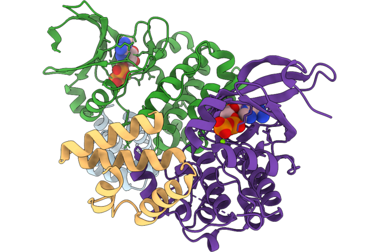
Crystal Structure Of Aurora-A Bound To Dbs5
Organism: Homo sapiens, Unidentified
Method: X-RAY DIFFRACTION Resolution:2.28 Å Release Date: 2026-07-22 Classification: TRANSFERASE Ligands: ADP, TAM, MG, CA |
Organism: Homo sapiens, Unidentified
Method: X-RAY DIFFRACTION
Release Date: 2026-07-22
Ligands: ADP, TAM, MG, CA
 |
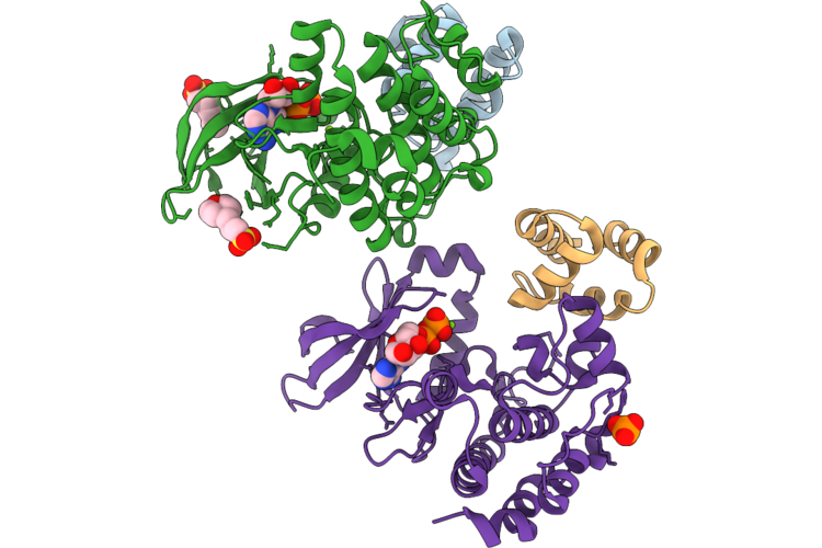
Crystal Structure Of Aurora-A Bound To Dbs2
Organism: Synthetic construct, Homo sapiens
Method: X-RAY DIFFRACTION Resolution:2.90 Å Release Date: 2026-07-22 Classification: TRANSFERASE Ligands: ADP, MG |
Organism: Synthetic construct, Homo sapiens
Method: X-RAY DIFFRACTION
Release Date: 2026-07-22
Ligands: ADP, MG
 |
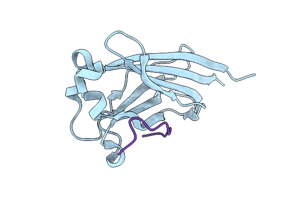
Crystal Structure Of Aurora-A Bound To Dbs4
Organism: Synthetic construct, Homo sapiens
Method: X-RAY DIFFRACTION Resolution:2.23 Å Release Date: 2026-07-22 Classification: TRANSFERASE Ligands: ADP, MPO, MG, PO4 |
Organism: Synthetic construct, Homo sapiens
Method: X-RAY DIFFRACTION
Release Date: 2026-07-22
Ligands: ADP, MPO, MG, PO4
 |
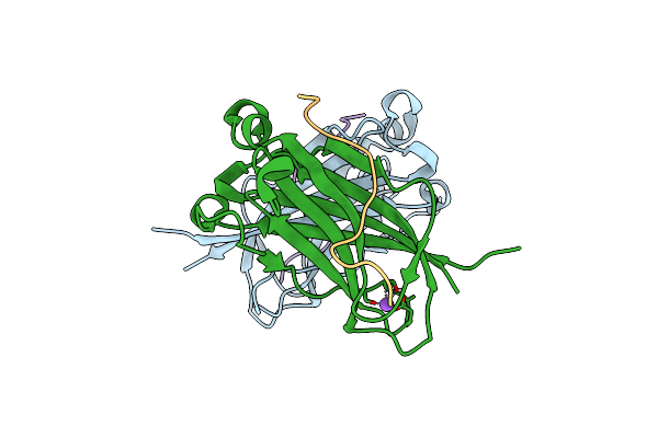
Myd88 Peptide_2 Bound To Spop Math Domain
Organism: Homo sapiens
Method: X-RAY DIFFRACTION Resolution:1.45 Å Release Date: 2025-06-18 Classification: LIGASE |
Organism: Homo sapiens
Method: X-RAY DIFFRACTION
Release Date: 2025-06-18
 |
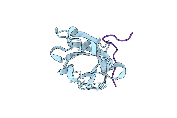
Caprin1 Peptide Bound To Spop Math Domain
Organism: Homo sapiens
Method: X-RAY DIFFRACTION Resolution:1.70 Å Release Date: 2025-04-23 Classification: LIGASE |
Organism: Homo sapiens
Method: X-RAY DIFFRACTION
Release Date: 2025-04-23
 |
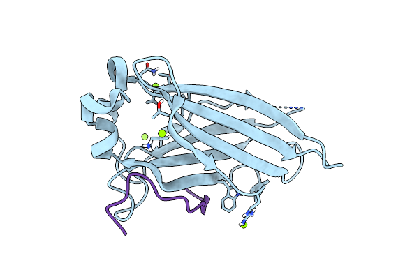
Src-3 (Ncoa-3) Peptide Bound To Spop Math Domain
Organism: Homo sapiens
Method: X-RAY DIFFRACTION Resolution:1.70 Å Release Date: 2025-04-23 Classification: LIGASE |
Organism: Homo sapiens
Method: X-RAY DIFFRACTION
Release Date: 2025-04-23
 |
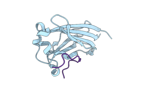
Setd2 Peptide Bound To Spop Math Domain
Organism: Homo sapiens
Method: X-RAY DIFFRACTION Resolution:1.90 Å Release Date: 2025-04-23 Classification: LIGASE Ligands: MG |
Organism: Homo sapiens
Method: X-RAY DIFFRACTION
Release Date: 2025-04-23
Ligands: MG
 |
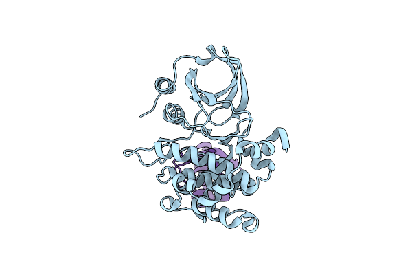
Myd88 Peptide_1 Bound To Spop Math Domain
Organism: Homo sapiens
Method: X-RAY DIFFRACTION Resolution:1.90 Å Release Date: 2025-04-23 Classification: LIGASE |
Organism: Homo sapiens
Method: X-RAY DIFFRACTION
Release Date: 2025-04-23
 |
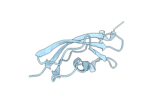
Identification Of An Intermediate Activation State Of Pak5 Reveals A Novel Mechanism Of Kinase Inhibition.
Organism: Homo sapiens, Synthetic construct
Method: X-RAY DIFFRACTION Resolution:1.55 Å Release Date: 2023-10-25 Classification: TRANSFERASE |
Organism: Homo sapiens, Synthetic construct
Method: X-RAY DIFFRACTION
Release Date: 2023-10-25
 |
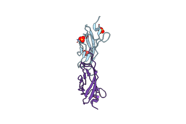
Structure Of Rib Standard, A Rib Domain From Lactobacillus Acidophilus
Organism: Lactobacillus acidophilus (strain atcc 700396 / nck56 / n2 / ncfm)
Method: X-RAY DIFFRACTION Resolution:1.25 Å Release Date: 2019-12-18 Classification: STRUCTURAL PROTEIN |
 |
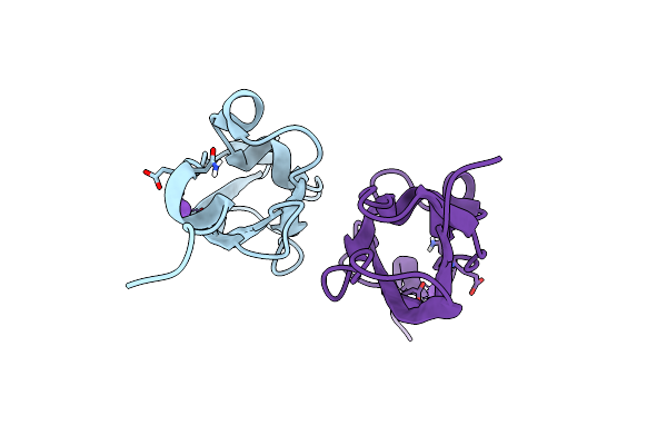
Structure Of Rib Domain 'Rib Long' From Lactobacillus Acidophilus
Organism: Lactobacillus acidophilus
Method: X-RAY DIFFRACTION Resolution:1.07 Å Release Date: 2019-12-11 Classification: STRUCTURAL PROTEIN Ligands: SO4, NA |
Organism: Lactobacillus acidophilus
Method: X-RAY DIFFRACTION
Release Date: 2019-12-11
Ligands: SO4, NA
 |
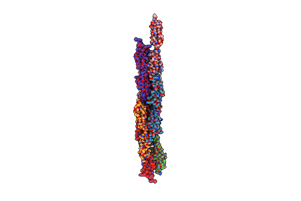
Structure Of Ribr, The Most N-Terminal Rib Domain From Group B Streptococcus Species Streptococcus Agalactiae
Organism: Streptococcus agalactiae
Method: X-RAY DIFFRACTION Resolution:1.70 Å Release Date: 2019-12-11 Classification: STRUCTURAL PROTEIN |
Organism: Streptococcus agalactiae
Method: X-RAY DIFFRACTION
Release Date: 2019-12-11
 |
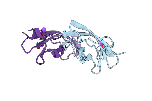
Structure Of Tandemly Arrayed Consecutive Rib Domains (Rib2R) From Group B Streptococcal Species Streptococcus Agalactiae
Organism: Streptococcus agalactiae
Method: X-RAY DIFFRACTION Resolution:2.30 Å Release Date: 2019-12-11 Classification: STRUCTURAL PROTEIN |
Organism: Streptococcus agalactiae
Method: X-RAY DIFFRACTION
Release Date: 2019-12-11
 |
Structure Of Rib R28N From Streptococcus Pyogenes
Organism: Streptococcus pyogenes
Method: X-RAY DIFFRACTION Resolution:1.80 Å Release Date: 2019-12-11 Classification: STRUCTURAL PROTEIN |
Organism: Streptococcus pyogenes
Method: X-RAY DIFFRACTION
Release Date: 2019-12-11