

Search Count: 24
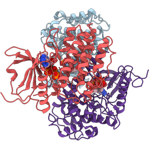 |
Crystal Structure Of C-Terminal Truncated Calcium-Dependent Protein Kinase 3 (Cpk3) From Arabidopsis Thaliana
Organism: Arabidopsis thaliana
Method: X-RAY DIFFRACTION Resolution:2.10 Å Release Date: 2026-10-07 Classification: TRANSFERASE Ligands: ANP, MG |
Organism: Arabidopsis thaliana
Method: X-RAY DIFFRACTION
Release Date: 2026-10-07
Ligands: ANP, MG
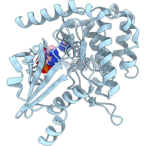 |
Crystal Structure Of Nucleotide-Bound Pak2 Kinase Domain Containing K278R Mutant
Organism: Homo sapiens
Method: X-RAY DIFFRACTION Resolution:1.90 Å Release Date: 2026-09-16 Classification: TRANSFERASE Ligands: ANP, GAI, CL |
Organism: Homo sapiens
Method: X-RAY DIFFRACTION
Release Date: 2026-09-16
Ligands: ANP, GAI, CL
 |
Crystal Structure Of Phosphorylated Pak2 Kinase Domain Containing K278R Mutant
Organism: Homo sapiens
Method: X-RAY DIFFRACTION Resolution:1.85 Å Release Date: 2026-09-16 Classification: TRANSFERASE Ligands: GAI, CL |
Organism: Homo sapiens
Method: X-RAY DIFFRACTION
Release Date: 2026-09-16
Ligands: GAI, CL
 |
Crystal Structure Of Pak2 Kinase Domain Containing D368N Mutant
Organism: Homo sapiens
Method: X-RAY DIFFRACTION Resolution:2.40 Å Release Date: 2026-09-16 Classification: TRANSFERASE Ligands: CIT, SO4 |
Organism: Homo sapiens
Method: X-RAY DIFFRACTION
Release Date: 2026-09-16
Ligands: CIT, SO4
 |
Crystal Structure Of Amp-Pnp-Bound Pak2 Kinase Domain Containing D368N Mutant
Organism: Homo sapiens
Method: X-RAY DIFFRACTION Resolution:2.00 Å Release Date: 2026-09-16 Classification: TRANSFERASE Ligands: ANP, MG, CL |
Organism: Homo sapiens
Method: X-RAY DIFFRACTION
Release Date: 2026-09-16
Ligands: ANP, MG, CL
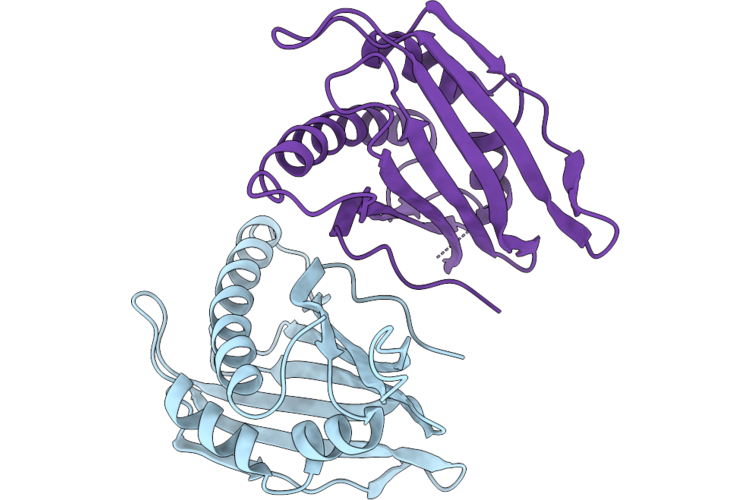 |
Crystal Structure Of The First Start-Like Domain Of Arabidopsis C2 And Gram Domain-Containing (C2Gr) Protein In Apo Form
Organism: Arabidopsis thaliana
Method: X-RAY DIFFRACTION Resolution:2.10 Å Release Date: 2026-07-22 Classification: LIPID BINDING PROTEIN |
Organism: Arabidopsis thaliana
Method: X-RAY DIFFRACTION
Release Date: 2026-07-22
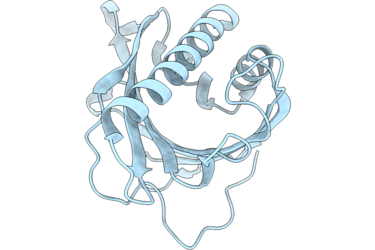 |
Crystal Structure Of The Second Start-Like Domain From Arabidopsis C2 And Gram Domain-Containing (C2Gr) Protein
Organism: Arabidopsis thaliana
Method: X-RAY DIFFRACTION Resolution:1.70 Å Release Date: 2026-07-22 Classification: LIPID BINDING PROTEIN |
Organism: Arabidopsis thaliana
Method: X-RAY DIFFRACTION
Release Date: 2026-07-22
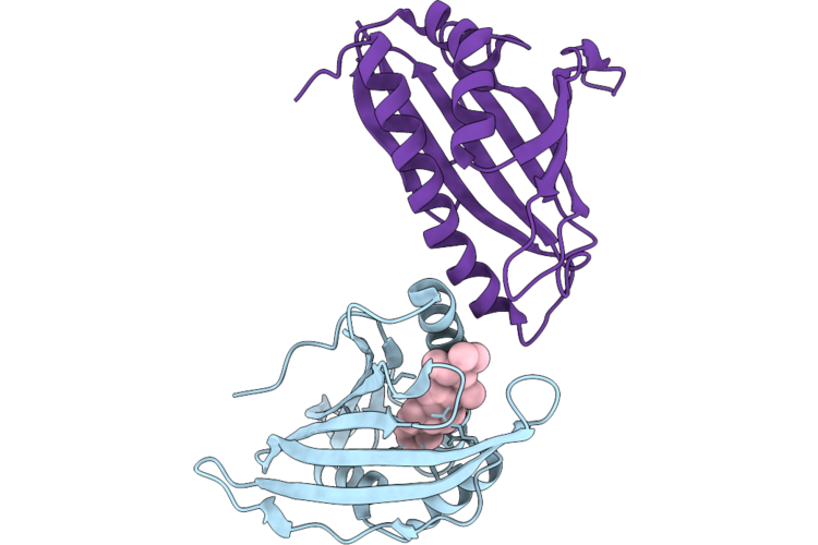 |
Crystal Structure Of The First Start-Like Domain Of Arabidopsis C2 And Gram Domain-Containing (C2Gr) Protein In Complex With Beta-Sitosterol
Organism: Arabidopsis thaliana
Method: X-RAY DIFFRACTION Resolution:2.79 Å Release Date: 2026-07-22 Classification: LIPID BINDING PROTEIN Ligands: A1LXL |
Organism: Arabidopsis thaliana
Method: X-RAY DIFFRACTION
Release Date: 2026-07-22
Ligands: A1LXL
 |
Crystal Structure Of Inactive Calcium-Dependent Protein Kinase 3 (Cpk3) From Arabidopsis Thaliana
Organism: Arabidopsis thaliana
Method: X-RAY DIFFRACTION Resolution:3.05 Å Release Date: 2026-06-24 Classification: TRANSFERASE Ligands: ANP, MG, PEG |
Organism: Arabidopsis thaliana
Method: X-RAY DIFFRACTION
Release Date: 2026-06-24
Ligands: ANP, MG, PEG
 |
Calcium-Bound Structure Of Calcium-Dependent Protein Kinase 3 (Cpk3) From Arabidopsis Thaliana
Organism: Arabidopsis thaliana
Method: X-RAY DIFFRACTION Resolution:2.11 Å Release Date: 2026-06-24 Classification: TRANSFERASE Ligands: CA |
Organism: Arabidopsis thaliana
Method: X-RAY DIFFRACTION
Release Date: 2026-06-24
Ligands: CA
 |
Structure Of The P38Alpha-Pepheptpm(16-31)(V31C ) Complex
Organism: Mus musculus, Homo sapiens
Method: X-RAY DIFFRACTION Resolution:2.14 Å Release Date: 2025-03-19 Classification: TRANSFERASE |
Organism: Mus musculus, Homo sapiens
Method: X-RAY DIFFRACTION
Release Date: 2025-03-19
 |
The Crystal Structure Of Inactive P38 Complexed With A Atf2 From 46 To 80
Organism: Homo sapiens, Mus musculus
Method: X-RAY DIFFRACTION Resolution:1.95 Å Release Date: 2025-03-19 Classification: TRANSFERASE |
Organism: Homo sapiens, Mus musculus
Method: X-RAY DIFFRACTION
Release Date: 2025-03-19
 |
The Crystal Structure Of Inactive P38 Complexed With A Atf2 From 46 To 90
Organism: Mus musculus, Homo sapiens
Method: X-RAY DIFFRACTION Resolution:2.00 Å Release Date: 2025-03-19 Classification: TRANSFERASE |
Organism: Mus musculus, Homo sapiens
Method: X-RAY DIFFRACTION
Release Date: 2025-03-19
 |
The Crystal Structure Of Cd163 Srcr5-9
Organism: Sus scrofa
Method: X-RAY DIFFRACTION Resolution:2.50 Å Release Date: 2023-10-25 Classification: ENDOCYTOSIS Ligands: NAG |
Organism: Sus scrofa
Method: X-RAY DIFFRACTION
Release Date: 2023-10-25
Ligands: NAG
 |
Crystal Structure Of Hsa-Myr Complex Soaked With Cisplatin For One Week
Organism: Homo sapiens
Method: X-RAY DIFFRACTION Resolution:2.89 Å Release Date: 2022-07-20 Classification: TRANSPORT PROTEIN Ligands: CPT, MYR |
Organism: Homo sapiens
Method: X-RAY DIFFRACTION
Release Date: 2022-07-20
Ligands: CPT, MYR
 |
Crystal Structure Of Hsa Soaked With Cisplatin For One Week
Organism: Homo sapiens
Method: X-RAY DIFFRACTION Resolution:2.90 Å Release Date: 2022-07-20 Classification: TRANSPORT PROTEIN Ligands: CPT, PO4 |
Organism: Homo sapiens
Method: X-RAY DIFFRACTION
Release Date: 2022-07-20
Ligands: CPT, PO4
 |
Plsy Grown In Lcp Soaked With Selenourea For 22 Min
Organism: Aquifex aeolicus vf5
Method: X-RAY DIFFRACTION Resolution:1.80 Å Release Date: 2021-08-11 Classification: TRANSFERASE Ligands: SEY, SO4, 79M |
Organism: Aquifex aeolicus vf5
Method: X-RAY DIFFRACTION
Release Date: 2021-08-11
Ligands: SEY, SO4, 79M
 |
Lysozyme Grown In Lcp Soaked With Selenourea For 6 Min
Organism: Gallus gallus
Method: X-RAY DIFFRACTION Resolution:1.71 Å Release Date: 2021-08-11 Classification: HYDROLASE Ligands: SEY, NA, CL |
Organism: Gallus gallus
Method: X-RAY DIFFRACTION
Release Date: 2021-08-11
Ligands: SEY, NA, CL
 |
Crystal Structure Of Atptp1 At 1.4 Angstrom
Organism: Arabidopsis thaliana
Method: X-RAY DIFFRACTION Resolution:1.40 Å Release Date: 2020-08-26 Classification: HYDROLASE Ligands: IOD, FLC, PEG |
Organism: Arabidopsis thaliana
Method: X-RAY DIFFRACTION
Release Date: 2020-08-26
Ligands: IOD, FLC, PEG
 |
Crystal Structure Of Atptp1 At 1.7 Angstrom
Organism: Arabidopsis thaliana
Method: X-RAY DIFFRACTION Resolution:1.70 Å Release Date: 2020-08-26 Classification: HYDROLASE Ligands: IOD, FLC |
Organism: Arabidopsis thaliana
Method: X-RAY DIFFRACTION
Release Date: 2020-08-26
Ligands: IOD, FLC