

Search Count: 234
 |
The Solution Structure Of The Cyanobacterial Calcium Binding Protein Cse At 293 K
Organism: Nostoc sp. pcc 7120 = fachb-418
Method: SOLUTION NMR Release Date: 2026-07-29 Classification: METAL BINDING PROTEIN Ligands: CA |
Organism: Nostoc sp. pcc 7120 = fachb-418
Method: SOLUTION NMR
Release Date: 2026-07-29
Ligands: CA
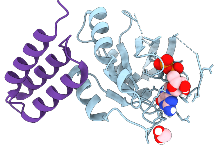 |
Crystal Structure Of Hras In Complex With N-Lhy Affibody
Organism: Homo sapiens, Unidentified
Method: X-RAY DIFFRACTION Resolution:1.80 Å Release Date: 2026-06-03 Classification: ONCOPROTEIN Ligands: GDP, EDO |
Organism: Homo sapiens, Unidentified
Method: X-RAY DIFFRACTION
Release Date: 2026-06-03
Ligands: GDP, EDO
 |
Ternary Cryo-Em Structure Of Yeast Alg3 With Dol25-Pp-Glcnac2Man5, Dol25-P-Man, And Fab
Organism: Synthetic construct, Saccharomyces cerevisiae
Method: ELECTRON MICROSCOPY Release Date: 2026-03-18 Classification: TRANSFERASE Ligands: A1JMA, Y01, IZY |
Organism: Synthetic construct, Saccharomyces cerevisiae
Method: ELECTRON MICROSCOPY
Release Date: 2026-03-18
Ligands: A1JMA, Y01, IZY
 |
Ternary Cryo-Em Structure Of Human Alg9 With Dol25-Pp-Glcnac2Man6, Dol25-P-Man And Fab
Organism: Homo sapiens, Synthetic construct
Method: ELECTRON MICROSCOPY Release Date: 2026-03-18 Classification: TRANSFERASE Ligands: A1JMT, Y01, NAG |
Organism: Homo sapiens, Synthetic construct
Method: ELECTRON MICROSCOPY
Release Date: 2026-03-18
Ligands: A1JMT, Y01, NAG
 |
Ternary Cryo-Em Structure Of Chicken Alg12 With Dol25-Pp-Glcnac2Man7, Dol25-P-Man, And Fab
Organism: Gallus gallus, Synthetic construct
Method: ELECTRON MICROSCOPY Release Date: 2026-03-18 Classification: TRANSFERASE Ligands: A1JMT, A1JMC, Y01, AV0 |
Organism: Gallus gallus, Synthetic construct
Method: ELECTRON MICROSCOPY
Release Date: 2026-03-18
Ligands: A1JMT, A1JMC, Y01, AV0
 |
Ternary Cryo-Em Structure Of Human Alg9 With Dol25-Pp-Glcnac2Man8, Dol25-P-Man And Fab
Organism: Homo sapiens, Synthetic construct
Method: ELECTRON MICROSCOPY Release Date: 2026-03-18 Classification: TRANSFERASE Ligands: NAG, A1JMT, A1JMB, Y01 |
Organism: Homo sapiens, Synthetic construct
Method: ELECTRON MICROSCOPY
Release Date: 2026-03-18
Ligands: NAG, A1JMT, A1JMB, Y01
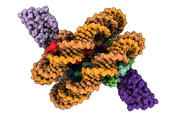 |
Competition For Different Elements Of The Nucleosome Acidic Patch Yields Distinct Functional Outcomes. Vhh 1B2
Organism: Homo sapiens, Artificial sequences
Method: ELECTRON MICROSCOPY Resolution:3.17 Å Release Date: 2026-03-04 Classification: DE NOVO PROTEIN |
Organism: Homo sapiens, Artificial sequences
Method: ELECTRON MICROSCOPY
Release Date: 2026-03-04
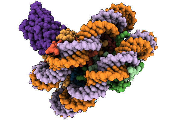 |
Competition For Different Elements Of The Nucleosome Acidic Patch Yields Distinct Functional Outcomes. Vhh 1G1
Organism: Artificial sequences, Homo sapiens
Method: ELECTRON MICROSCOPY Resolution:3.10 Å Release Date: 2026-03-04 Classification: DE NOVO PROTEIN |
Organism: Artificial sequences, Homo sapiens
Method: ELECTRON MICROSCOPY
Release Date: 2026-03-04
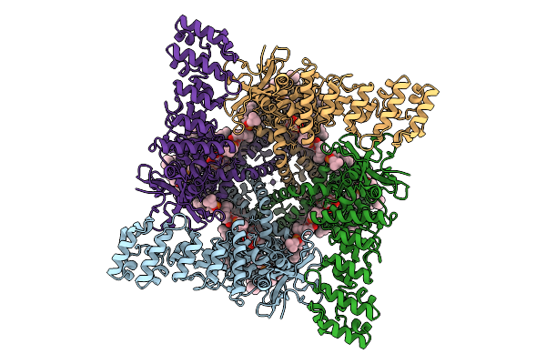 |
Cryo-Em Structure Of Full-Length Human Trpv1 In The Presence Of Alpha-Humulene
Organism: Homo sapiens
Method: ELECTRON MICROSCOPY Release Date: 2025-07-23 Classification: MEMBRANE PROTEIN Ligands: POV, 8IJ, TRD, NA |
Organism: Homo sapiens
Method: ELECTRON MICROSCOPY
Release Date: 2025-07-23
Ligands: POV, 8IJ, TRD, NA
 |
Structure Of Zincore (Sephs1:Qrich1) Binding To Zfp91 On Dna
Organism: Homo sapiens
Method: ELECTRON MICROSCOPY Resolution:3.26 Å Release Date: 2025-07-09 Classification: STRUCTURAL PROTEIN Ligands: ZN |
Organism: Homo sapiens
Method: ELECTRON MICROSCOPY
Release Date: 2025-07-09
Ligands: ZN
 |
Structure Of 2X Zincore (Sephs1:Qrich1) Binding To Zfp91 On Dna
Organism: Homo sapiens
Method: ELECTRON MICROSCOPY Resolution:3.16 Å Release Date: 2025-07-09 Classification: STRUCTURAL PROTEIN Ligands: ZN |
Organism: Homo sapiens
Method: ELECTRON MICROSCOPY
Release Date: 2025-07-09
Ligands: ZN
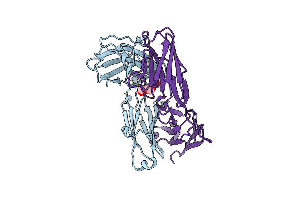 |
Crystal Structure Of Adi-64596 (Human Fab, With Substituted Igg1-Ch1 (Hc-L145Q, K147E, And S181E) And Substituted Kappa Constant Domain (Lc-T129R, T178R, And T180Q))
Organism: Homo sapiens
Method: X-RAY DIFFRACTION Resolution:2.35 Å Release Date: 2025-04-02 Classification: IMMUNE SYSTEM Ligands: BR, PEG |
Organism: Homo sapiens
Method: X-RAY DIFFRACTION
Release Date: 2025-04-02
Ligands: BR, PEG
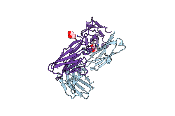 |
Crystal Structure Of Adi-64597 ((Human Fab, With Substituted Igg1-Ch1 (Hc-L128R And K147R) And Substituted Kappa Constant Domain (Lc-Q124E, V133Q, And T178R))
Organism: Homo sapiens
Method: X-RAY DIFFRACTION Resolution:2.22 Å Release Date: 2025-04-02 Classification: IMMUNE SYSTEM Ligands: GOL, CL |
Organism: Homo sapiens
Method: X-RAY DIFFRACTION
Release Date: 2025-04-02
Ligands: GOL, CL
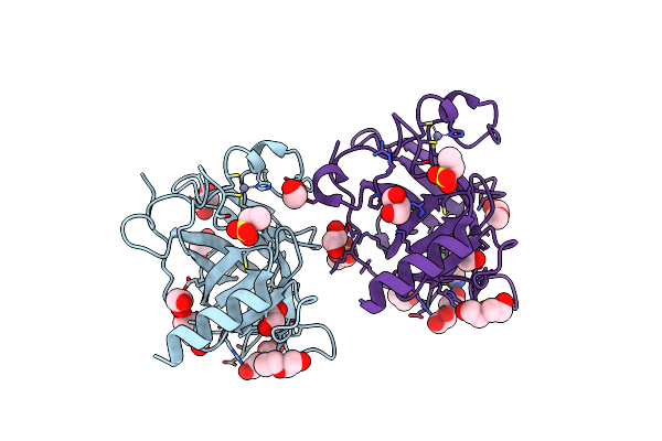 |
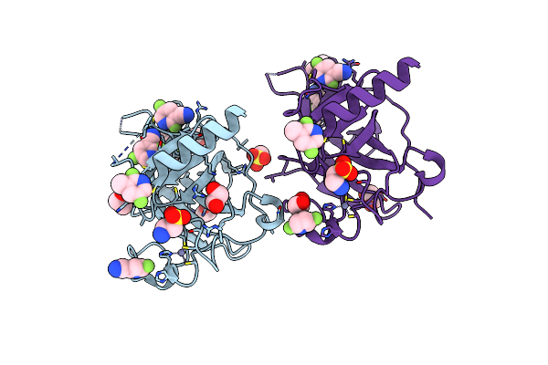
P53-Y220C Core Domain Covalently Bound To 2,5,6-Trifluoropyridine-3-Carbonitrile Soaked At 5 Mm
Organism: Homo sapiens
Method: X-RAY DIFFRACTION Resolution:1.57 Å Release Date: 2024-11-27 Classification: CELL CYCLE Ligands: YLW, PEG, EDO, EPE, ZN, GOL |
Organism: Homo sapiens
Method: X-RAY DIFFRACTION
Release Date: 2024-11-27
Ligands: YLW, PEG, EDO, EPE, ZN, GOL
 |
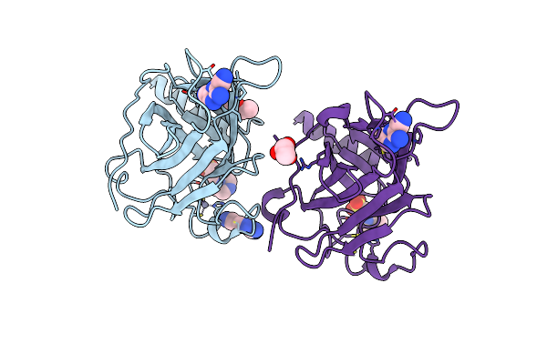
P53-Y220C Core Domain Covalently Bound To 2,5,6-Trifluoropyridine-3-Carbonitrile Soaked At 40 Mm
Organism: Homo sapiens
Method: X-RAY DIFFRACTION Resolution:1.69 Å Release Date: 2024-11-27 Classification: CELL CYCLE Ligands: YLW, GOL, EDO, EPE, ZN |
Organism: Homo sapiens
Method: X-RAY DIFFRACTION
Release Date: 2024-11-27
Ligands: YLW, GOL, EDO, EPE, ZN
 |
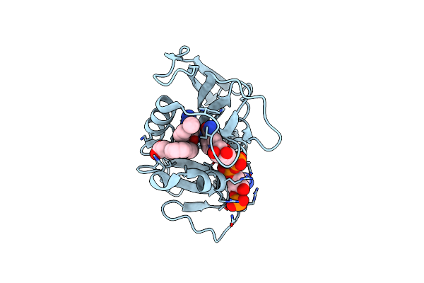
P53-Y220C Core Domain Covalently Bound To 3-Amino-5-Chloropyrazine-2,6-Dicarbonitrile Soaked At 5 Mm
Organism: Homo sapiens
Method: X-RAY DIFFRACTION Resolution:2.06 Å Release Date: 2024-11-27 Classification: CELL CYCLE Ligands: YM0, EPE, PEG, ZN |
Organism: Homo sapiens
Method: X-RAY DIFFRACTION
Release Date: 2024-11-27
Ligands: YM0, EPE, PEG, ZN
 |
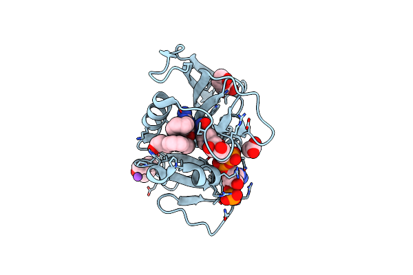
Crystal Structure Of Dihydrofolate Reductase (Dhfr) From Mycobacterium Ulcerans Agy99 In Complex With Nadp And Inhibitor Mam881
Organism: Mycobacterium ulcerans
Method: X-RAY DIFFRACTION Resolution:1.95 Å Release Date: 2024-08-28 Classification: OXIDOREDUCTASE/INHIBITOR Ligands: DQI, NAP |
Organism: Mycobacterium ulcerans
Method: X-RAY DIFFRACTION
Release Date: 2024-08-28
Ligands: DQI, NAP
 |
Crystal Structure Of Dihydrofolate Reductase (Dhfr) From Mycobacterium Ulcerans Agy99 In Complex With Nadp And Inhibitor Mam907
Organism: Mycobacterium ulcerans
Method: X-RAY DIFFRACTION Resolution:1.50 Å Release Date: 2024-08-28 Classification: OXIDOREDUCTASE/INHIBITOR |
Organism: Mycobacterium ulcerans
Method: X-RAY DIFFRACTION
Release Date: 2024-08-28
 |
Crystal Structure Of Dihydrofolate Reductase (Dhfr) From Mycobacterium Ulcerans Agy99 In Complex With Nadp And Inhibitor Mam758
Organism: Mycobacterium ulcerans
Method: X-RAY DIFFRACTION Resolution:1.75 Å Release Date: 2024-08-28 Classification: OXIDOREDUCTASE/INHIBITOR Ligands: NAP, HIJ |
Organism: Mycobacterium ulcerans
Method: X-RAY DIFFRACTION
Release Date: 2024-08-28
Ligands: NAP, HIJ
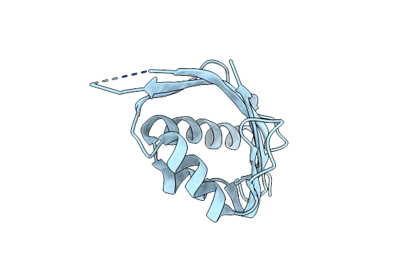 |
Crystal Structure Of Sam0.2
Organism: Synthetic construct
Method: X-RAY DIFFRACTION Resolution:2.65 Å Release Date: 2024-05-22 Classification: DE NOVO PROTEIN |
Organism: Synthetic construct
Method: X-RAY DIFFRACTION
Release Date: 2024-05-22