9S6T image
Deposition Date 2025-08-01
Release Date 2026-03-18
Last Version Date 2026-03-25
Entry Detail
PDB ID:
9S6T
Keywords:
Title:
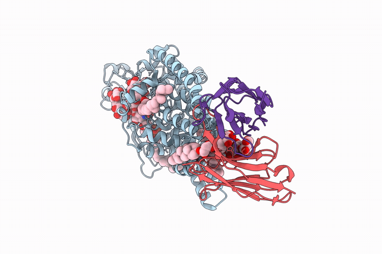
Ternary cryo-EM structure of chicken ALG12 with Dol25-PP-GlcNAc2Man7, Dol25-P-Man, and Fab
Biological Source:
Source Organism(s):
Method Details:
Experimental Method:
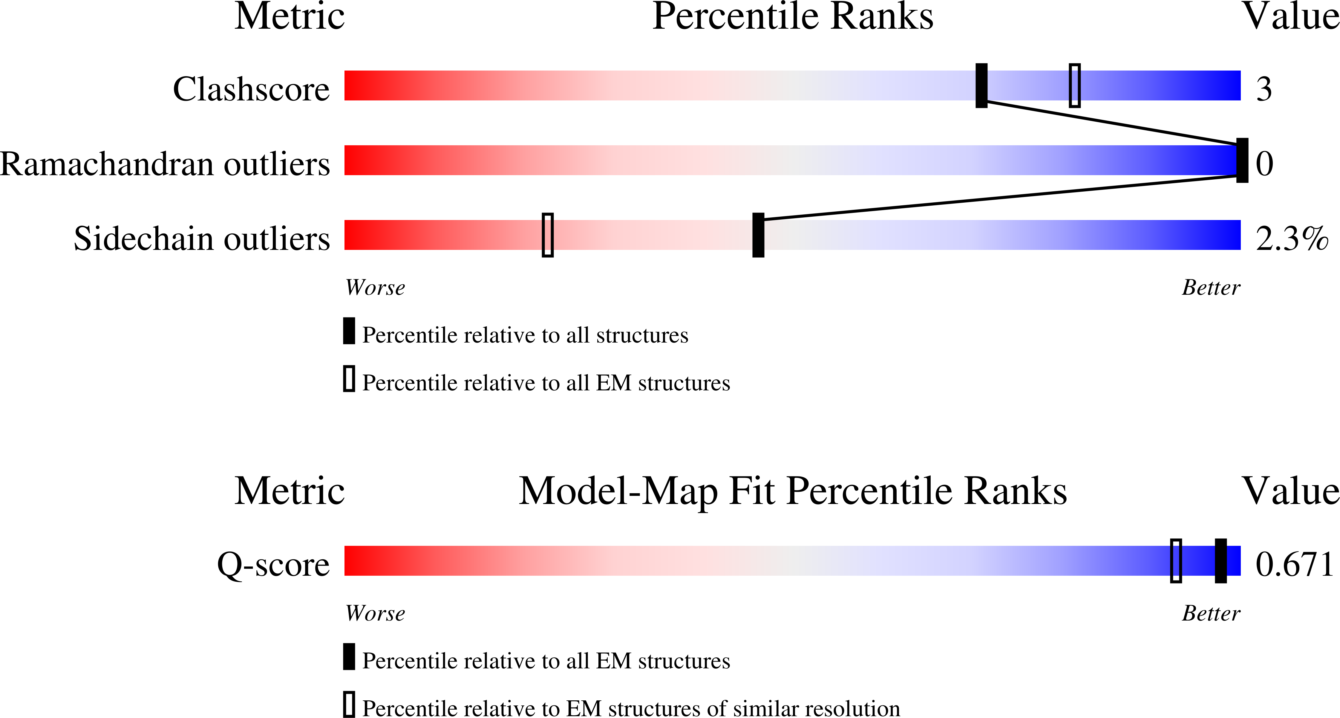
Resolution:
2.42 Å
Aggregation State:
PARTICLE
Reconstruction Method:
SINGLE PARTICLE
Macromolecular Entities
Structures with similar UniProt ID
Protein Blast
Polymer Type:polypeptide(L)
Molecule:Mannosyltransferase
Mutagens:E35Q
Chain IDs:A
Chain Length:496
Number of Molecules:1
Biological Source:Gallus gallus
Protein Blast
Polymer Type:polypeptide(L)
Molecule:Gg12-11 Fab heavy chain
Chain IDs:B (auth: H)
Chain Length:234
Number of Molecules:1
Biological Source:synthetic construct
Protein Blast
Polymer Type:polypeptide(L)
Molecule:Gg12-11 Fab light chain
Chain IDs:C (auth: L)
Chain Length:215
Number of Molecules:1
Biological Source:synthetic construct
Primary Citation
Structures of ALG3/9/12 reveal the assembly logic of the N-glycan oligomannose core.
Nat.Chem.Biol. ? ? ? (2026)
PMID: 41807832 DOI: 10.1038/s41589-026-02164-7

Abstact

Asparagine-linked glycans are essential for the maturation and function of most eukaryotic secretory proteins. The biosynthesis and transfer of dolichylpyrophosphate-anchored GlcNAc(2)Man(9)Glc(3) glycan is a highly conserved process occurring in the endoplasmic reticulum (ER) membrane and involving over a dozen membrane proteins whose dysfunction is linked to congenital disorders of glycosylation (CDGs). Three membrane-integral mannosyltransferases, ALG3, ALG9 and ALG12, mediate four consecutive mannosylation reactions that convert GlcNAc(2)Man(5) to GlcNAc(2)Man(9). Here, using chemoenzymatically synthesized lipid-linked glycan donor and acceptor analogs, we recapitulated this biosynthetic pathway in vitro. High-resolution cryo-electron microscopy structures of pseudo-Michaelis complexes of each step revealed how the branched glycan is accurately synthesized and unwanted side products are averted. Molecular dynamics simulations and mutagenesis studies uncovered a subtle but precise mechanism selecting the dolichylphosphomannose donor substrate over dolichylphosphoglucose, which is also present in the ER membrane. Our results also provide mechanistic explanations for enzyme dysfunction in CDGs and offer opportunities for N-glycan engineering.

Legend

Protein

Chemical

Disease

Primary Citation of related structures
Feedback Form
Name
Email
Institute
Feedback