

Search Count: 23
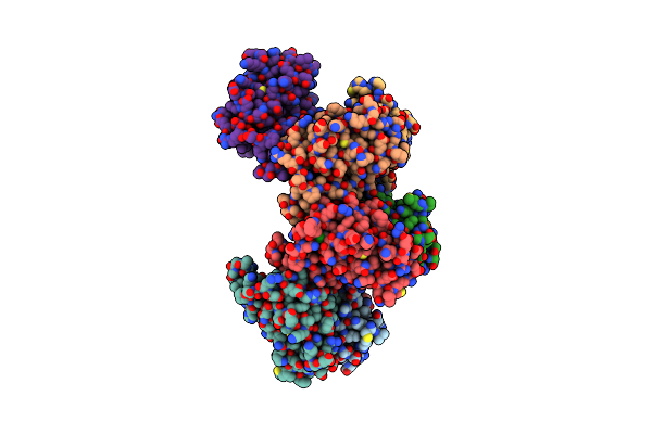 |
Structure Of Thr354Asn, Glu355Gln, Thr412Asn, Ile414Met, Ile464His, And Phe467Met Mutant Human Camkii Alpha Hub Bound To Pipa
Organism: Homo sapiens
Method: X-RAY DIFFRACTION Resolution:2.10 Å Release Date: 2024-09-25 Classification: SIGNALING PROTEIN Ligands: ACT, A1H6K, PG4 |
Organism: Homo sapiens
Method: X-RAY DIFFRACTION
Release Date: 2024-09-25
Ligands: ACT, A1H6K, PG4
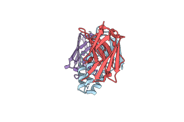 |
Structure Of Api M1 In Complex With Two Nanobodies
Organism: Lama glama, Apis mellifera
Method: X-RAY DIFFRACTION Resolution:1.76 Å Release Date: 2024-05-08 Classification: ALLERGEN Ligands: CL |
Organism: Lama glama, Apis mellifera
Method: X-RAY DIFFRACTION
Release Date: 2024-05-08
Ligands: CL
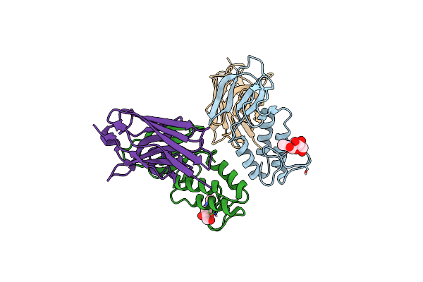 |
Structure Of Api M1 In Complex With The Am1-4 Nanobody
Organism: Lama glama, Apis mellifera
Method: X-RAY DIFFRACTION Resolution:4.10 Å Release Date: 2024-05-08 Classification: ALLERGEN Ligands: NAG |
Organism: Lama glama, Apis mellifera
Method: X-RAY DIFFRACTION
Release Date: 2024-05-08
Ligands: NAG
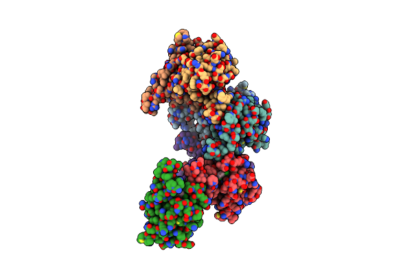 |
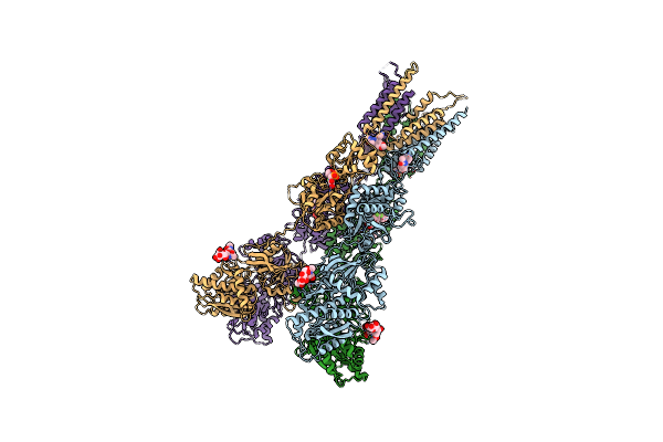
Structure Of Thr354Asn, Glu355Gln, Thr412Asn, Ile414Met, Ile464His, And Phe467Met Mutant Human Camkii Alpha Hub Bound To 5-Hdc
Organism: Homo sapiens
Method: X-RAY DIFFRACTION Resolution:2.20 Å Release Date: 2021-07-21 Classification: SIGNALING PROTEIN Ligands: 7ZV, NA |
Organism: Homo sapiens
Method: X-RAY DIFFRACTION
Release Date: 2021-07-21
Ligands: 7ZV, NA
 |
Structure Of Glua2Cryst In Complex The Antagonist Zk200775 And The Negative Allosteric Modulator Gyki53655 At 4.65 A Resolution
Organism: Rattus norvegicus
Method: X-RAY DIFFRACTION Resolution:4.65 Å Release Date: 2020-06-24 Classification: MEMBRANE PROTEIN Ligands: GYK, ZK1 |
Organism: Rattus norvegicus
Method: X-RAY DIFFRACTION
Release Date: 2020-06-24
Ligands: GYK, ZK1
 |
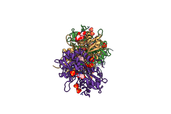
Characterization Of An Intertidal Zone Metagenome Oligoribonuclease And The Role Of The Intermolecular Disulfide Bond For Homodimer Formation And Nuclease Activity.
Organism: Metagenome
Method: X-RAY DIFFRACTION Resolution:3.15 Å Release Date: 2019-08-28 Classification: HYDROLASE Ligands: MN |
Organism: Metagenome
Method: X-RAY DIFFRACTION
Release Date: 2019-08-28
Ligands: MN
 |
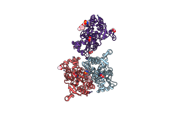
Crystal Structure Of The Glua2 Ligand-Binding Domain (S1S2J-L504Y-N775S) In Complex With Glutamate And Bpam121 At 1.78 A Resolution
Organism: Rattus norvegicus
Method: X-RAY DIFFRACTION Resolution:1.78 Å Release Date: 2018-01-03 Classification: MEMBRANE PROTEIN Ligands: SO4, 7M6, CL, GOL, GLU, EDO, CIT, PEG |
Organism: Rattus norvegicus
Method: X-RAY DIFFRACTION
Release Date: 2018-01-03
Ligands: SO4, 7M6, CL, GOL, GLU, EDO, CIT, PEG
 |
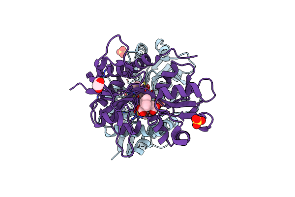
Crystal Structure Of The Glua2 Ligand-Binding Domain (S1S2J) In Complex With Glutamate And Positive Allosteric Modulator Bpam538
Organism: Rattus norvegicus
Method: X-RAY DIFFRACTION Resolution:2.00 Å Release Date: 2018-01-03 Classification: MEMBRANE PROTEIN Ligands: GLU, ZN, ACT, 9TE |
Organism: Rattus norvegicus
Method: X-RAY DIFFRACTION
Release Date: 2018-01-03
Ligands: GLU, ZN, ACT, 9TE
 |
Crystal Structure Of The Gluk1 Ligand-Binding Domain In Complex With Kainate And Bpam-344 At 1.90 A Resolution
Organism: Rattus norvegicus
Method: X-RAY DIFFRACTION Resolution:1.90 Å Release Date: 2017-04-12 Classification: MEMBRANE PROTEIN Ligands: 2J9, KAI, CL, GOL, SO4 |
Organism: Rattus norvegicus
Method: X-RAY DIFFRACTION
Release Date: 2017-04-12
Ligands: 2J9, KAI, CL, GOL, SO4
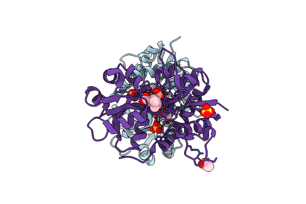 |
Crystal Structure Of The Gluk1 Ligand-Binding Domain In Complex With Kainate And Bpam-521 At 2.18 A Resolution
Organism: Rattus norvegicus
Method: X-RAY DIFFRACTION Resolution:2.18 Å Release Date: 2017-04-12 Classification: MEMBRANE PROTEIN Ligands: 5PX, KAI, CL, SO4, GOL, ACT |
Organism: Rattus norvegicus
Method: X-RAY DIFFRACTION
Release Date: 2017-04-12
Ligands: 5PX, KAI, CL, SO4, GOL, ACT
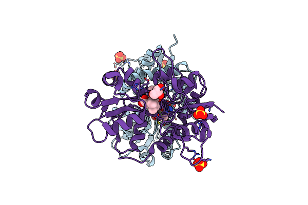 |
Crystal Structure Of The Gluk1 Ligand-Binding Domain In Complex With Kainate And Bpam-121 At 2.10 A Resolution
Organism: Rattus norvegicus
Method: X-RAY DIFFRACTION Resolution:2.10 Å Release Date: 2017-04-12 Classification: MEMBRANE PROTEIN Ligands: 7M6, CL, KAI, SO4, GOL, ACT |
Organism: Rattus norvegicus
Method: X-RAY DIFFRACTION
Release Date: 2017-04-12
Ligands: 7M6, CL, KAI, SO4, GOL, ACT
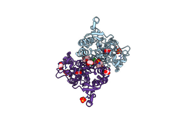 |
Crystal Structure Of The Glua2 Ligand-Binding Domain (L483Y-N754S) In Complex With Glutamate And Bpam-321 At 2.07 A Resolution
Organism: Rattus norvegicus
Method: X-RAY DIFFRACTION Resolution:2.07 Å Release Date: 2016-02-17 Classification: MEMBRANE PROTEIN Ligands: 4V6, GLU, SO4, EDO |
Organism: Rattus norvegicus
Method: X-RAY DIFFRACTION
Release Date: 2016-02-17
Ligands: 4V6, GLU, SO4, EDO
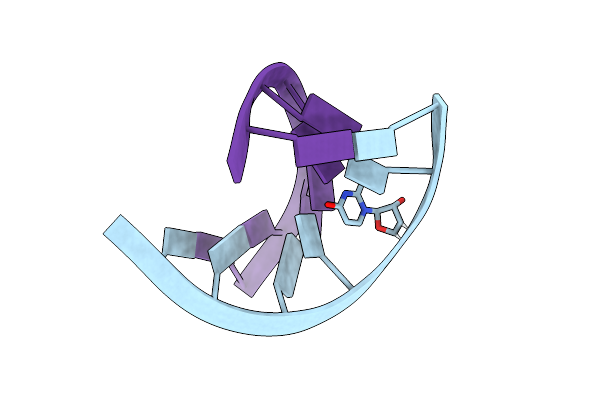 |
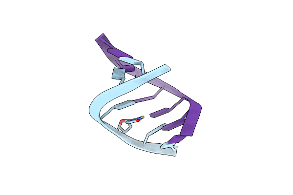
Crystal Structures Of Rna Duplexes Containing 2-Thio-Uridine
Organism: Synthetic construct
Method: X-RAY DIFFRACTION Resolution:1.35 Å Release Date: 2014-08-13 Classification: RNA |
Organism: Synthetic construct
Method: X-RAY DIFFRACTION
Release Date: 2014-08-13
 |
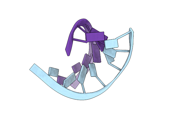
Crystal Structures Of Rna Duplexes Containing 2-Thio-Uridine
Organism: Synthetic construct
Method: X-RAY DIFFRACTION Resolution:1.55 Å Release Date: 2014-08-13 Classification: RNA |
Organism: Synthetic construct
Method: X-RAY DIFFRACTION
Release Date: 2014-08-13
 |
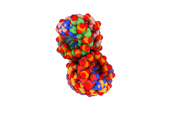
Native 7Mer-Rna Duplex
Organism: Synthetic construct
Method: X-RAY DIFFRACTION Resolution:1.55 Å Release Date: 2014-08-13 Classification: RNA |
Organism: Synthetic construct
Method: X-RAY DIFFRACTION
Release Date: 2014-08-13
 |
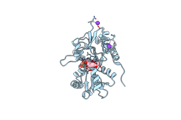
Rna Duplex Containing Uu Mispair
Organism: Synthetic construct
Method: X-RAY DIFFRACTION Resolution:1.80 Å Release Date: 2014-08-13 Classification: RNA |
Organism: Synthetic construct
Method: X-RAY DIFFRACTION
Release Date: 2014-08-13
 |
Crystal Structure Of The Gluk3 Ligand-Binding Domain (S1S2) In Complex With The Agonist (2S,4R)-4-(3-Methoxy-3-Oxopropyl)Glutamic Acid At 2.01 A Resolution.
Organism: Rattus norvegicus
Method: X-RAY DIFFRACTION Resolution:2.01 Å Release Date: 2014-08-06 Classification: MEMBRANE PROTEIN, RECEPTOR/Agonist Ligands: K, PO4, CL, 2QE |
Organism: Rattus norvegicus
Method: X-RAY DIFFRACTION
Release Date: 2014-08-06
Ligands: K, PO4, CL, 2QE
 |
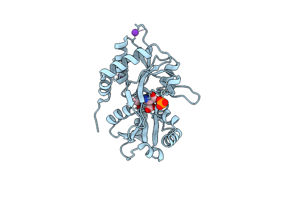
Crystal Structure Of The Kainate Receptor Gluk3 Ligand-Binding Domain In Complex With The Agonist (2S,4R)-4-(3-Methylamino-3-Oxopropyl)Glutamic Acid At 2.6 A Resolution
Organism: Rattus norvegicus
Method: X-RAY DIFFRACTION Resolution:2.60 Å Release Date: 2014-08-06 Classification: MEMBRANE PROTEIN/AGONIST Ligands: 2QD, K, CL |
Organism: Rattus norvegicus
Method: X-RAY DIFFRACTION
Release Date: 2014-08-06
Ligands: 2QD, K, CL
 |
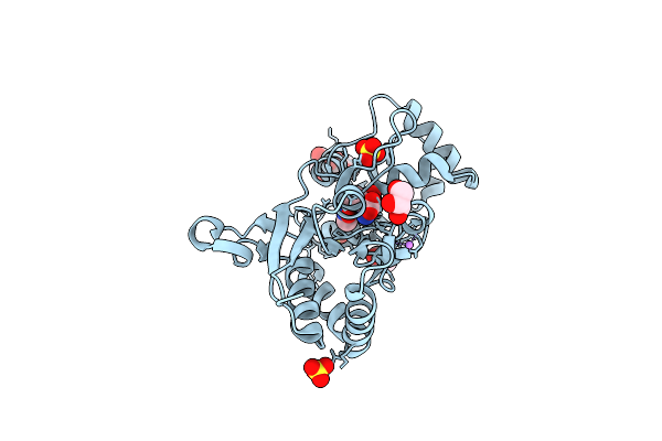
Crystal Structure Of The Kainate Receptor Gluk3 Ligand-Binding Domain In Complex With The Agonist Za302
Organism: Rattus norvegicus
Method: X-RAY DIFFRACTION Resolution:2.65 Å Release Date: 2013-03-06 Classification: MEMBRANE PROTEIN Ligands: 3ZA, K, CL, PO4 |
Organism: Rattus norvegicus
Method: X-RAY DIFFRACTION
Release Date: 2013-03-06
Ligands: 3ZA, K, CL, PO4
 |
Crystal Structure Of The Glua2 Ligand-Binding Domain (S1S2J) In Complex With The Agonist Za302 At 1.24A Resolution
Organism: Rattus norvegicus
Method: X-RAY DIFFRACTION Resolution:1.24 Å Release Date: 2013-03-06 Classification: MEMBRANE PROTEIN Ligands: 3ZA, SO4, LI, GOL |
Organism: Rattus norvegicus
Method: X-RAY DIFFRACTION
Release Date: 2013-03-06
Ligands: 3ZA, SO4, LI, GOL