
Search Count: 55,760
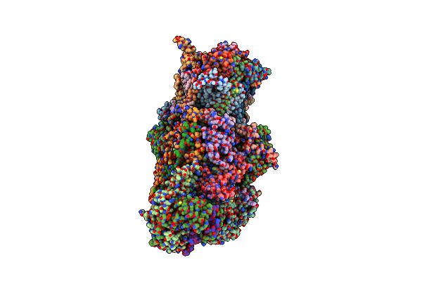 |
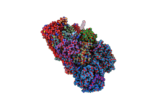
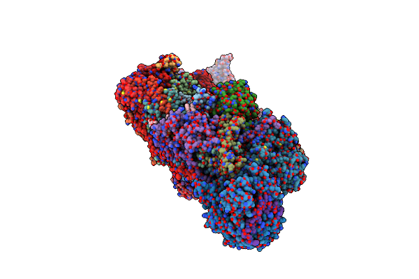
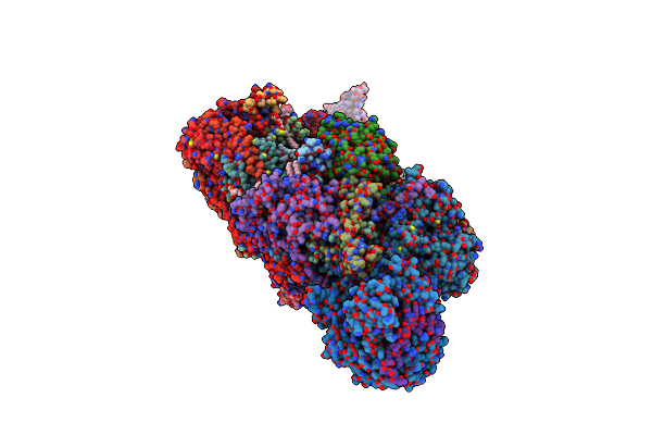
Organism: Chaetomium thermophilum var. thermophilum dsm 1495
Method: ELECTRON MICROSCOPY Release Date: 2022-11-30 Classification: OXIDOREDUCTASE Ligands: PC1, LMT, CDL, 3PE, FES, SF4, FMN, NDP, ZN, ZMP |
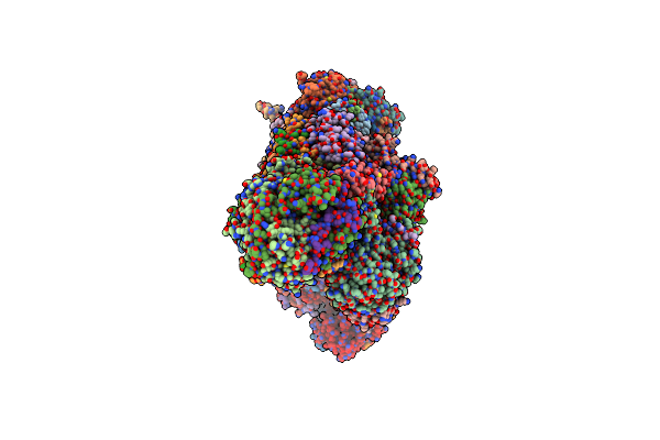 |
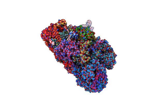
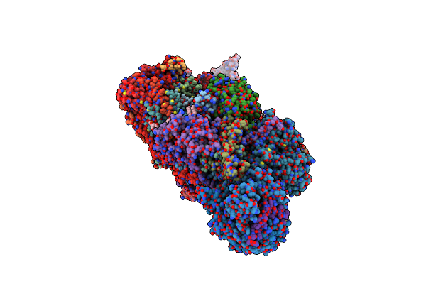
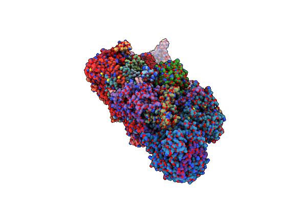
Organism: Chaetomium thermophilum var. thermophilum dsm 1495
Method: ELECTRON MICROSCOPY Release Date: 2022-11-30 Classification: OXIDOREDUCTASE Ligands: 3PE, PC1, CDL, LMN, FES, SF4, FMN, NDP, ZN, ZMP |
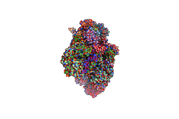 |
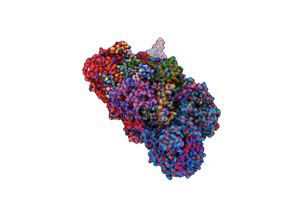
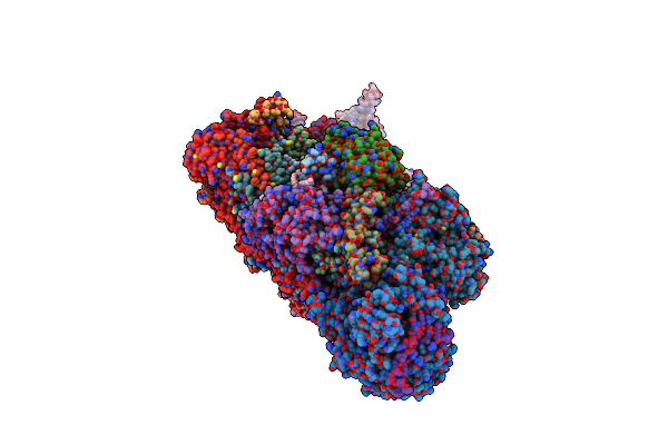
Organism: Chaetomium thermophilum var. thermophilum dsm 1495
Method: ELECTRON MICROSCOPY Release Date: 2022-11-30 Classification: OXIDOREDUCTASE Ligands: 3PE, PC1, CDL, LMN, FES, SF4, FMN, NDP, ZN, ZMP |
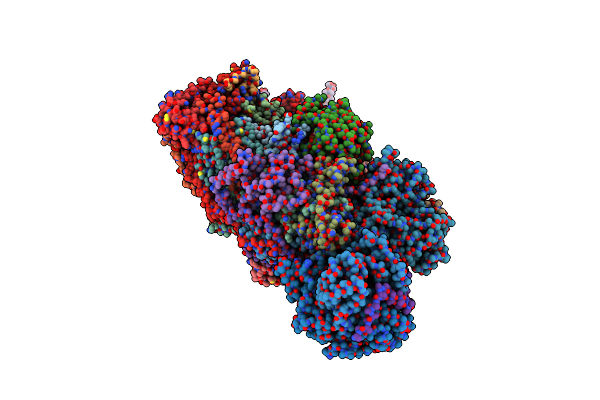 |
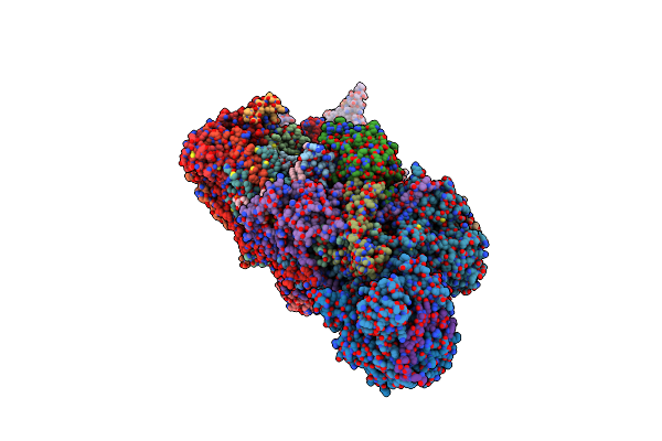
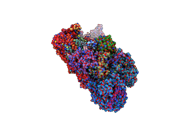
Organism: Bos taurus
Method: ELECTRON MICROSCOPY Resolution:4.35 Å Release Date: 2016-09-07 Classification: OXIDOREDUCTASE Ligands: SF4, FES, FMN, NAP, ZN |
 |
Organism: Bos taurus
Method: ELECTRON MICROSCOPY Release Date: 2016-09-14 Classification: OXIDOREDUCTASE Ligands: SF4, FES, FMN, NAP, ZN |
 |
Organism: Mus musculus
Method: ELECTRON MICROSCOPY Release Date: 2023-08-09 Classification: OXIDOREDUCTASE Ligands: LMT, PC1, SF4, FES, FMN, NA, CDL, 3PE, MG, DGT, NDP, ZN, EHZ, MYR |
 |
Organism: Bos taurus
Method: ELECTRON MICROSCOPY Release Date: 2022-05-25 Classification: OXIDOREDUCTASE Ligands: PC1, 3PE, SF4, FES, FMN, K, CDL, CHD, GTP, MG, NDP, ZN, EHZ, MYR |
 |
Organism: Bos taurus
Method: ELECTRON MICROSCOPY Release Date: 2022-05-25 Classification: OXIDOREDUCTASE Ligands: 3PE, PC1, SF4, U10, FES, FMN, K, CDL, GTP, MG, NDP, ZN, EHZ, CHD, MYR |
 |
Organism: Bos taurus
Method: ELECTRON MICROSCOPY Release Date: 2022-05-25 Classification: OXIDOREDUCTASE Ligands: 3PE, PC1, SF4, FES, FMN, K, GOL, LMT, CDL, GTP, MG, NDP, ZN, EHZ, CHD, MYR |
 |
Organism: Bos taurus
Method: ELECTRON MICROSCOPY Resolution:2.76 Å Release Date: 2022-05-25 Classification: OXIDOREDUCTASE Ligands: 3PE, PC1, SF4, FES, FMN, K, CDL, GTP, MG, NDP, ZN, EHZ, CHD, MYR |
 |
Organism: Bos taurus
Method: ELECTRON MICROSCOPY Release Date: 2023-02-08 Classification: OXIDOREDUCTASE Ligands: LMT, 3PE, SF4, PC1, FES, FMN, K, I49, CDL, GTP, MG, NDP, ZN, EHZ, MYR |
 |
Organism: Bos taurus
Method: ELECTRON MICROSCOPY Release Date: 2023-02-08 Classification: OXIDOREDUCTASE Ligands: LMT, SF4, PC1, FES, FMN, K, 3PE, CDL, I49, GTP, MG, NDP, ZN, EHZ, MYR |
 |
Organism: Bos taurus
Method: ELECTRON MICROSCOPY Release Date: 2023-02-08 Classification: OXIDOREDUCTASE Ligands: LMT, 3PE, SF4, FES, FMN, K, I49, CDL, PC1, GTP, MG, NDP, ZN, EHZ, MYR |
 |
Organism: Bos taurus
Method: ELECTRON MICROSCOPY Release Date: 2022-03-02 Classification: OXIDOREDUCTASE Ligands: PC1, 3PE, SF4, FES, FMN, CDL, LMT, GTP, MG, NDP, ZN, EHZ |
 |
Organism: Bos taurus
Method: ELECTRON MICROSCOPY Release Date: 2023-02-08 Classification: OXIDOREDUCTASE Ligands: LMT, 3PE, SF4, PC1, FES, FMN, K, CDL, I49, GTP, MG, NDP, ZN, EHZ, MYR |
 |
Organism: Bos taurus
Method: ELECTRON MICROSCOPY Release Date: 2023-02-08 Classification: OXIDOREDUCTASE Ligands: LMT, PC1, SF4, FES, FMN, K, 3PE, CDL, I49, GTP, MG, NDP, ZN, EHZ, MYR |
 |
Organism: Bos taurus
Method: ELECTRON MICROSCOPY Release Date: 2023-02-08 Classification: OXIDOREDUCTASE Ligands: LMT, PC1, SF4, FES, FMN, K, 3PE, CDL, I49, GTP, MG, NDP, ZN, EHZ, MYR |
 |
Organism: Bos taurus
Method: ELECTRON MICROSCOPY Release Date: 2023-02-08 Classification: OXIDOREDUCTASE Ligands: LMT, 3PE, SF4, PC1, FES, FMN, K, I49, CDL, GTP, MG, NDP, ZN, EHZ, MYR |
 |
Organism: Bos taurus
Method: ELECTRON MICROSCOPY Release Date: 2023-02-08 Classification: OXIDOREDUCTASE Ligands: LMT, SF4, PC1, FES, FMN, K, 3PE, I49, CDL, GTP, MG, NDP, ZN, EHZ, MYR |
