

Search Count: 23
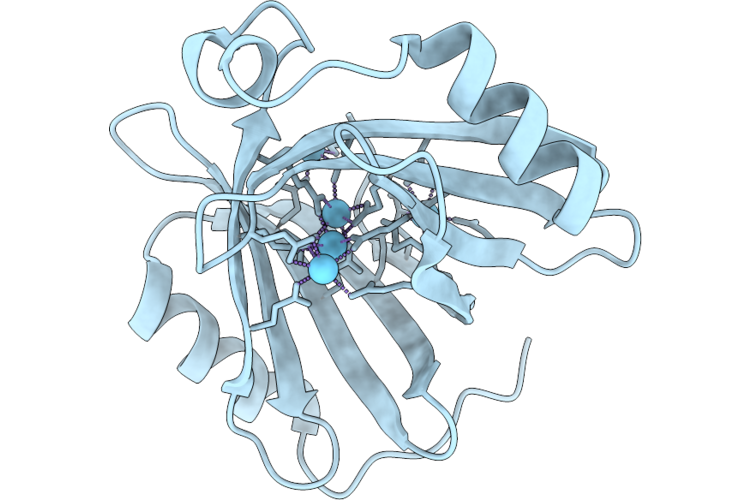 |
Structure Of Mif Binding With Lanthanum Ions
Organism: Methylobacillus flagellatus kt
Method: X-RAY DIFFRACTION Resolution:1.25 Å Release Date: 2026-07-22 Classification: METAL BINDING PROTEIN Ligands: LA |
Organism: Methylobacillus flagellatus kt
Method: X-RAY DIFFRACTION
Release Date: 2026-07-22
Ligands: LA
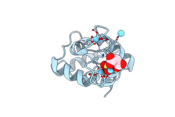 |
Crystal Structure Of Cod Allergen Gad M 1.0201 Coordinating Lanthanum
Organism: Gadus morhua
Method: X-RAY DIFFRACTION Resolution:1.65 Å Release Date: 2024-12-04 Classification: ALLERGEN Ligands: LA, MLI, CL, SO4 |
Organism: Gadus morhua
Method: X-RAY DIFFRACTION
Release Date: 2024-12-04
Ligands: LA, MLI, CL, SO4
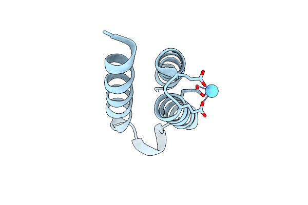 |
X-Ray Crystal Structure Of Methylorubrum Extorquens La(Iii)-Bound Land
Organism: Methylorubrum extorquens
Method: X-RAY DIFFRACTION Resolution:1.90 Å Release Date: 2024-10-16 Classification: METAL BINDING PROTEIN Ligands: LA |
Organism: Methylorubrum extorquens
Method: X-RAY DIFFRACTION
Release Date: 2024-10-16
Ligands: LA
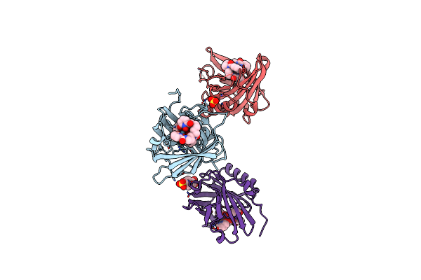 |
Fundamental Characterization Of Chelated And Crystallized Actinium In A Macromolecular Host
Organism: Homo sapiens
Method: X-RAY DIFFRACTION Resolution:2.00 Å Release Date: 2024-09-25 Classification: METAL BINDING PROTEIN Ligands: LA, 4OL, SO4, PIN |
Organism: Homo sapiens
Method: X-RAY DIFFRACTION
Release Date: 2024-09-25
Ligands: LA, 4OL, SO4, PIN
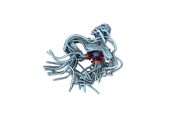 |
Lanthanide Binding Tag Complex Lbt3-Nh2:La3+
Organism: Homo sapiens
Method: SOLUTION NMR Release Date: 2024-07-17 Classification: DE NOVO PROTEIN Ligands: LA |
Organism: Homo sapiens
Method: SOLUTION NMR
Release Date: 2024-07-17
Ligands: LA
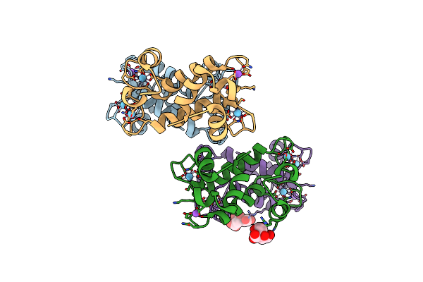 |
X-Ray Crystal Structure Of Hansschlegelia Quercus Lanmodulin (Lanm) With Lanthanum (Iii) Bound At Ph 7
Organism: Hansschlegelia quercus
Method: X-RAY DIFFRACTION Resolution:1.80 Å Release Date: 2023-06-07 Classification: METAL BINDING PROTEIN Ligands: LA, NA, CIT |
Organism: Hansschlegelia quercus
Method: X-RAY DIFFRACTION
Release Date: 2023-06-07
Ligands: LA, NA, CIT
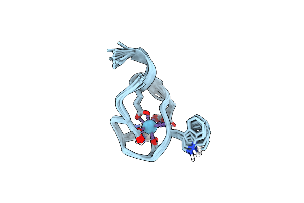 |
The Binding Structure Of A Lanthanide Binding Tag (Lbt3) With Lanthanum Ion (La3+)
Organism: Homo sapiens
Method: SOLUTION NMR Release Date: 2021-04-28 Classification: DE NOVO PROTEIN Ligands: LA |
Organism: Homo sapiens
Method: SOLUTION NMR
Release Date: 2021-04-28
Ligands: LA
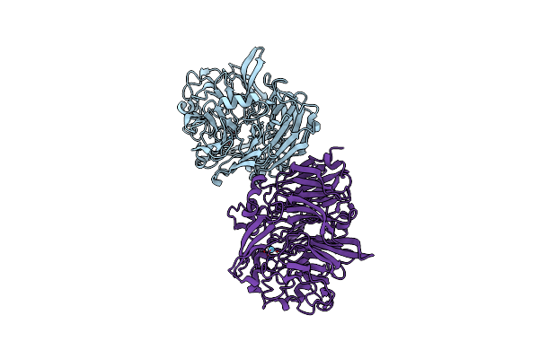 |
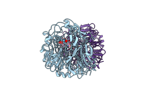
Lanthanide-Dependent Methanol Dehydrogenase Xoxf From Methylobacterium Extorquens, In Complex With Lanthanum
Organism: Methylorubrum extorquens am1
Method: X-RAY DIFFRACTION Resolution:2.80 Å Release Date: 2020-05-13 Classification: OXIDOREDUCTASE Ligands: LA |
Organism: Methylorubrum extorquens am1
Method: X-RAY DIFFRACTION
Release Date: 2020-05-13
Ligands: LA
 |
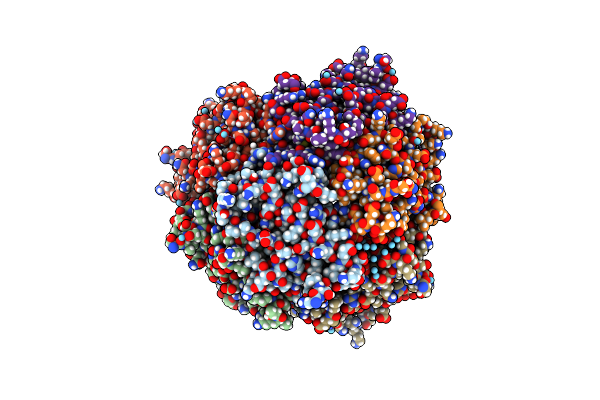
Lanthanide-Dependent Methanol Dehydrogenase Xoxf From Methylobacterium Extorquens, In Complex With Lanthanum And Pyrroloquinoline Quinone
Organism: Methylorubrum extorquens am1
Method: X-RAY DIFFRACTION Resolution:2.89 Å Release Date: 2020-05-13 Classification: OXIDOREDUCTASE Ligands: LA, PQQ |
Organism: Methylorubrum extorquens am1
Method: X-RAY DIFFRACTION
Release Date: 2020-05-13
Ligands: LA, PQQ
 |
The Structure Of Dps From Listeria Innocua Soaked Before Soaking Experiments With Zn, Co And La
Organism: Listeria innocua serovar 6a (strain atcc baa-680 / clip 11262)
Method: X-RAY DIFFRACTION Resolution:1.90 Å Release Date: 2019-09-04 Classification: METAL BINDING PROTEIN Ligands: LA |
Organism: Listeria innocua serovar 6a (strain atcc baa-680 / clip 11262)
Method: X-RAY DIFFRACTION
Release Date: 2019-09-04
Ligands: LA
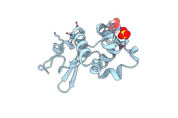 |
Crystal Structure Of Lanthanum Ion (La3+) Bound Bovine Alpha-Lactalbumin
Organism: Bos taurus
Method: X-RAY DIFFRACTION Resolution:1.85 Å Release Date: 2019-02-20 Classification: METAL BINDING PROTEIN Ligands: SO4, GOL, LA |
Organism: Bos taurus
Method: X-RAY DIFFRACTION
Release Date: 2019-02-20
Ligands: SO4, GOL, LA
 |
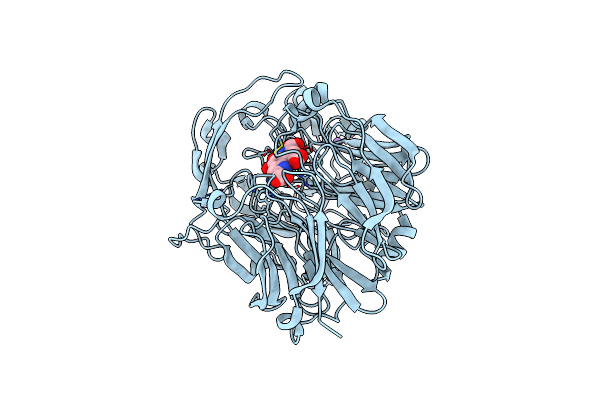
Crystal Structure Of Ttha0139 L34A With Lanthanum From Thermus Thermophilus Hb8
Organism: Thermus thermophilus (strain hb8 / atcc 27634 / dsm 579)
Method: X-RAY DIFFRACTION Resolution:2.39 Å Release Date: 2018-11-21 Classification: DNA BINDING PROTEIN Ligands: LA |
Organism: Thermus thermophilus (strain hb8 / atcc 27634 / dsm 579)
Method: X-RAY DIFFRACTION
Release Date: 2018-11-21
Ligands: LA
 |
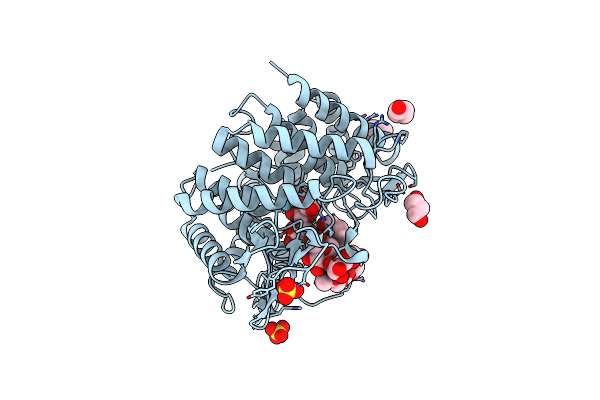
Crystal Structure Of Lanthanide-Dependent Methanol Dehydrogenase Xoxf From Methylomicrobium Buryatense 5G
Organism: Methylomicrobium buryatense 5g
Method: X-RAY DIFFRACTION Resolution:1.85 Å Release Date: 2018-09-05 Classification: OXIDOREDUCTASE Ligands: LA, PQQ, NA |
Organism: Methylomicrobium buryatense 5g
Method: X-RAY DIFFRACTION
Release Date: 2018-09-05
Ligands: LA, PQQ, NA
 |
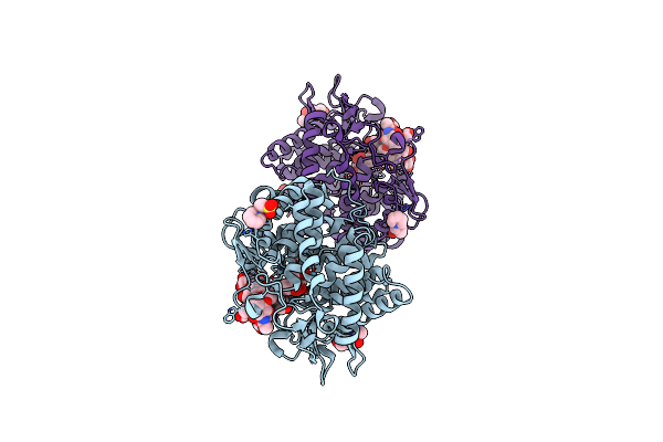
Crystal Structure Of The Class I Human Endoplasmic Reticulum 1,2-Alpha-Mannosidase And Man9Glcnac2-Pa Complex
Organism: Homo sapiens
Method: X-RAY DIFFRACTION Resolution:1.65 Å Release Date: 2017-05-10 Classification: HYDROLASE Ligands: LA, SO4, BU1, 1PS |
Organism: Homo sapiens
Method: X-RAY DIFFRACTION
Release Date: 2017-05-10
Ligands: LA, SO4, BU1, 1PS
 |
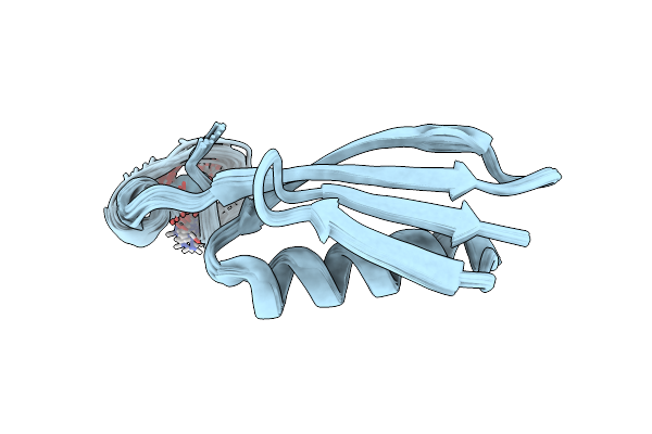
Structure Of Mouse Golgi Alpha-1,2-Mannosidase Ia And Man9Glcnac2-Pa Complex
Organism: Mus musculus
Method: X-RAY DIFFRACTION Resolution:1.77 Å Release Date: 2016-12-07 Classification: SUGAR BINDING PROTEIN Ligands: NAG, LA, 1PS, BU1 |
Organism: Mus musculus
Method: X-RAY DIFFRACTION
Release Date: 2016-12-07
Ligands: NAG, LA, 1PS, BU1
 |
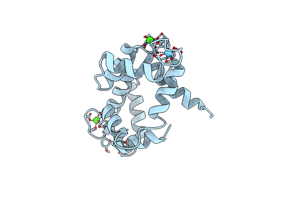
Solution Structure Of Gb1 With Lbt Probe
Organism: Streptococcus sp. group g
Method: SOLUTION NMR Release Date: 2009-09-15 Classification: IMMUNE SYSTEM Ligands: LA |
Organism: Streptococcus sp. group g
Method: SOLUTION NMR
Release Date: 2009-09-15
Ligands: LA
 |
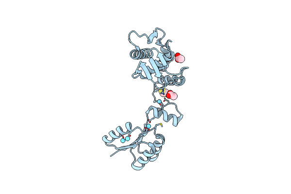
Solution Structure Of Cam Complexed To Drp1P
Organism: Homo sapiens
Method: SOLUTION NMR Release Date: 2009-03-10 Classification: METAL BINDING PROTEIN Ligands: CA, LA |
Organism: Homo sapiens
Method: SOLUTION NMR
Release Date: 2009-03-10
Ligands: CA, LA
 |
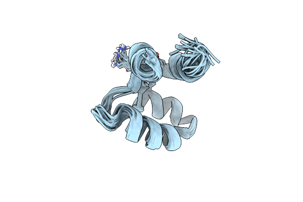
The Structure Of The Response Regulator Regx3 From Mycobacterium Tuberculosis
Organism: Mycobacterium tuberculosis h37rv
Method: X-RAY DIFFRACTION Resolution:2.03 Å Release Date: 2007-10-16 Classification: TRANSCRIPTION,SIGNALING PROTEIN Ligands: ACT, LA, BME |
Organism: Mycobacterium tuberculosis h37rv
Method: X-RAY DIFFRACTION
Release Date: 2007-10-16
Ligands: ACT, LA, BME
 |
The Refined Structure Of C-Terminal Domain Of An Ef-Hand Calcium Binding Protein From Entamoeba Histolytica
Organism: Entamoeba histolytica
Method: SOLUTION NMR Release Date: 2006-08-22 Classification: METAL BINDING PROTEIN Ligands: LA |
Organism: Entamoeba histolytica
Method: SOLUTION NMR
Release Date: 2006-08-22
Ligands: LA
 |
Average Nmr Solution Structure Of Ca Ln Calbindin D9K
Organism: Bos taurus
Method: SOLUTION NMR Release Date: 2002-01-23 Classification: METAL BINDING PROTEIN Ligands: LA |
Organism: Bos taurus
Method: SOLUTION NMR
Release Date: 2002-01-23
Ligands: LA