
Search Count: 746
All
Selected
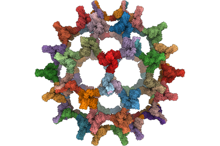 |
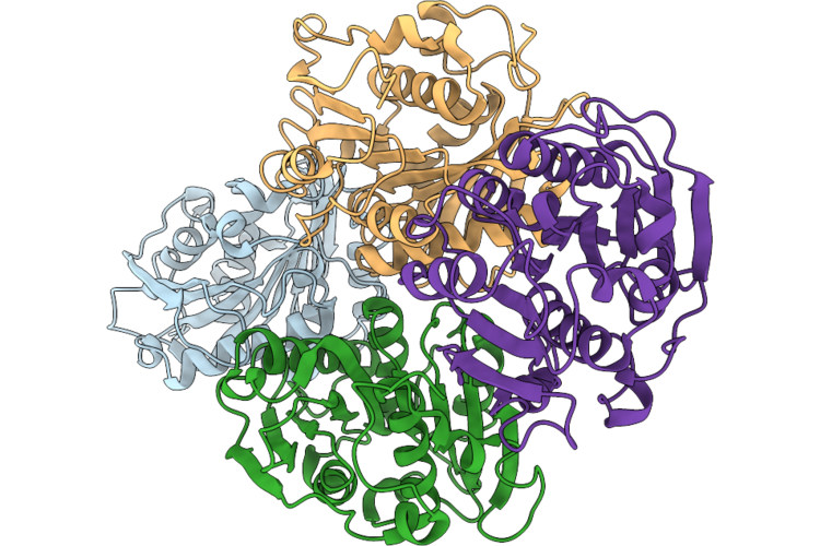
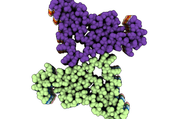
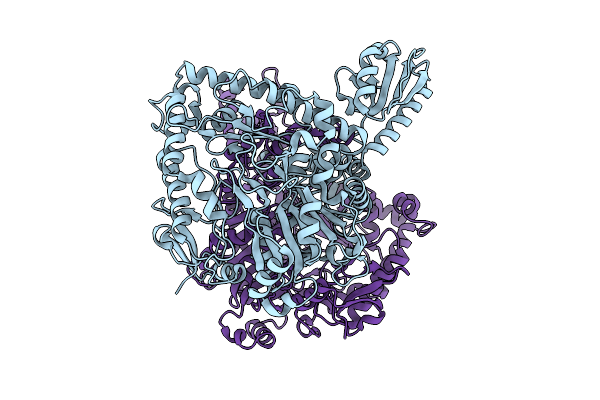
Two Component Protein Nano-Particle (T=3). De Novo Design, Computationally Relaxed Into Low Resolution Single Particle Cryoem Map With Icosahedral Symmetry Applied
Organism: Synthetic construct
Method: ELECTRON MICROSCOPY Release Date: 2026-05-20 Classification: DE NOVO PROTEIN |
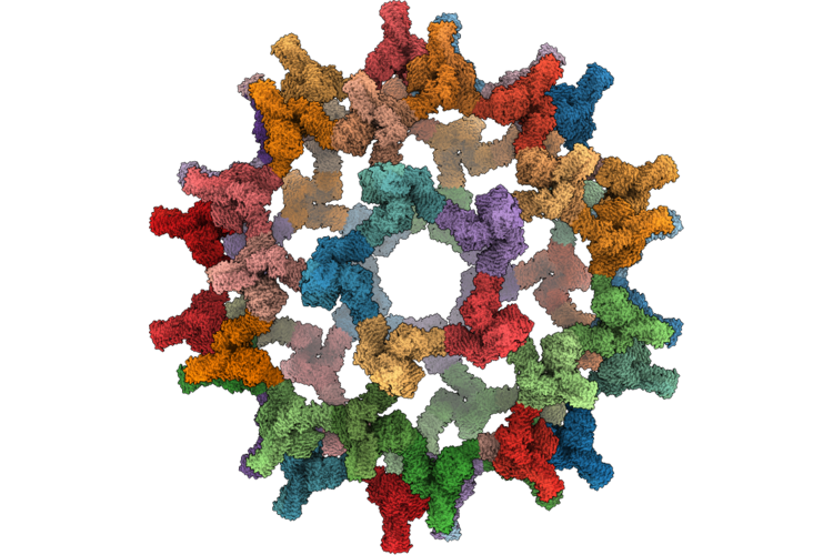 |
Two Component Protein Nano-Particle (T=3). De Novo Design, Computationally Relaxed Into Low Resolution Subtomogram Averaged Cryoem Map With Icosahedral Symmetry Applied
Organism: Synthetic construct
Method: ELECTRON MICROSCOPY Release Date: 2026-05-20 Classification: DE NOVO PROTEIN |
 |
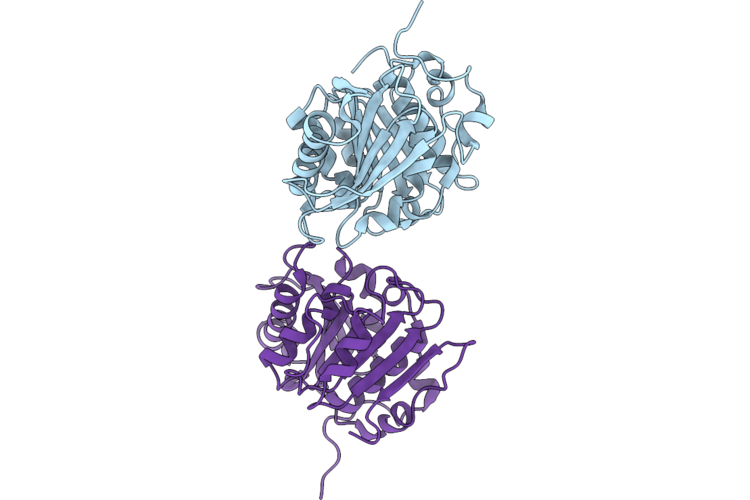
Organism: Cryptosporangium aurantiacum
Method: X-RAY DIFFRACTION Resolution:2.01 Å Release Date: 2026-04-29 Classification: HYDROLASE |
 |
Organism: Cryptosporangium aurantiacum
Method: X-RAY DIFFRACTION Resolution:1.63 Å Release Date: 2026-04-29 Classification: HYDROLASE |
 |
Organism: Cryptosporangium aurantiacum
Method: X-RAY DIFFRACTION Resolution:1.73 Å Release Date: 2026-04-29 Classification: HYDROLASE |
 |
Organism: Cryptosporangium aurantiacum
Method: X-RAY DIFFRACTION Resolution:1.66 Å Release Date: 2026-04-29 Classification: HYDROLASE |
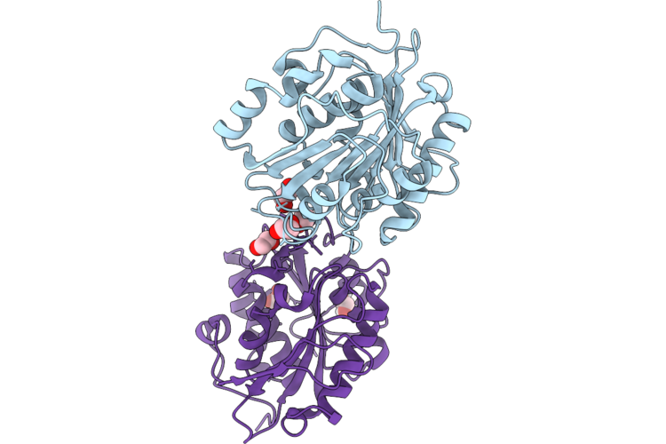 |
Organism: Cryptosporangium aurantiacum
Method: X-RAY DIFFRACTION Resolution:1.26 Å Release Date: 2026-04-29 Classification: HYDROLASE Ligands: EDO |
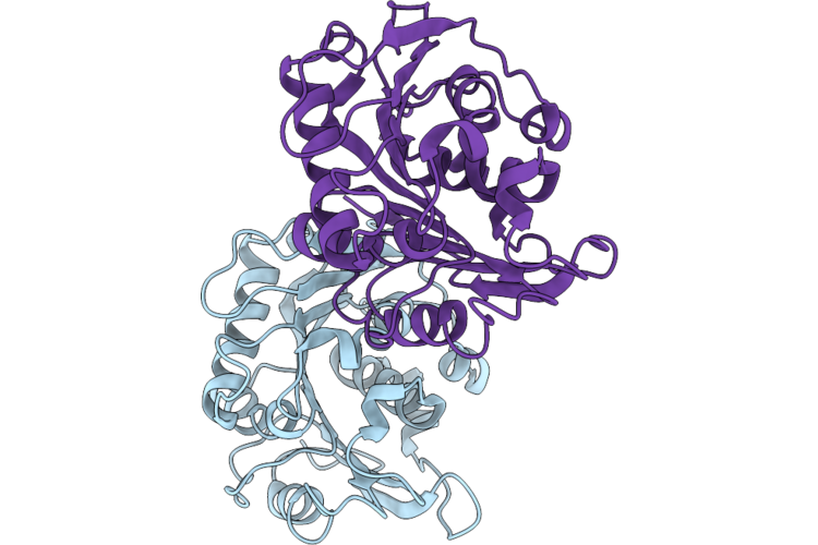 |
Organism: Cryptosporangium aurantiacum
Method: X-RAY DIFFRACTION Resolution:1.22 Å Release Date: 2026-04-29 Classification: HYDROLASE |
 |
Structure Of Amplified Asyn Filament By Using Seed Amplification Assay (Saa) From Msa Patient Csf.
Organism: Homo sapiens
Method: ELECTRON MICROSCOPY Resolution:3.18 Å Release Date: 2026-02-25 Classification: PROTEIN FIBRIL |
 |
Organism: Homo sapiens
Method: ELECTRON MICROSCOPY Resolution:2.75 Å Release Date: 2026-02-25 Classification: PROTEIN FIBRIL |
 |
Organism: Corynebacterium glutamicum
Method: ELECTRON MICROSCOPY Resolution:3.09 Å Release Date: 2026-02-18 Classification: TRANSPORT PROTEIN |
 |
Organism: Corynebacterium glutamicum
Method: ELECTRON MICROSCOPY Resolution:3.31 Å Release Date: 2026-02-18 Classification: TRANSPORT PROTEIN Ligands: PGT, CDL |
 |
Organism: Corynebacterium glutamicum
Method: ELECTRON MICROSCOPY Resolution:3.25 Å Release Date: 2026-02-18 Classification: TRANSPORT PROTEIN Ligands: PGT, BET |
 |
Organism: Corynebacterium glutamicum
Method: ELECTRON MICROSCOPY Resolution:3.57 Å Release Date: 2026-02-18 Classification: TRANSPORT PROTEIN Ligands: PGT, CDL |
 |
Organism: Corynebacterium glutamicum
Method: ELECTRON MICROSCOPY Resolution:3.33 Å Release Date: 2026-02-18 Classification: TRANSPORT PROTEIN Ligands: K |
 |
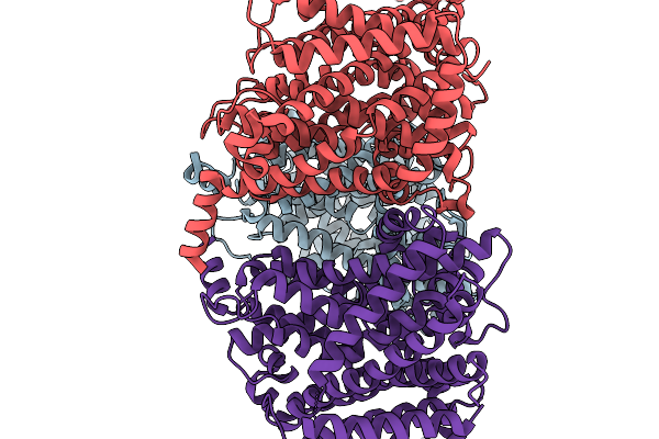
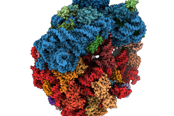
Cryo-Em Structure Of Skm-70S Ribosomal Stalled Complex In The Major State (Vacant A-Site, Canon)
Organism: Streptococcus sanguinis, Escherichia coli b
Method: ELECTRON MICROSCOPY Resolution:2.26 Å Release Date: 2026-02-18 Classification: RIBOSOME Ligands: ZN, A1JA7, MG |
 |
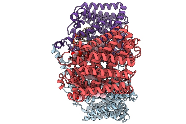
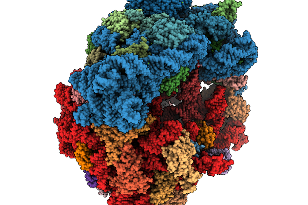
Cryo-Em Structure Of Skm-70S Ribosomal Stalled Complex In The A-Trna Positioned (Body Open) State.
Organism: Streptococcus sanguinis, Escherichia coli b
Method: ELECTRON MICROSCOPY Resolution:2.62 Å Release Date: 2026-02-18 Classification: RIBOSOME Ligands: ZN, A1JAI, MG |
 |
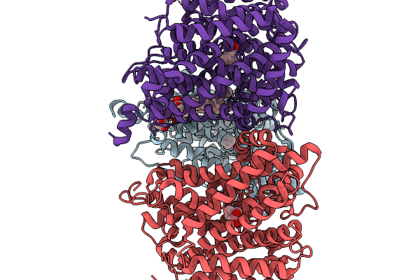
Cryo-Em Structure Of Skm-70S Ribosomal Stalled Complex In The Rotated State With Hybrid Trnas
Organism: Streptococcus sanguinis, Escherichia coli
Method: ELECTRON MICROSCOPY Resolution:2.60 Å Release Date: 2026-02-18 Classification: RIBOSOME Ligands: ZN, MG, A1JAI |
 |
Organism: Lacticaseibacillus rhamnosus
Method: X-RAY DIFFRACTION Resolution:2.09 Å Release Date: 2025-12-24 Classification: LYASE |
 |
Organism: Lacticaseibacillus rhamnosus
Method: X-RAY DIFFRACTION Resolution:2.64 Å Release Date: 2025-12-24 Classification: LYASE |