

Search Count: 136
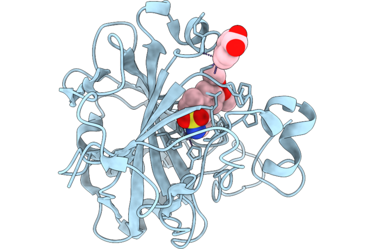 |
Crystal Structure Of Hca Ii In Complex With 1-Aryl-6,7-Dimethoxy-3,4-Dihydroisoquinoline-2(1H)-Sulfonamide Inhibitor
Organism: Homo sapiens
Method: X-RAY DIFFRACTION Resolution:1.05 Å Release Date: 2026-08-05 Classification: LYASE Ligands: ZN, MBO, A1J8Z |
Organism: Homo sapiens
Method: X-RAY DIFFRACTION
Release Date: 2026-08-05
Ligands: ZN, MBO, A1J8Z
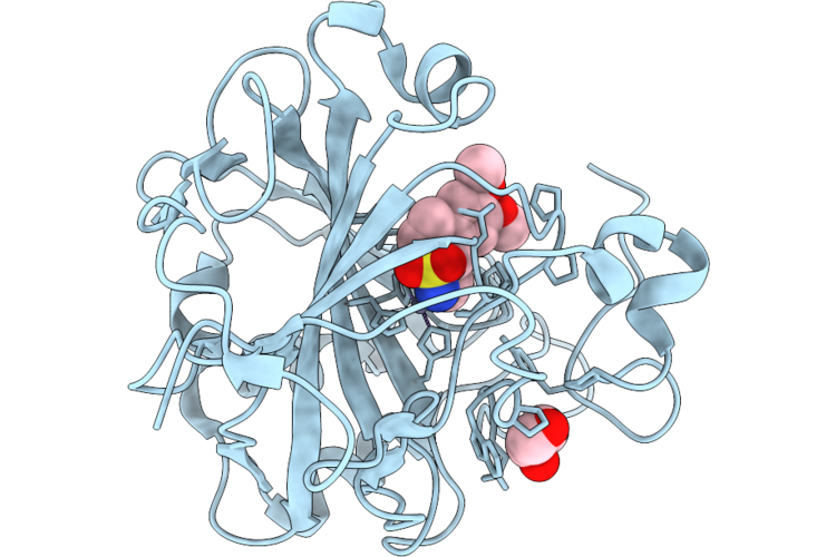 |
Crystal Structure Of Hca Ii In Complex With An 1-Aryl-6,7-Dimethoxy-3,4-Dihydroisoquinoline-2(1H)-Sulfonamide Inhibitor
Organism: Homo sapiens
Method: X-RAY DIFFRACTION Resolution:1.15 Å Release Date: 2026-08-05 Classification: LYASE Ligands: ZN, A1J8Y, GOL |
Organism: Homo sapiens
Method: X-RAY DIFFRACTION
Release Date: 2026-08-05
Ligands: ZN, A1J8Y, GOL
 |
Structure Of Ptp1B Complexed With Difluoromethylphosphonate Inhibitor Compound 2
Organism: Homo sapiens
Method: X-RAY DIFFRACTION Resolution:1.73 Å Release Date: 2026-05-27 Classification: HYDROLASE/INHIBITOR Ligands: A1C3A |
Organism: Homo sapiens
Method: X-RAY DIFFRACTION
Release Date: 2026-05-27
Ligands: A1C3A
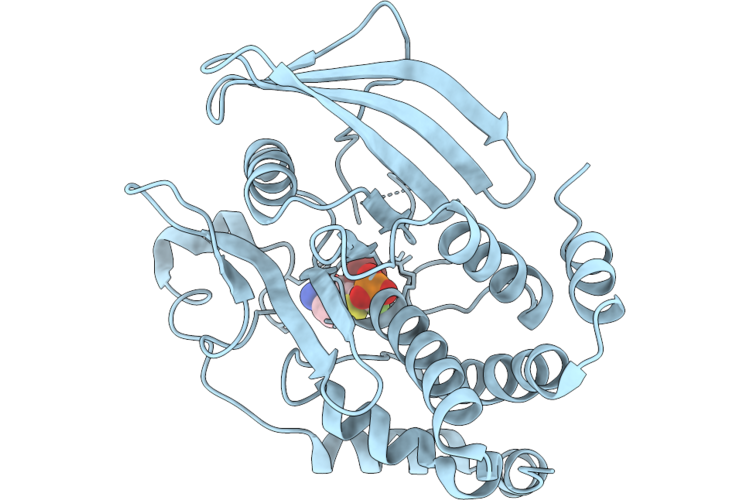 |
Structure Of Ptp1B Complexed With Difluoromethylphosphonate Inhibitor Compound 10
Organism: Homo sapiens
Method: X-RAY DIFFRACTION Resolution:2.51 Å Release Date: 2026-05-27 Classification: HYDROLASE/INHIBITOR Ligands: A1C3B |
Organism: Homo sapiens
Method: X-RAY DIFFRACTION
Release Date: 2026-05-27
Ligands: A1C3B
 |
Structure Of Ptp1B Complexed With Difluoromethylphosphonate Inhibitor Compound 15
Organism: Homo sapiens
Method: X-RAY DIFFRACTION Resolution:2.15 Å Release Date: 2026-05-27 Classification: HYDROLASE/INHIBITOR Ligands: A1C3C, DMS |
Organism: Homo sapiens
Method: X-RAY DIFFRACTION
Release Date: 2026-05-27
Ligands: A1C3C, DMS
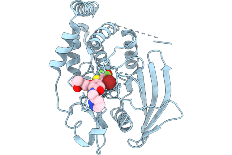 |
Structure Of Ptp1B Complexed With Difluoromethylphosphonate Inhibitor Compound 30
Organism: Homo sapiens
Method: X-RAY DIFFRACTION Resolution:1.94 Å Release Date: 2026-05-27 Classification: HYDROLASE/INHIBITOR Ligands: A1C3D |
Organism: Homo sapiens
Method: X-RAY DIFFRACTION
Release Date: 2026-05-27
Ligands: A1C3D
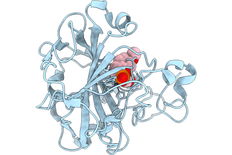 |
Crystal Structure Of A Phosphocoumarin Derivative In Complex With Human Carbonic Anhydrase Ii
Organism: Homo sapiens
Method: X-RAY DIFFRACTION Resolution:1.65 Å Release Date: 2026-05-13 Classification: LYASE Ligands: ZN, A1JX9, GOL |
Organism: Homo sapiens
Method: X-RAY DIFFRACTION
Release Date: 2026-05-13
Ligands: ZN, A1JX9, GOL
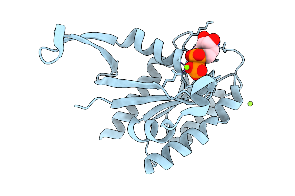 |
Crystal Structure Of Gdp-Bound Kras G12D/I55E: Suppressing G12D Oncogenicity Via Second-Site I55E Mutation
Organism: Homo sapiens
Method: X-RAY DIFFRACTION Resolution:1.23 Å Release Date: 2026-03-25 Classification: ONCOPROTEIN Ligands: GDP, MG, CL |
Organism: Homo sapiens
Method: X-RAY DIFFRACTION
Release Date: 2026-03-25
Ligands: GDP, MG, CL
 |
Crystal Structure Of The Doc Domain Of The E3 Ligase Hectd3
Organism: Homo sapiens
Method: X-RAY DIFFRACTION Resolution:1.61 Å Release Date: 2026-01-28 Classification: LIGASE Ligands: MG |
Organism: Homo sapiens
Method: X-RAY DIFFRACTION
Release Date: 2026-01-28
Ligands: MG
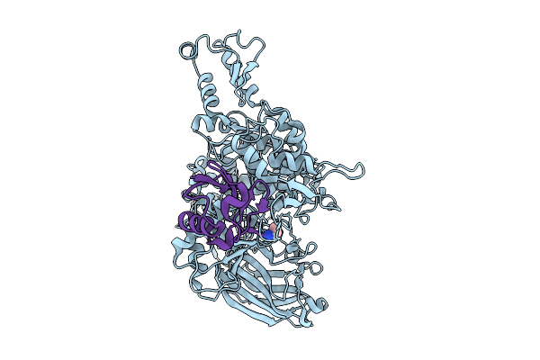 |
Cryo-Em Structure Of The E3 Ligase Hectd3 Conjugated To Ubiquitin
Organism: Homo sapiens
Method: ELECTRON MICROSCOPY Release Date: 2026-01-28 Classification: LIGASE Ligands: AYE |
Organism: Homo sapiens
Method: ELECTRON MICROSCOPY
Release Date: 2026-01-28
Ligands: AYE
 |
Cryo-Em Structure Of The E3 Ligase Hectd3
|
Organism: Homo sapiens
Method: ELECTRON MICROSCOPY
Release Date: 2026-01-28
 |
Cryo-Em Structure Of The E3 Ligase Hectd3
|
Organism: Homo sapiens
Method: ELECTRON MICROSCOPY
Release Date: 2026-01-28
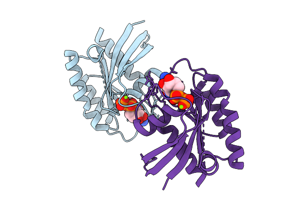 |
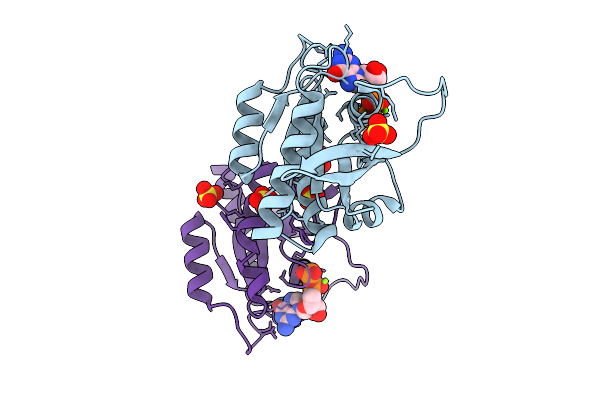
Crystal Structure Of Gdp-Bound Kras G12D/M67R: Suppressing G12D Oncogenicity Via Second-Site M67R Mutation
Organism: Homo sapiens
Method: X-RAY DIFFRACTION Resolution:1.70 Å Release Date: 2025-10-08 Classification: ONCOPROTEIN Ligands: GDP, MG |
Organism: Homo sapiens
Method: X-RAY DIFFRACTION
Release Date: 2025-10-08
Ligands: GDP, MG
 |
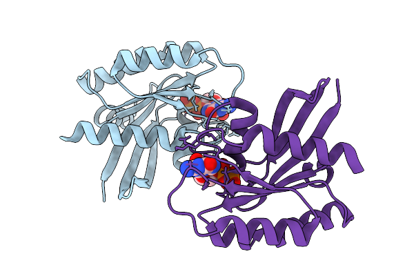
Crystal Structure Of Gdp-Bound Kras G12D/F28K: Suppressing G12D Oncogenicity Via Second-Site F28K Mutation
Organism: Homo sapiens
Method: X-RAY DIFFRACTION Resolution:1.74 Å Release Date: 2025-10-08 Classification: ONCOPROTEIN Ligands: GDP, MG, SO4 |
Organism: Homo sapiens
Method: X-RAY DIFFRACTION
Release Date: 2025-10-08
Ligands: GDP, MG, SO4
 |
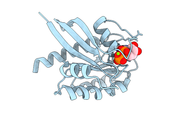
Crystal Structure Of Gdp-Bound Kras G12D/P34R: Suppressing G12D Oncogenicity Via Second-Site P34R Mutation
Organism: Homo sapiens
Method: X-RAY DIFFRACTION Resolution:1.50 Å Release Date: 2025-10-08 Classification: ONCOPROTEIN Ligands: MG, GDP |
Organism: Homo sapiens
Method: X-RAY DIFFRACTION
Release Date: 2025-10-08
Ligands: MG, GDP
 |
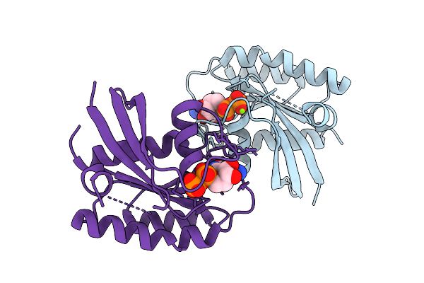
Crystal Structure Of Gdp-Bound Kras G12D/R41Q: Suppressing G12D Oncogenicity Via Second-Site R41Q Mutation
Organism: Homo sapiens
Method: X-RAY DIFFRACTION Resolution:1.22 Å Release Date: 2025-10-08 Classification: ONCOPROTEIN Ligands: GDP, MG |
Organism: Homo sapiens
Method: X-RAY DIFFRACTION
Release Date: 2025-10-08
Ligands: GDP, MG
 |
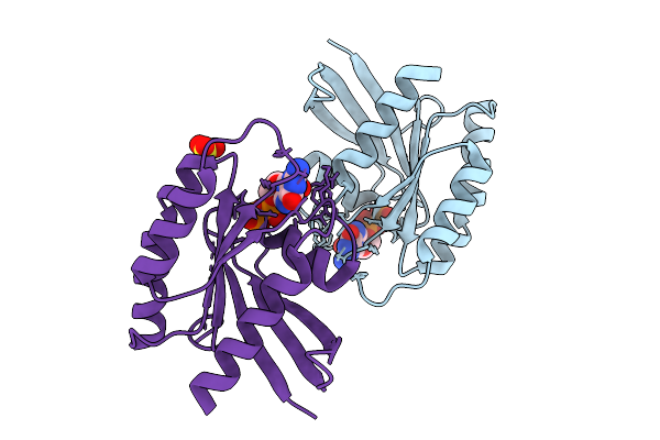
Crystal Structure Of Gdp-Bound Kras G12D/V45E: Suppressing G12D Oncogenicity Via Second-Site V45E Mutation
Organism: Homo sapiens
Method: X-RAY DIFFRACTION Resolution:2.20 Å Release Date: 2025-10-08 Classification: ONCOPROTEIN Ligands: GDP, MG |
Organism: Homo sapiens
Method: X-RAY DIFFRACTION
Release Date: 2025-10-08
Ligands: GDP, MG
 |
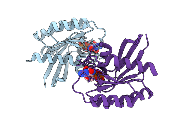
Crystal Structure Of Gdp-Bound Kras G12D/D54R: Suppressing G12D Oncogenicity Via Second-Site D54R Mutation
Organism: Homo sapiens
Method: X-RAY DIFFRACTION Resolution:2.51 Å Release Date: 2025-10-08 Classification: ONCOPROTEIN Ligands: GDP, MG, SO4 |
Organism: Homo sapiens
Method: X-RAY DIFFRACTION
Release Date: 2025-10-08
Ligands: GDP, MG, SO4
 |
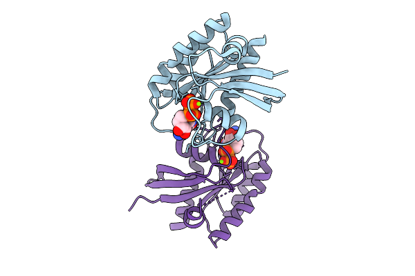
Crystal Structure Of Gdp-Bound Kras G12D/G60R: Suppressing G12D Oncogenicity Via Second-Site G60R Mutation
Organism: Homo sapiens
Method: X-RAY DIFFRACTION Resolution:1.80 Å Release Date: 2025-10-08 Classification: ONCOPROTEIN Ligands: MG, GDP |
Organism: Homo sapiens
Method: X-RAY DIFFRACTION
Release Date: 2025-10-08
Ligands: MG, GDP
 |
Crystal Structure Of Gdp-Bound Kras G12D/V103Y: Suppressing G12D Oncogenicity Via Second-Site V103Y Mutation
Organism: Homo sapiens
Method: X-RAY DIFFRACTION Resolution:1.80 Å Release Date: 2025-10-08 Classification: ONCOPROTEIN Ligands: MG, GDP |
Organism: Homo sapiens
Method: X-RAY DIFFRACTION
Release Date: 2025-10-08
Ligands: MG, GDP