

Search Count: 88
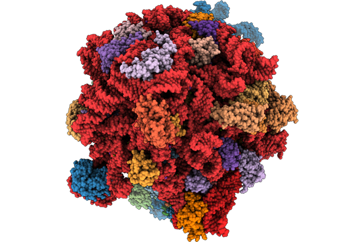 |
Doxycycline Bound E. Coli Ribosome With Rearranged Peptidyl Transferase Centre
Organism: Escherichia coli bl21(de3)
Method: ELECTRON MICROSCOPY Release Date: 2026-04-22 Classification: RIBOSOME Ligands: ZN, MG, K, DXT |
Organism: Escherichia coli bl21(de3)
Method: ELECTRON MICROSCOPY
Release Date: 2026-04-22
Ligands: ZN, MG, K, DXT
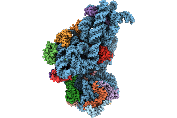 |
30S Doxycycline Bound E. Coli Ribosome
Organism: Escherichia coli bl21(de3)
Method: ELECTRON MICROSCOPY Release Date: 2026-04-22 Classification: RIBOSOME Ligands: ZN, DXT, MG, K |
Organism: Escherichia coli bl21(de3)
Method: ELECTRON MICROSCOPY
Release Date: 2026-04-22
Ligands: ZN, DXT, MG, K
 |
Hpfcold Bound Hibernating C. Burnetii 30S Ribosome
Organism: Coxiella burnetii
Method: ELECTRON MICROSCOPY Resolution:2.87 Å Release Date: 2026-04-22 Classification: RIBOSOME Ligands: ZN, K, MG |
Organism: Coxiella burnetii
Method: ELECTRON MICROSCOPY
Release Date: 2026-04-22
Ligands: ZN, K, MG
 |
Doxycycline Bound C. Burnetii 30S Ribosome
Organism: Coxiella burnetii
Method: ELECTRON MICROSCOPY Release Date: 2026-04-22 Classification: RIBOSOME Ligands: DXT, K, MG, ZN |
Organism: Coxiella burnetii
Method: ELECTRON MICROSCOPY
Release Date: 2026-04-22
Ligands: DXT, K, MG, ZN
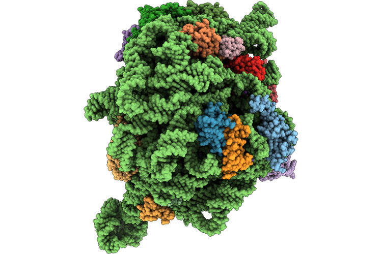 |
Triple Stack Doxycycline Bound 50S Subunit Of The Coxiella Burnetii Ribosome
Organism: Coxiella burnetii
Method: ELECTRON MICROSCOPY Release Date: 2026-04-22 Classification: ANTIBIOTIC Ligands: ZN, MG, K, DXT |
Organism: Coxiella burnetii
Method: ELECTRON MICROSCOPY
Release Date: 2026-04-22
Ligands: ZN, MG, K, DXT
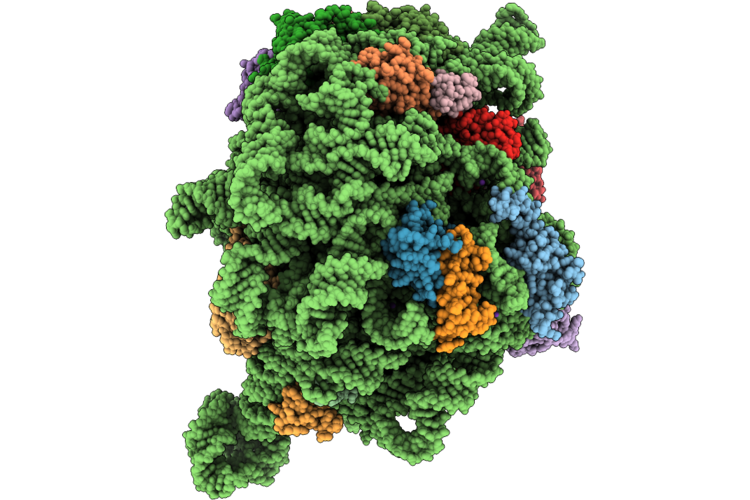 |
Empty 50S Subunit Of The Coxiella Burnetii Ribosome
Organism: Coxiella burnetii
Method: ELECTRON MICROSCOPY Release Date: 2026-04-22 Classification: RIBOSOME Ligands: ZN, MG, K |
Organism: Coxiella burnetii
Method: ELECTRON MICROSCOPY
Release Date: 2026-04-22
Ligands: ZN, MG, K
 |
70S Coxiella Burnetii Ribosome With Doxycycline And Hpfcold
Organism: Coxiella burnetii
Method: ELECTRON MICROSCOPY Release Date: 2026-04-22 Classification: RIBOSOME Ligands: MG, K, ZN, DXT |
Organism: Coxiella burnetii
Method: ELECTRON MICROSCOPY
Release Date: 2026-04-22
Ligands: MG, K, ZN, DXT
 |
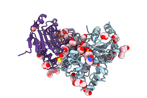
Crystal Structure Of Oxa-48 Apoenzyme
Organism: Klebsiella pneumoniae
Method: X-RAY DIFFRACTION Resolution:1.38 Å Release Date: 2025-09-03 Classification: ANTIMICROBIAL PROTEIN Ligands: CL, GOL |
Organism: Klebsiella pneumoniae
Method: X-RAY DIFFRACTION
Release Date: 2025-09-03
Ligands: CL, GOL
 |
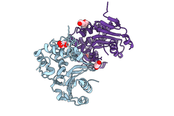
Crystal Structure Of Oxa-48 In Complex With Nacubactam
Organism: Klebsiella pneumoniae
Method: X-RAY DIFFRACTION Resolution:1.51 Å Release Date: 2025-09-03 Classification: ANTIMICROBIAL PROTEIN Ligands: CL, PGE, PEG, EDO, OP0, DMS, GOL, PG4 |
Organism: Klebsiella pneumoniae
Method: X-RAY DIFFRACTION
Release Date: 2025-09-03
Ligands: CL, PGE, PEG, EDO, OP0, DMS, GOL, PG4
 |
Crystal Structure Of Oxa-163 Apoenzyme
Organism: Enterobacter cloacae
Method: X-RAY DIFFRACTION Resolution:1.44 Å Release Date: 2025-09-03 Classification: ANTIMICROBIAL PROTEIN Ligands: CL, GOL, EDO, PEG |
Organism: Enterobacter cloacae
Method: X-RAY DIFFRACTION
Release Date: 2025-09-03
Ligands: CL, GOL, EDO, PEG
 |
Crystal Structure Of Oxa-163 In Complex With Avibactam
Organism: Enterobacter cloacae
Method: X-RAY DIFFRACTION Resolution:1.56 Å Release Date: 2025-09-03 Classification: ANTIMICROBIAL PROTEIN Ligands: NXL, A1IY4, CL, EDO, 2PE |
Organism: Enterobacter cloacae
Method: X-RAY DIFFRACTION
Release Date: 2025-09-03
Ligands: NXL, A1IY4, CL, EDO, 2PE
 |
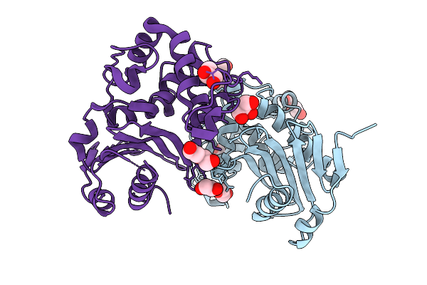
Crystal Structure Of Oxa-163 In Complex With Nacubactam
Organism: Enterobacter cloacae
Method: X-RAY DIFFRACTION Resolution:2.18 Å Release Date: 2025-09-03 Classification: ANTIMICROBIAL PROTEIN Ligands: OP0, EDO, GOL, PEG, CL, DMS |
Organism: Enterobacter cloacae
Method: X-RAY DIFFRACTION
Release Date: 2025-09-03
Ligands: OP0, EDO, GOL, PEG, CL, DMS
 |
Crystal Structure Of Oxa-405 Apoenzyme
Organism: Serratia marcescens
Method: X-RAY DIFFRACTION Resolution:1.56 Å Release Date: 2025-09-03 Classification: ANTIMICROBIAL PROTEIN Ligands: PEG, CL, GOL, PG4, EDO |
Organism: Serratia marcescens
Method: X-RAY DIFFRACTION
Release Date: 2025-09-03
Ligands: PEG, CL, GOL, PG4, EDO
 |
Crystal Structure Of Oxa-405 In Complex With Avibactam
Organism: Serratia marcescens
Method: X-RAY DIFFRACTION Resolution:1.46 Å Release Date: 2025-09-03 Classification: ANTIMICROBIAL PROTEIN Ligands: GOL, EDO, NXL, A1IY4, CL, PGE, PEG |
Organism: Serratia marcescens
Method: X-RAY DIFFRACTION
Release Date: 2025-09-03
Ligands: GOL, EDO, NXL, A1IY4, CL, PGE, PEG
 |
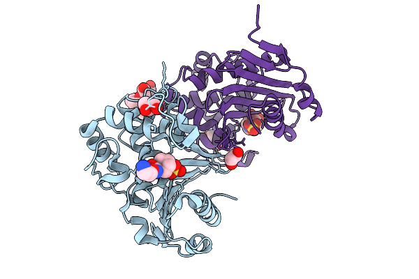
Crystal Structure Of Oxa-405 In Complex With Nacubactam
Organism: Serratia marcescens
Method: X-RAY DIFFRACTION Resolution:1.33 Å Release Date: 2025-09-03 Classification: ANTIMICROBIAL PROTEIN Ligands: OP0, PEG, EDO, A1IYS, CL, 2PE |
Organism: Serratia marcescens
Method: X-RAY DIFFRACTION
Release Date: 2025-09-03
Ligands: OP0, PEG, EDO, A1IYS, CL, 2PE
 |
Crystal Structure Of Oxa-163 In Complex With Nacubactam (16 Hour Soak)
Organism: Enterobacter cloacae
Method: X-RAY DIFFRACTION Resolution:1.41 Å Release Date: 2025-09-03 Classification: ANTIMICROBIAL PROTEIN Ligands: OP0, EDO, GOL, A1IYS, CL, 2PE |
Organism: Enterobacter cloacae
Method: X-RAY DIFFRACTION
Release Date: 2025-09-03
Ligands: OP0, EDO, GOL, A1IYS, CL, 2PE
 |
Crystal Structure Of The Coxiella Burnetii 2-Methylisocitrate Lyase
Organism: Coxiella burnetii
Method: X-RAY DIFFRACTION Resolution:1.60 Å Release Date: 2025-08-13 Classification: CYTOSOLIC PROTEIN Ligands: EPE, EDO, PEG, CL |
Organism: Coxiella burnetii
Method: X-RAY DIFFRACTION
Release Date: 2025-08-13
Ligands: EPE, EDO, PEG, CL
 |
Crystal Structure Of The Coxiella Burnetii E110Q Mutant 2-Methylisocitrate Lyase
Organism: Coxiella burnetii
Method: X-RAY DIFFRACTION Resolution:1.82 Å Release Date: 2025-08-13 Classification: CYTOSOLIC PROTEIN Ligands: EDO, CL |
Organism: Coxiella burnetii
Method: X-RAY DIFFRACTION
Release Date: 2025-08-13
Ligands: EDO, CL
 |
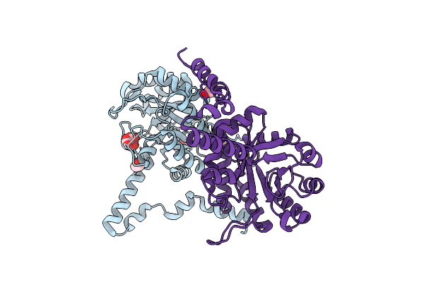
Crystal Structure Of The Coxiella Burnetii 2-Methylisocitrate Lyase Bound To Substrate 2-Mic
Organism: Coxiella burnetii
Method: X-RAY DIFFRACTION Resolution:1.90 Å Release Date: 2025-08-13 Classification: CYTOSOLIC PROTEIN Ligands: MIC, EOH, EDO, MG, CL |
Organism: Coxiella burnetii
Method: X-RAY DIFFRACTION
Release Date: 2025-08-13
Ligands: MIC, EOH, EDO, MG, CL
 |
Crystal Structure Of The Coxiella Burnetii R152Q Mutant 2-Methylisocitrate Lyase
Organism: Coxiella burnetii
Method: X-RAY DIFFRACTION Resolution:1.70 Å Release Date: 2025-08-13 Classification: CYTOSOLIC PROTEIN Ligands: CIT, EDO, CL |
Organism: Coxiella burnetii
Method: X-RAY DIFFRACTION
Release Date: 2025-08-13
Ligands: CIT, EDO, CL