

Search Count: 6,505
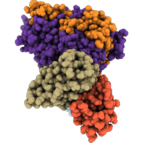 |
Cryo-Em Structure Of Mouse Myeloperoxidase In Complex With Fab Fragments Of Antibodies Mab-A24 And Mab-A46
Organism: Mus musculus
Method: ELECTRON MICROSCOPY Release Date: 2026-09-16 Classification: IMMUNE SYSTEM Ligands: HEM, NAG, CA |
Organism: Mus musculus
Method: ELECTRON MICROSCOPY
Release Date: 2026-09-16
Ligands: HEM, NAG, CA
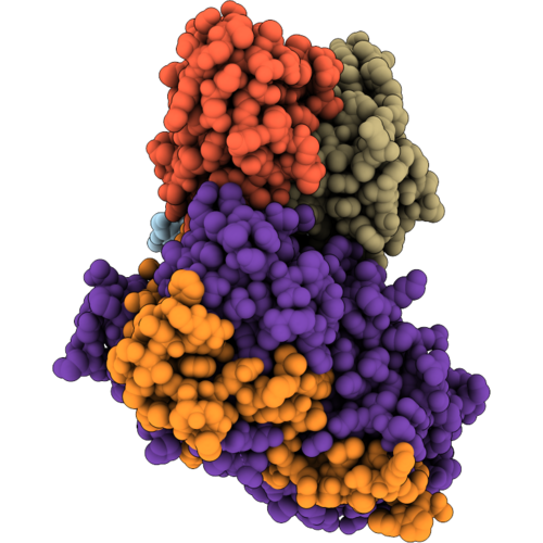 |
Cryo-Em Structure Of Mouse Myeloperoxidase In Complex With Fab Fragments Of Antibodies Mab-A46 And Mab-B88
Organism: Mus musculus
Method: ELECTRON MICROSCOPY Release Date: 2026-09-16 Classification: IMMUNE SYSTEM Ligands: HEM, NAG, CA |
Organism: Mus musculus
Method: ELECTRON MICROSCOPY
Release Date: 2026-09-16
Ligands: HEM, NAG, CA
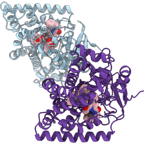 |
Crystal Structure Of Cyai In Complex With The Substrate Marinacarbolines E
Organism: Actinoalloteichus caeruleus dsm 43889
Method: X-RAY DIFFRACTION Resolution:1.50 Å Release Date: 2026-09-16 Classification: OXIDOREDUCTASE Ligands: G5I, HEM, NA |
Organism: Actinoalloteichus caeruleus dsm 43889
Method: X-RAY DIFFRACTION
Release Date: 2026-09-16
Ligands: G5I, HEM, NA
 |
Bacteroides Thetaiotaomicron Ccsba Mutant W703C
Organism: Bacteroides thetaiotaomicron vpi-5482
Method: ELECTRON MICROSCOPY Resolution:2.91 Å Release Date: 2026-09-09 Classification: MEMBRANE PROTEIN Ligands: HEM, PTY |
Organism: Bacteroides thetaiotaomicron vpi-5482
Method: ELECTRON MICROSCOPY
Release Date: 2026-09-09
Ligands: HEM, PTY
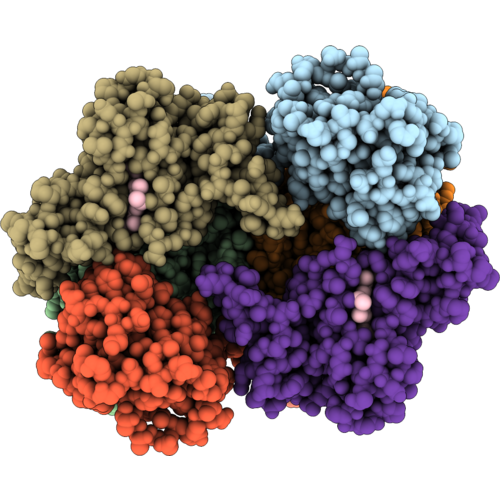 |
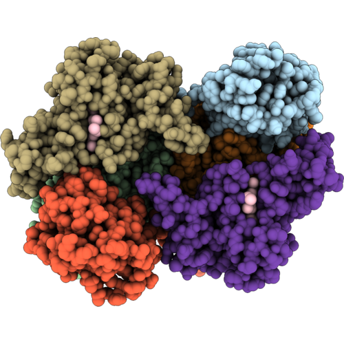
Cryo-Em Structure Of Rhodobacter Capsulatus Cytochrome Bc1 Dimer At 2.16 A Resolution With Water Molecules And Quinone In The Quinone Reduction Site
Organism: Rhodobacter capsulatus sb 1003
Method: ELECTRON MICROSCOPY Release Date: 2026-09-09 Classification: ELECTRON TRANSPORT Ligands: FES, UMQ, HEM, UQ1, HEC |
Organism: Rhodobacter capsulatus sb 1003
Method: ELECTRON MICROSCOPY
Release Date: 2026-09-09
Ligands: FES, UMQ, HEM, UQ1, HEC
 |
Cryo-Em Structure Of Rhodobacter Capsulatus Cytochrome Bc1 Dimer With One Rieske Protein In The C Position And One In The B Position And Quinone In The Quinone Oxidation Site
Organism: Rhodobacter capsulatus sb 1003
Method: ELECTRON MICROSCOPY Resolution:2.69 Å Release Date: 2026-09-09 Classification: ELECTRON TRANSPORT Ligands: FES, UMQ, HEM, UQ1, HEC, U10 |
Organism: Rhodobacter capsulatus sb 1003
Method: ELECTRON MICROSCOPY
Release Date: 2026-09-09
Ligands: FES, UMQ, HEM, UQ1, HEC, U10
 |
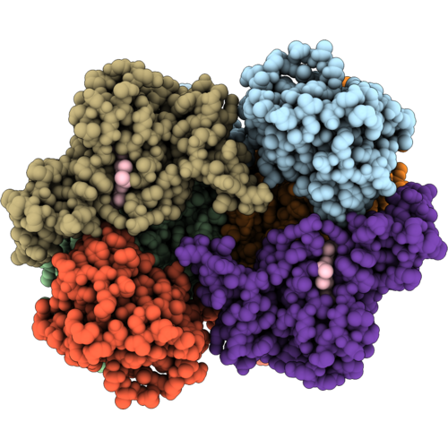
Cryo-Em Structure Of Rhodobacter Capsulatus Cytochrome Bc1 Dimer With Both Rieske Proteins In The B Position
Organism: Rhodobacter capsulatus sb 1003
Method: ELECTRON MICROSCOPY Release Date: 2026-09-09 Classification: ELECTRON TRANSPORT Ligands: FES, UMQ, HEM, UQ1, HEC |
Organism: Rhodobacter capsulatus sb 1003
Method: ELECTRON MICROSCOPY
Release Date: 2026-09-09
Ligands: FES, UMQ, HEM, UQ1, HEC
 |
Cryo-Em Structure Of Rhodobacter Capsulatus Cytochrome Bc1 Dimer With Both Rieske Proteins In The C Position
Organism: Rhodobacter capsulatus sb 1003
Method: ELECTRON MICROSCOPY Resolution:2.43 Å Release Date: 2026-09-09 Classification: ELECTRON TRANSPORT Ligands: FES, UMQ, HEM, UQ1, HEC |
Organism: Rhodobacter capsulatus sb 1003
Method: ELECTRON MICROSCOPY
Release Date: 2026-09-09
Ligands: FES, UMQ, HEM, UQ1, HEC
 |
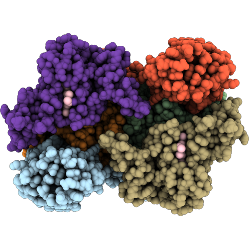
Cryo-Em Structure Of Rhodobacter Capsulatus Cytochrome Bc1 Dimer With One Rieske Protein In The C Position And One In An Intermediate Position
Organism: Rhodobacter capsulatus sb 1003
Method: ELECTRON MICROSCOPY Release Date: 2026-09-09 Classification: ELECTRON TRANSPORT Ligands: FES, UMQ, HEM, UQ1, HEC |
Organism: Rhodobacter capsulatus sb 1003
Method: ELECTRON MICROSCOPY
Release Date: 2026-09-09
Ligands: FES, UMQ, HEM, UQ1, HEC
 |
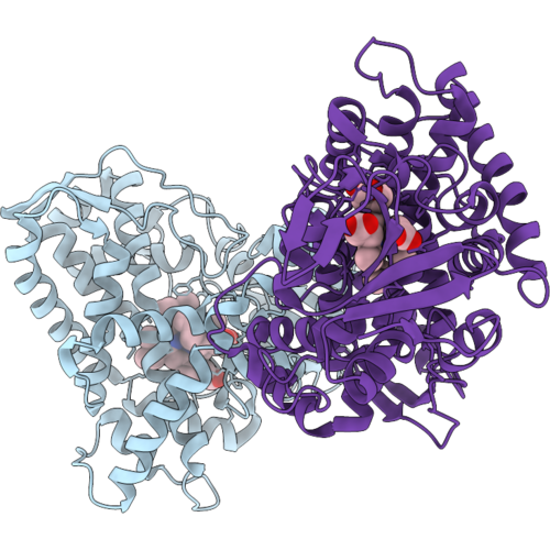
Cryo-Em Structure Of +2Ala Insertion Mutant Of Rhodobacter Capsulatus Cytochrome Bc1 Dimer At 2.41 A With Quinone In Quinone Oxidation And Reduction Sites
Organism: Rhodobacter capsulatus sb 1003
Method: ELECTRON MICROSCOPY Resolution:2.41 Å Release Date: 2026-09-09 Classification: ELECTRON TRANSPORT Ligands: FES, LMT, HEM, U10, UQ1, HEC |
Organism: Rhodobacter capsulatus sb 1003
Method: ELECTRON MICROSCOPY
Release Date: 2026-09-09
Ligands: FES, LMT, HEM, U10, UQ1, HEC
 |
Crystal Structure Of P450Bsbeta 5M Variant In Complex With Octanoic Acid
Organism: Bacillus subtilis (strain 168)
Method: X-RAY DIFFRACTION Resolution:2.30 Å Release Date: 2026-09-09 Classification: HYDROLASE Ligands: HEM, OCA |
Organism: Bacillus subtilis (strain 168)
Method: X-RAY DIFFRACTION
Release Date: 2026-09-09
Ligands: HEM, OCA
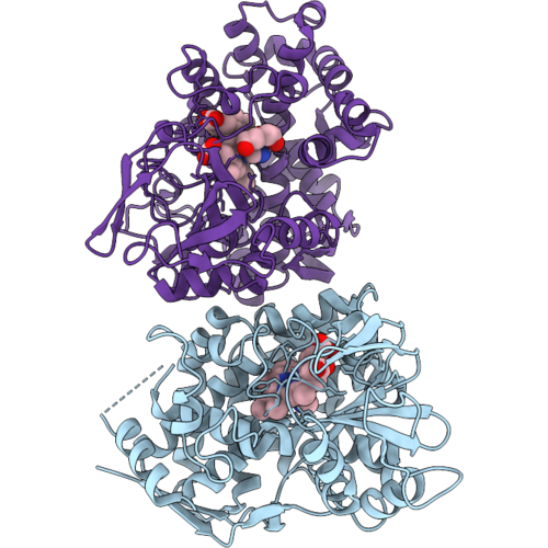 |
The Crystal Structure Of Ssbcmd
Organism: Streptomyces cinnamoneus
Method: X-RAY DIFFRACTION Resolution:1.91 Å Release Date: 2026-09-09 Classification: OXIDOREDUCTASE Ligands: HEM, IMD, PEG |
Organism: Streptomyces cinnamoneus
Method: X-RAY DIFFRACTION
Release Date: 2026-09-09
Ligands: HEM, IMD, PEG
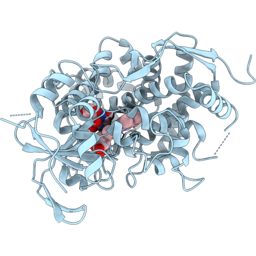 |
The Complex Structure Of Sobcmd And Its Substrate
Organism: Streptomyces ossamyceticus
Method: X-RAY DIFFRACTION Resolution:1.41 Å Release Date: 2026-09-09 Classification: OXIDOREDUCTASE Ligands: CL, HEM, 8KI |
Organism: Streptomyces ossamyceticus
Method: X-RAY DIFFRACTION
Release Date: 2026-09-09
Ligands: CL, HEM, 8KI
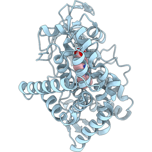 |
Cytochrome P450 158A2 (Cyp158A2) Variant P354L
Organism: Streptomyces coelicolor
Method: X-RAY DIFFRACTION Resolution:1.88 Å Release Date: 2026-09-09 Classification: OXIDOREDUCTASE Ligands: HEM |
Organism: Streptomyces coelicolor
Method: X-RAY DIFFRACTION
Release Date: 2026-09-09
Ligands: HEM
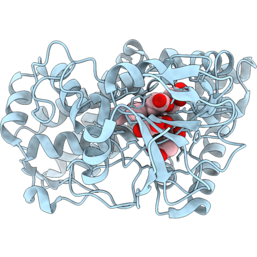 |
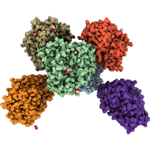
Cytochrome P450 158A2 (Cyp158A2) Variant P354L Bound To Substrate Flaviolin
Organism: Streptomyces coelicolor
Method: X-RAY DIFFRACTION Resolution:2.20 Å Release Date: 2026-09-09 Classification: OXIDOREDUCTASE Ligands: HEM, FLV |
Organism: Streptomyces coelicolor
Method: X-RAY DIFFRACTION
Release Date: 2026-09-09
Ligands: HEM, FLV
 |
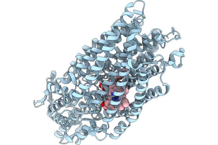
Olep Triple Mutant F84Q/S240A/V291G In Complex With Lca
Organism: Streptomyces antibioticus
Method: X-RAY DIFFRACTION Resolution:2.70 Å Release Date: 2026-09-02 Classification: OXIDOREDUCTASE Ligands: HEM, 4OA, NA, FMT |
Organism: Streptomyces antibioticus
Method: X-RAY DIFFRACTION
Release Date: 2026-09-02
Ligands: HEM, 4OA, NA, FMT
 |
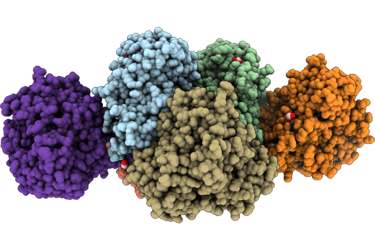
Cryo-Em Structure Of Native Monomeric Quinol-Dependent Nitric Oxide Reductase From Achromobacter Xylosoxidans.
Organism: Achromobacter xylosoxidans
Method: ELECTRON MICROSCOPY Release Date: 2026-08-26 Classification: MEMBRANE PROTEIN Ligands: CA, HEM, CU, FE |
Organism: Achromobacter xylosoxidans
Method: ELECTRON MICROSCOPY
Release Date: 2026-08-26
Ligands: CA, HEM, CU, FE
 |
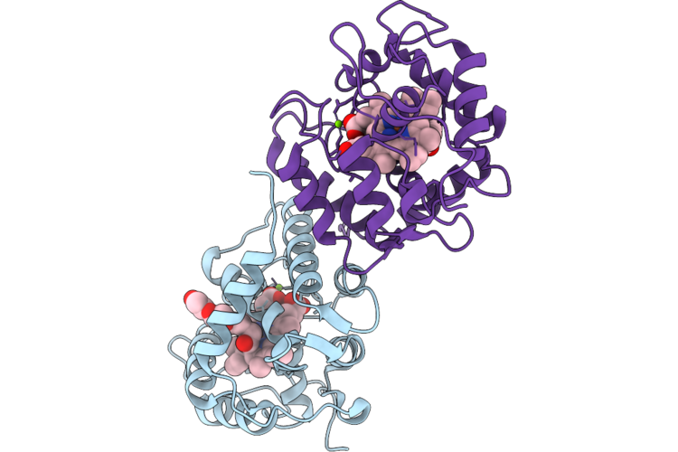
Olep Mutant F84Q In Complex With Lca
Organism: Streptomyces antibioticus
Method: X-RAY DIFFRACTION Resolution:2.78 Å Release Date: 2026-08-26 Classification: OXIDOREDUCTASE Ligands: HEM, 4OA, NA, FMT |
Organism: Streptomyces antibioticus
Method: X-RAY DIFFRACTION
Release Date: 2026-08-26
Ligands: HEM, 4OA, NA, FMT
 |
Unspecific Peroxygenase From Daldinia Sp. Ec12 In Complex With Pg4
Organism: Daldinia sp. ec12
Method: X-RAY DIFFRACTION Resolution:1.89 Å Release Date: 2026-08-26 Classification: OXIDOREDUCTASE Ligands: MG, PG4, HEM |
Organism: Daldinia sp. ec12
Method: X-RAY DIFFRACTION
Release Date: 2026-08-26
Ligands: MG, PG4, HEM
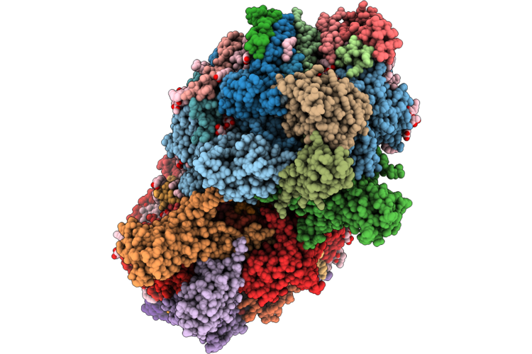 |
Rt Xfel Structure Of Dark-Stable State Of Photosystem Ii (0F, S1 Rich) At 1.97 Angstrom Obtained From Anomalous Data At 9.5 Kev
Organism: Thermosynechococcus vestitus bp-1
Method: X-RAY DIFFRACTION Resolution:1.97 Å Release Date: 2026-08-26 Classification: PHOTOSYNTHESIS Ligands: OEX, FE2, CL, CLA, PHO, BCR, PL9, STE, SQD, DGD, LMG, BCT, LHG, HEM, HEC |