

Search Count: 45
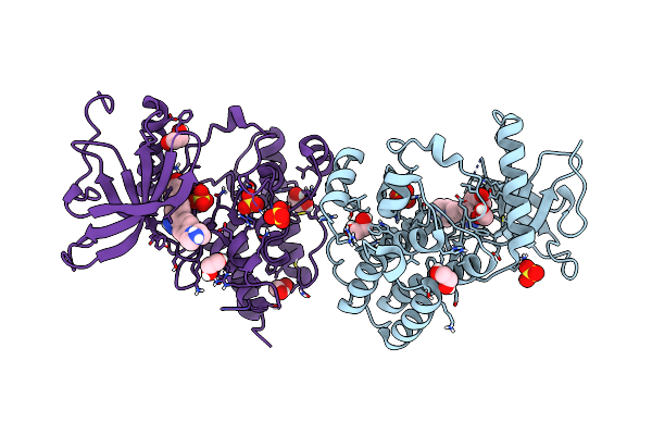 |
Fgfr1 Kinase Domain (Residues 458-765) With Mutations C488A, C584S In Complex With 38.
Organism: Homo sapiens
Method: X-RAY DIFFRACTION Resolution:1.71 Å Release Date: 2021-12-01 Classification: TRANSFERASE Ligands: 47I, SO4, EDO |
Organism: Homo sapiens
Method: X-RAY DIFFRACTION
Release Date: 2021-12-01
Ligands: 47I, SO4, EDO
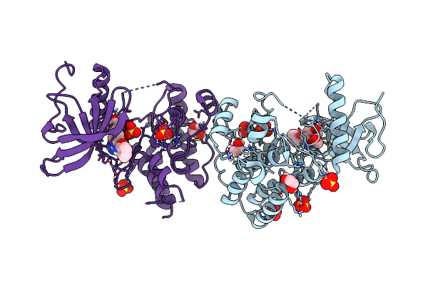 |
Fgfr1 Kinase Domain (Residues 458-765) With Mutations C488A, C584S In Complex With 34.
Organism: Homo sapiens
Method: X-RAY DIFFRACTION Resolution:1.82 Å Release Date: 2021-12-01 Classification: TRANSFERASE Ligands: 42I, SO4, EDO |
Organism: Homo sapiens
Method: X-RAY DIFFRACTION
Release Date: 2021-12-01
Ligands: 42I, SO4, EDO
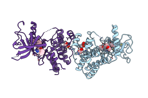 |
Fgfr1 Kinase Domain (Residues 458-765) With Mutations C488A, C584S In Complex With 19.
Organism: Homo sapiens
Method: X-RAY DIFFRACTION Resolution:1.82 Å Release Date: 2021-12-01 Classification: TRANSFERASE Ligands: 466, SO4, EDO, CL |
Organism: Homo sapiens
Method: X-RAY DIFFRACTION
Release Date: 2021-12-01
Ligands: 466, SO4, EDO, CL
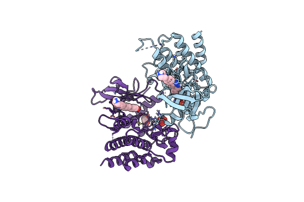 |
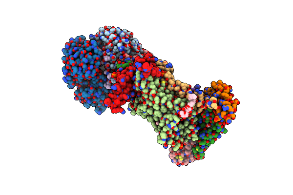
Fgfr2 Kinase Domain (Residues 461-763) In Complex With 38.
Organism: Homo sapiens
Method: X-RAY DIFFRACTION Resolution:2.28 Å Release Date: 2021-12-01 Classification: TRANSFERASE Ligands: 47I, SO4, EDO |
Organism: Homo sapiens
Method: X-RAY DIFFRACTION
Release Date: 2021-12-01
Ligands: 47I, SO4, EDO
 |
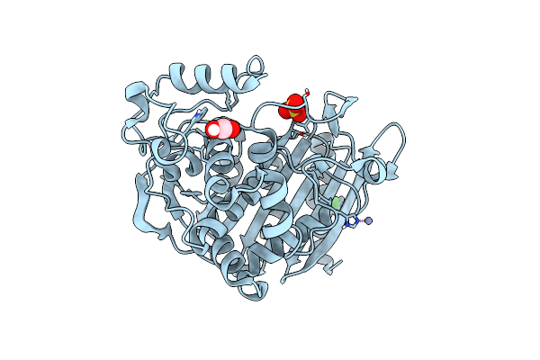
Crystal Structure Of Bovine Cytochrome Bc1 In Complex With Tetrahydro-Quinolone Inhibitor Jag021
Organism: Bos taurus
Method: X-RAY DIFFRACTION Resolution:3.50 Å Release Date: 2020-07-22 Classification: MEMBRANE PROTEIN Ligands: PG4, 6PE, PO4, CDL, HEM, LMT, PEE, JAG, HEC, FES, PX4 |
Organism: Bos taurus
Method: X-RAY DIFFRACTION
Release Date: 2020-07-22
Ligands: PG4, 6PE, PO4, CDL, HEM, LMT, PEE, JAG, HEC, FES, PX4
 |
Crystal Structure Of Ampc From E.Coli
Organism: Escherichia coli k-12
Method: X-RAY DIFFRACTION Resolution:1.50 Å Release Date: 2020-06-24 Classification: HYDROLASE Ligands: EDO, ZN, SO4, CL |
Organism: Escherichia coli k-12
Method: X-RAY DIFFRACTION
Release Date: 2020-06-24
Ligands: EDO, ZN, SO4, CL
 |
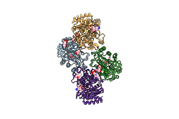
Crystal Structure Of Ampc From E. Coli With Taniborbactam (Vnrx-5133)
Organism: Escherichia coli k-12
Method: X-RAY DIFFRACTION Resolution:1.42 Å Release Date: 2020-06-24 Classification: HYDROLASE Ligands: KJK, MES, PEG, EDO, K9K, ZN, CL, NA |
Organism: Escherichia coli k-12
Method: X-RAY DIFFRACTION
Release Date: 2020-06-24
Ligands: KJK, MES, PEG, EDO, K9K, ZN, CL, NA
 |
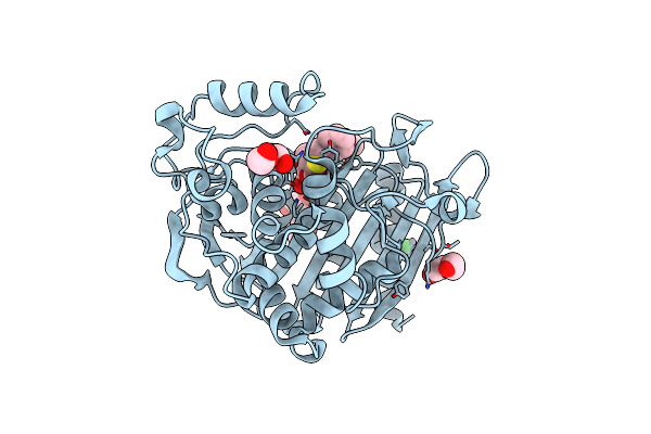
Crystal Structure Of Ampc From E. Coli With Cyclic Boronate 2
Organism: Escherichia coli (strain k12)
Method: X-RAY DIFFRACTION Resolution:2.04 Å Release Date: 2020-06-24 Classification: HYDROLASE Ligands: OK3, MPD, EPE, NA, CL, GOL |
Organism: Escherichia coli (strain k12)
Method: X-RAY DIFFRACTION
Release Date: 2020-06-24
Ligands: OK3, MPD, EPE, NA, CL, GOL
 |
Crystal Structure Of Ampc From E. Coli With Cyclic Boronate 3 (Cb3 / Apc308)
Organism: Escherichia coli k-12
Method: X-RAY DIFFRACTION Resolution:1.60 Å Release Date: 2020-06-24 Classification: HYDROLASE Ligands: KL8, PEG, DMS, EDO, CL, ZN |
Organism: Escherichia coli k-12
Method: X-RAY DIFFRACTION
Release Date: 2020-06-24
Ligands: KL8, PEG, DMS, EDO, CL, ZN
 |
Crystal Structure Of E. Coli Seryl-Trna Synthetase Complexed To Seryl Sulfamoyl Adenosine
Organism: Escherichia coli
Method: X-RAY DIFFRACTION Resolution:1.50 Å Release Date: 2020-01-22 Classification: LIGASE Ligands: SSA, PO4, EDO |
Organism: Escherichia coli
Method: X-RAY DIFFRACTION
Release Date: 2020-01-22
Ligands: SSA, PO4, EDO
 |
Crystal Structure Of S. Aureus Seryl-Trna Synthetase Complexed To Seryl Sulfamoyl Adenosine
Organism: Staphylococcus aureus
Method: X-RAY DIFFRACTION Resolution:2.03 Å Release Date: 2020-01-22 Classification: LIGASE Ligands: SSA, EDO, PEG, MG |
Organism: Staphylococcus aureus
Method: X-RAY DIFFRACTION
Release Date: 2020-01-22
Ligands: SSA, EDO, PEG, MG
 |
Crystal Structure Of E. Coli Seryl-Trna Synthetase Complexed To A Seryl Sulfamoyl Adenosine Derivative
Organism: Escherichia coli
Method: X-RAY DIFFRACTION Resolution:2.60 Å Release Date: 2020-01-22 Classification: LIGASE Ligands: JPE, CL, SO4, DMS, EDO, TRS |
Organism: Escherichia coli
Method: X-RAY DIFFRACTION
Release Date: 2020-01-22
Ligands: JPE, CL, SO4, DMS, EDO, TRS
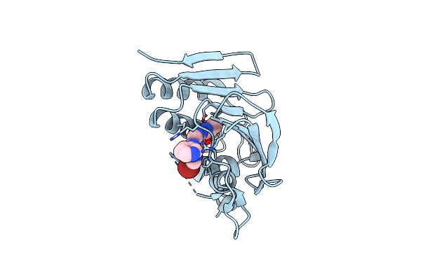 |
Crystal Structure Of E. Coli Gyraseb 24Kda In Complex With 4-(4-Bromo-1H-Pyrazol-1-Yl)-6-[(Ethylcarbamoyl)Amino]-N-(Pyridin-3-Yl)Pyridine-3-Carboxamide
Organism: Escherichia coli o157:h7
Method: X-RAY DIFFRACTION Resolution:1.90 Å Release Date: 2019-06-26 Classification: ISOMERASE Ligands: CWW |
Organism: Escherichia coli o157:h7
Method: X-RAY DIFFRACTION
Release Date: 2019-06-26
Ligands: CWW
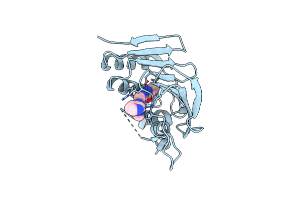 |
Crystal Structure Of E. Coli Gyraseb 24Kda In Complex With 6-[(Ethylcarbamoyl)Amino]-4-(1H-Pyrazol-1-Yl)-N-(Pyridin-3-Yl)Pyridine-3-Carboxamide
Organism: Escherichia coli o157:h7
Method: X-RAY DIFFRACTION Resolution:1.95 Å Release Date: 2019-06-26 Classification: ISOMERASE Ligands: CZ5 |
Organism: Escherichia coli o157:h7
Method: X-RAY DIFFRACTION
Release Date: 2019-06-26
Ligands: CZ5
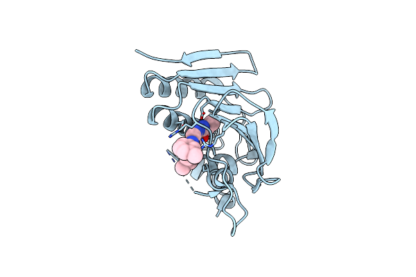 |
Crystal Structure Of E. Coli Gyraseb 24Kda In Complex With 6-[(Ethylcarbamoyl)Amino]-4-[(3-Methyphenyl)Amino]-N-(3-Methyphenyl)Pyridine-3-Carboxamide
Organism: Escherichia coli o157:h7
Method: X-RAY DIFFRACTION Resolution:2.35 Å Release Date: 2019-06-26 Classification: ISOMERASE Ligands: D0H |
Organism: Escherichia coli o157:h7
Method: X-RAY DIFFRACTION
Release Date: 2019-06-26
Ligands: D0H
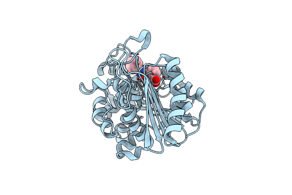 |
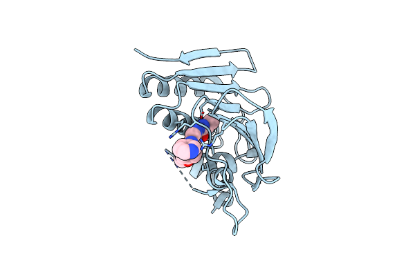
Crystal Structure Of The Ampc From Pseudomonas Aeruginosa With 1C
Organism: Pseudomonas aeruginosa
Method: X-RAY DIFFRACTION Resolution:2.21 Å Release Date: 2019-02-20 Classification: HYDROLASE Ligands: C6S |
Organism: Pseudomonas aeruginosa
Method: X-RAY DIFFRACTION
Release Date: 2019-02-20
Ligands: C6S
 |
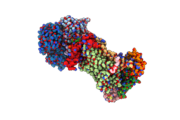
Crystal Structure Of E. Coli Gyraseb 24Kda In Complex With 6-[(Ethylcarbamoyl)Amino]-4-[(4-Methoxyphenyl)Amino]-N-(Pyridin-3-Yl)Pyridine-3-Carboxamide
Organism: Escherichia coli o157:h7
Method: X-RAY DIFFRACTION Resolution:2.50 Å Release Date: 2019-01-30 Classification: ISOMERASE Ligands: D0K |
Organism: Escherichia coli o157:h7
Method: X-RAY DIFFRACTION
Release Date: 2019-01-30
Ligands: D0K
 |
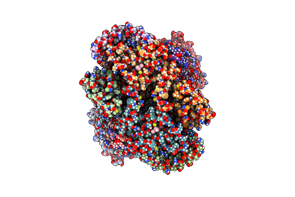
Crystal Structure Of Bovine Cytochrome Bc1 In Complex With Inhibitor Scr0911.
Organism: Bos taurus
Method: X-RAY DIFFRACTION Resolution:3.10 Å Release Date: 2018-02-28 Classification: ELECTRON TRANSPORT Ligands: PG4, 6PE, CDL, HEM, 9XE, LMT, PEE, HEC, PO4, FES, PX4 |
Organism: Bos taurus
Method: X-RAY DIFFRACTION
Release Date: 2018-02-28
Ligands: PG4, 6PE, CDL, HEM, 9XE, LMT, PEE, HEC, PO4, FES, PX4
 |
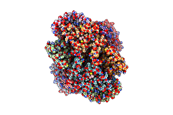
Cryoem Structure Of Bovine Cytochrome Bc1 In Complex With The Anti-Malarial Compound Gsk932121
Organism: Bos taurus
Method: ELECTRON MICROSCOPY Release Date: 2018-02-28 Classification: MEMBRANE PROTEIN Ligands: HEM, G8U, HEC |
Organism: Bos taurus
Method: ELECTRON MICROSCOPY
Release Date: 2018-02-28
Ligands: HEM, G8U, HEC
 |
Cryoem Structure Of Bovine Cytochrome Bc1 With No Ligand Bound
Organism: Bos taurus
Method: ELECTRON MICROSCOPY Release Date: 2018-02-28 Classification: MEMBRANE PROTEIN Ligands: HEM, HEC |
Organism: Bos taurus
Method: ELECTRON MICROSCOPY
Release Date: 2018-02-28
Ligands: HEM, HEC