

Search Count: 66
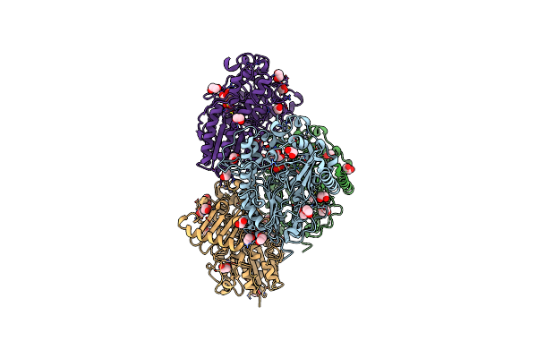 |
Bt1596 In Complex With Its Substrate 4,5 Unsaturated Uronic Acid Alpha 1,4 D-Glucosamine-2-N, 6-O-Disulfate
Organism: Bacteroides thetaiotaomicron
Method: X-RAY DIFFRACTION Resolution:1.90 Å Release Date: 2017-05-24 Classification: HYDROLASE Ligands: ZN, EDO, CL, UAP |
Organism: Bacteroides thetaiotaomicron
Method: X-RAY DIFFRACTION
Release Date: 2017-05-24
Ligands: ZN, EDO, CL, UAP
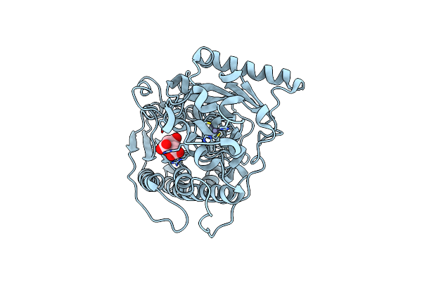 |
Structure Of Bt1596,A 2-O Gag Sulfatase
Organism: Bacteroides thetaiotaomicron
Method: X-RAY DIFFRACTION Resolution:1.43 Å Release Date: 2017-05-24 Classification: HYDROLASE Ligands: ZN, CIT |
Organism: Bacteroides thetaiotaomicron
Method: X-RAY DIFFRACTION
Release Date: 2017-05-24
Ligands: ZN, CIT
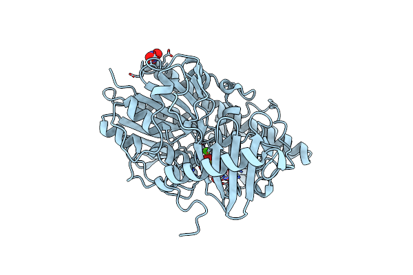 |
Structure Of Bt4656 In Complex With Its Substrate D-Glucosamine-2-N, 6-O-Disulfate.
Organism: Bacteroides thetaiotaomicron
Method: X-RAY DIFFRACTION Resolution:1.39 Å Release Date: 2017-05-24 Classification: HYDROLASE Ligands: CA, SGN, NO3 |
Organism: Bacteroides thetaiotaomicron
Method: X-RAY DIFFRACTION
Release Date: 2017-05-24
Ligands: CA, SGN, NO3
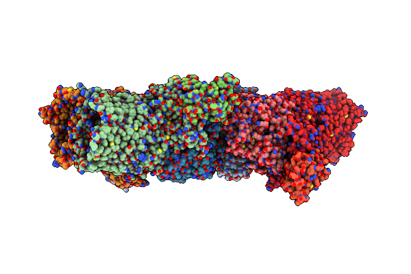 |
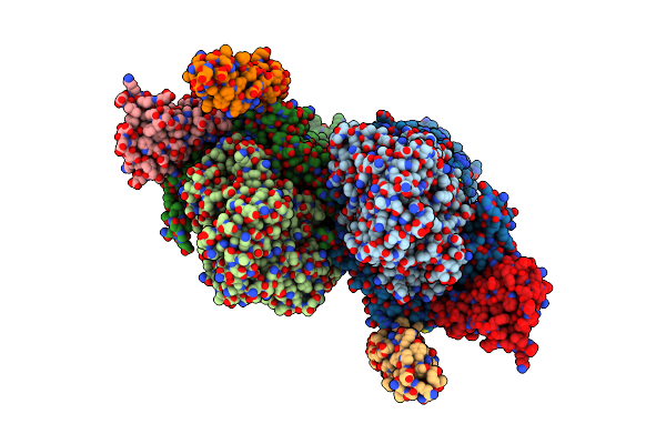
Crystal Structure Of The Suscd Complex Bt2261-2264 From Bacteroides Thetaiotaomicron
Organism: Bacteroides thetaiotaomicron
Method: X-RAY DIFFRACTION Resolution:2.80 Å Release Date: 2016-12-21 Classification: MEMBRANE PROTEIN Ligands: KR0, NA, CA |
Organism: Bacteroides thetaiotaomicron
Method: X-RAY DIFFRACTION
Release Date: 2016-12-21
Ligands: KR0, NA, CA
 |
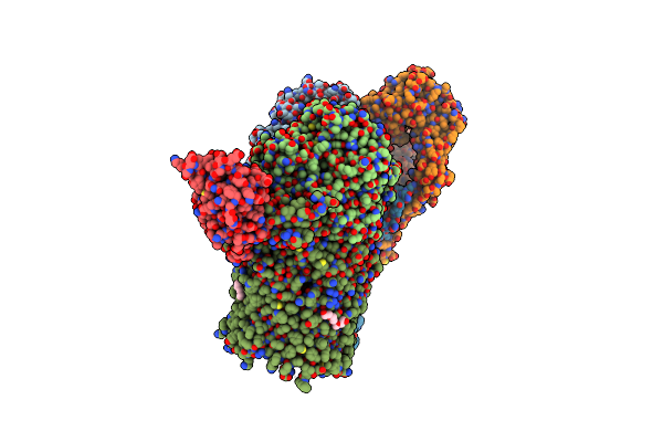
Crystal Structure Of The Suscd Complex Bt2261-2264 From Bacteroides Thetaiotaomicron
Organism: Bacteroides thetaiotaomicron
Method: X-RAY DIFFRACTION Resolution:3.40 Å Release Date: 2016-12-21 Classification: MEMBRANE PROTEIN Ligands: MG, NA |
Organism: Bacteroides thetaiotaomicron
Method: X-RAY DIFFRACTION
Release Date: 2016-12-21
Ligands: MG, NA
 |
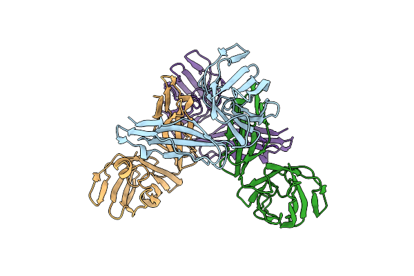
Crystal Structure Of The Suscd Complex Bt2261-2264 From Bacteroides Thetaiotaomicron
Organism: Bacteroides thetaiotaomicron
Method: X-RAY DIFFRACTION Resolution:2.75 Å Release Date: 2016-12-21 Classification: MEMBRANE PROTEIN Ligands: MG, KR0, CA, C8E |
Organism: Bacteroides thetaiotaomicron
Method: X-RAY DIFFRACTION
Release Date: 2016-12-21
Ligands: MG, KR0, CA, C8E
 |
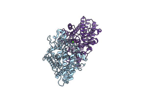
Crystal Structure Of The Lipoprotein Bt2262 From Bacteroides Thetaiotaomicron
Organism: Bacteroides thetaiotaomicron
Method: X-RAY DIFFRACTION Resolution:3.10 Å Release Date: 2016-12-14 Classification: MEMBRANE PROTEIN |
Organism: Bacteroides thetaiotaomicron
Method: X-RAY DIFFRACTION
Release Date: 2016-12-14
 |
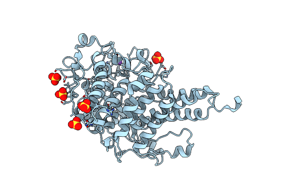
Crystal Structure Of The Lipoprotein Bt2263 From Bacteroides Thetaiotaomicron
Organism: Bacteroides thetaiotaomicron
Method: X-RAY DIFFRACTION Resolution:1.90 Å Release Date: 2016-12-14 Classification: MEMBRANE PROTEIN Ligands: CA |
Organism: Bacteroides thetaiotaomicron
Method: X-RAY DIFFRACTION
Release Date: 2016-12-14
Ligands: CA
 |
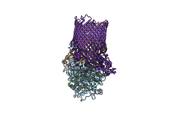
Crystal Structure Of Bt1762
Organism: Bacteroides thetaiotaomicron (strain atcc 29148 / dsm 2079 / nctc 10582 / e50 / vpi-5482)
Method: X-RAY DIFFRACTION Resolution:1.76 Å Release Date: 2016-12-14 Classification: TRANSPORT PROTEIN Ligands: SO4, NA |
Organism: Bacteroides thetaiotaomicron (strain atcc 29148 / dsm 2079 / nctc 10582 / e50 / vpi-5482)
Method: X-RAY DIFFRACTION
Release Date: 2016-12-14
Ligands: SO4, NA
 |
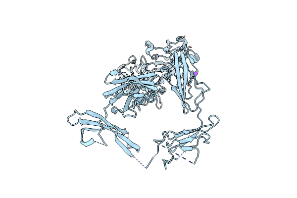
Crystal Structure Of Bt1762-1763
Organism: Bacteroides thetaiotaomicron (strain atcc 29148 / dsm 2079 / nctc 10582 / e50 / vpi-5482)
Method: X-RAY DIFFRACTION Resolution:3.10 Å Release Date: 2016-12-14 Classification: TRANSPORT PROTEIN Ligands: MG, NA |
Organism: Bacteroides thetaiotaomicron (strain atcc 29148 / dsm 2079 / nctc 10582 / e50 / vpi-5482)
Method: X-RAY DIFFRACTION
Release Date: 2016-12-14
Ligands: MG, NA
 |
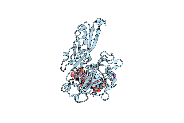
Crystal Structure Of Bt1762-1763
Organism: Bacteroides thetaiotaomicron (strain atcc 29148 / dsm 2079 / nctc 10582 / e50 / vpi-5482)
Method: X-RAY DIFFRACTION Resolution:3.10 Å Release Date: 2016-12-14 Classification: TRANSPORT PROTEIN Ligands: MG |
Organism: Bacteroides thetaiotaomicron (strain atcc 29148 / dsm 2079 / nctc 10582 / e50 / vpi-5482)
Method: X-RAY DIFFRACTION
Release Date: 2016-12-14
Ligands: MG
 |
Structure Of Bt4661, A Suse-Like Surface Located Polysaccharide Binding Protein From The Bacteroides Thetaiotaomicron Heparin Utilisation Locus
Organism: Bacteroides thetaiotaomicron
Method: X-RAY DIFFRACTION Resolution:1.95 Å Release Date: 2013-03-06 Classification: HEPARIN-BINDING PROTEIN |
Organism: Bacteroides thetaiotaomicron
Method: X-RAY DIFFRACTION
Release Date: 2013-03-06
 |
Structure Of Bt4661, A Suse-Like Surface Located Polysaccharide Binding Protein From The Bacteroides Thetaiotaomicron Heparin Utilisation Locus
Organism: Bacteroides thetaiotaomicron
Method: X-RAY DIFFRACTION Resolution:1.35 Å Release Date: 2013-03-06 Classification: HEPARIN-BINDING PROTEIN |
Organism: Bacteroides thetaiotaomicron
Method: X-RAY DIFFRACTION
Release Date: 2013-03-06
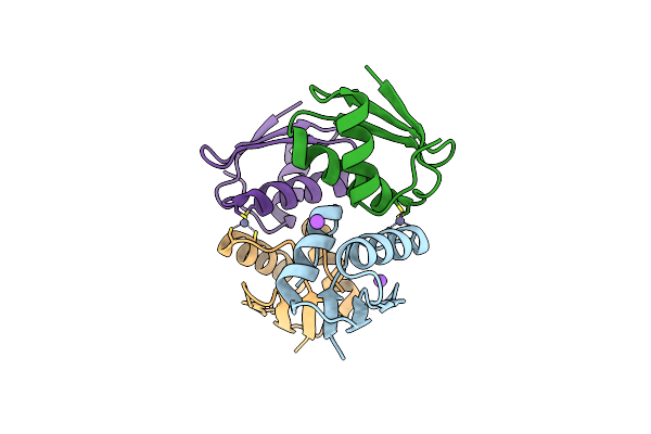 |
Crosstalk Between Cu(I) And Zn(Ii) Homeostasis
Organism: Synechocystis sp. pcc 6803
Method: X-RAY DIFFRACTION Resolution:1.85 Å Release Date: 2012-12-12 Classification: TRANSPORT PROTEIN Ligands: ZN, NA, CL |
Organism: Synechocystis sp. pcc 6803
Method: X-RAY DIFFRACTION
Release Date: 2012-12-12
Ligands: ZN, NA, CL
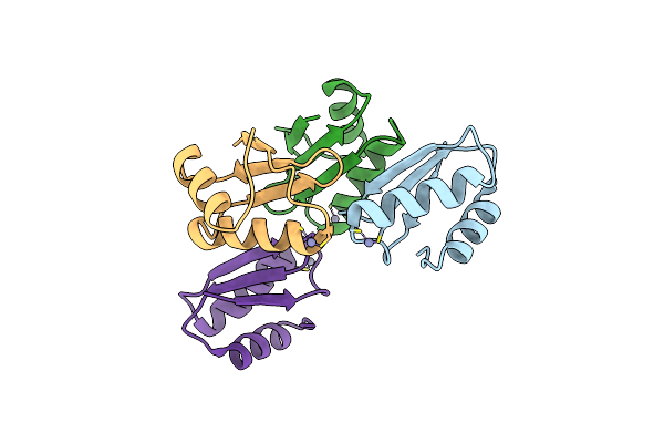 |
Crosstalk Between Cu(I) And Zn(Ii) Homeostasis
Organism: Synechocystis sp.
Method: X-RAY DIFFRACTION Resolution:1.90 Å Release Date: 2012-12-12 Classification: TRANSPORT PROTEIN Ligands: ZN |
Organism: Synechocystis sp.
Method: X-RAY DIFFRACTION
Release Date: 2012-12-12
Ligands: ZN
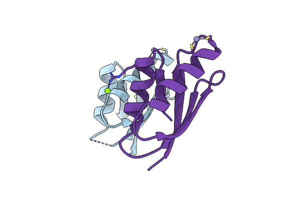 |
Crosstalk Between Cu(I) And Zn(Ii) Homeostasis
Organism: Synechocystis
Method: X-RAY DIFFRACTION Resolution:1.40 Å Release Date: 2012-12-12 Classification: TRANSPORT PROTEIN Ligands: ZN, MG |
Organism: Synechocystis
Method: X-RAY DIFFRACTION
Release Date: 2012-12-12
Ligands: ZN, MG
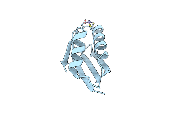 |
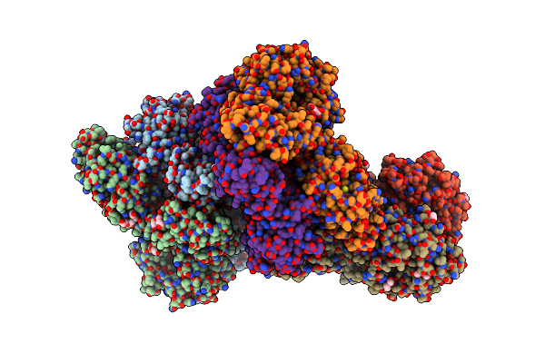
Crosstalk Between Cu(I) And Zn(Ii) Homeostasis
Organism: Synechocystis
Method: X-RAY DIFFRACTION Resolution:1.25 Å Release Date: 2012-12-12 Classification: HYDROLASE Ligands: ZN |
Organism: Synechocystis
Method: X-RAY DIFFRACTION
Release Date: 2012-12-12
Ligands: ZN
 |
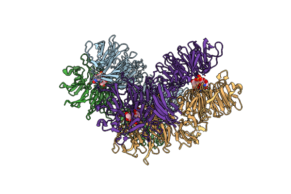
Structure Of The Periplasmic Domain Of The Heparin And Heparan Sulphate Sensing Hybrid Two Component System Bt4663 In Apo And Ligand Bound Forms
Organism: Bacteroides thetaiotaomicron
Method: X-RAY DIFFRACTION Resolution:2.60 Å Release Date: 2012-05-02 Classification: TRANSCRIPTION Ligands: PGE, EDO, PEG, PG4, MES, OAA, 2PE |
Organism: Bacteroides thetaiotaomicron
Method: X-RAY DIFFRACTION
Release Date: 2012-05-02
Ligands: PGE, EDO, PEG, PG4, MES, OAA, 2PE
 |
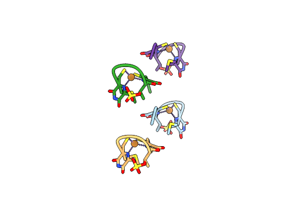
Structure Of The Periplasmic Domain Of The Heparin And Heparan Sulphate Sensing Hybrid Two Component System Bt4663 In Apo And Ligand Bound Forms
Organism: Bacteroides thetaiotaomicron
Method: X-RAY DIFFRACTION Resolution:3.40 Å Release Date: 2012-05-02 Classification: TRANSCRIPTION |
Organism: Bacteroides thetaiotaomicron
Method: X-RAY DIFFRACTION
Release Date: 2012-05-02
 |
Methanobactin Hm1
Organism: Methylocystis hirsuta
Method: X-RAY DIFFRACTION Resolution:0.80 Å Release Date: 2012-04-25 Classification: METAL TRANSPORT Ligands: CU |
Organism: Methylocystis hirsuta
Method: X-RAY DIFFRACTION
Release Date: 2012-04-25
Ligands: CU