

Search Count: 71
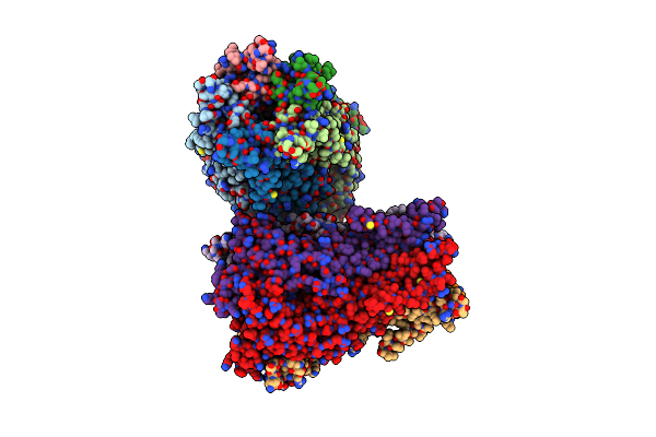 |
X-Ray Structure Of A Pentameric Ligand Gated Ion Channel From Erwinia Chrysanthemi (Elic) In Complex With Bromoform
Organism: Erwinia chrysanthemi
Method: X-RAY DIFFRACTION Resolution:3.65 Å Release Date: 2013-02-06 Classification: TRANSPORT PROTEIN Ligands: MBR |
Organism: Erwinia chrysanthemi
Method: X-RAY DIFFRACTION
Release Date: 2013-02-06
Ligands: MBR
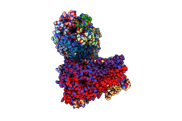 |
X-Ray Structure Of A Pentameric Ligand Gated Ion Channel From Erwinia Chrysanthemi (Elic) In Complex With Gaba And Flurazepam
Organism: Erwinia chrysanthemi
Method: X-RAY DIFFRACTION Resolution:3.90 Å Release Date: 2012-10-31 Classification: TRANSPORT PROTEIN Ligands: ABU, FL7 |
Organism: Erwinia chrysanthemi
Method: X-RAY DIFFRACTION
Release Date: 2012-10-31
Ligands: ABU, FL7
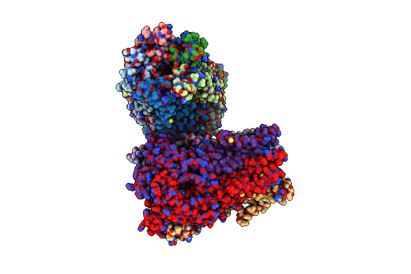 |
X-Ray Structure Of A Pentameric Ligand Gated Ion Channel From Erwinia Chrysanthemi (Elic) In Complex With Zopiclone
Organism: Erwinia chrysanthemi
Method: X-RAY DIFFRACTION Resolution:3.34 Å Release Date: 2012-10-17 Classification: TRANSPORT PROTEIN Ligands: ZPC |
Organism: Erwinia chrysanthemi
Method: X-RAY DIFFRACTION
Release Date: 2012-10-17
Ligands: ZPC
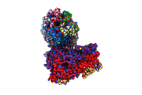 |
X-Ray Structure Of A Pentameric Ligand Gated Ion Channel From Erwinia Chrysanthemi (Elic) In Complex With Bromoflurazepam
Organism: Erwinia chrysanthemi
Method: X-RAY DIFFRACTION Resolution:3.61 Å Release Date: 2012-10-17 Classification: TRANSPORT PROTEIN Ligands: BFZ |
Organism: Erwinia chrysanthemi
Method: X-RAY DIFFRACTION
Release Date: 2012-10-17
Ligands: BFZ
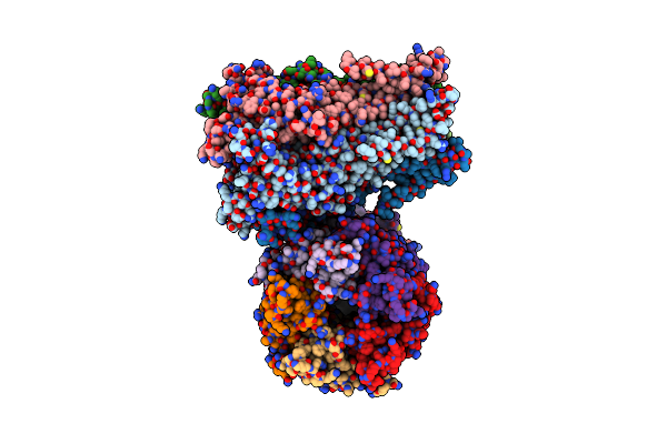 |
X-Ray Structure Of A Pentameric Ligand Gated Ion Channel From Erwinia Chrysanthemi (Elic) Mutant F247L (F16L)
Organism: Erwinia chrysanthemi
Method: X-RAY DIFFRACTION Resolution:3.50 Å Release Date: 2012-04-04 Classification: TRANSPORT PROTEIN |
Organism: Erwinia chrysanthemi
Method: X-RAY DIFFRACTION
Release Date: 2012-04-04
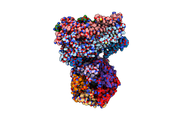 |
X-Ray Structure Of A Pentameric Ligand Gated Ion Channel From Erwinia Chrysanthemi (Elic) Mutant L240A F247L (L9A F16L) In The Presence Of 10 Mm Cysteamine
Organism: Erwinia chrysanthemi
Method: X-RAY DIFFRACTION Resolution:4.20 Å Release Date: 2012-04-04 Classification: TRANSPORT PROTEIN |
Organism: Erwinia chrysanthemi
Method: X-RAY DIFFRACTION
Release Date: 2012-04-04
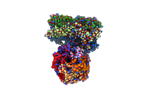 |
X-Ray Structure Of A Pentameric Ligand Gated Ion Channel From Erwinia Chrysanthemi (Elic) Mutant L240S F247L (L9S F16L) In Presence Of 10 Mm Cysteamine
Organism: Erwinia chrysanthemi
Method: X-RAY DIFFRACTION Resolution:3.80 Å Release Date: 2012-04-04 Classification: TRANSPORT PROTEIN |
Organism: Erwinia chrysanthemi
Method: X-RAY DIFFRACTION
Release Date: 2012-04-04
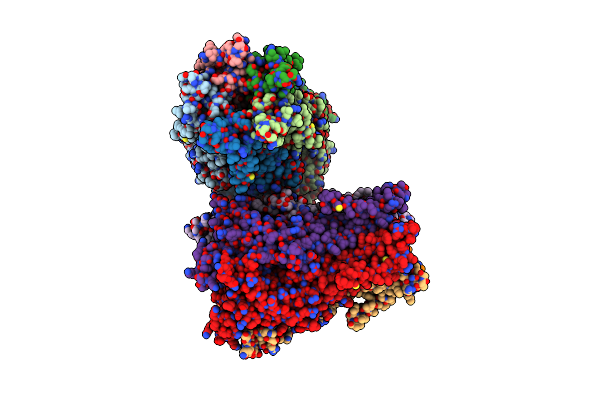 |
Pentameric Ligand Gated Ion Channel Elic Mutant F246A
Organism: Erwinia chrysanthemi
Method: X-RAY DIFFRACTION Resolution:3.30 Å Release Date: 2011-07-06 Classification: TRANSPORT PROTEIN |
Organism: Erwinia chrysanthemi
Method: X-RAY DIFFRACTION
Release Date: 2011-07-06
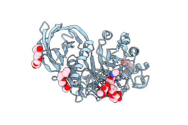 |
Structural Basis For Substrate Recognition By Erwinia Chrysanthemi Gh5 Glucuronoxylanase
Organism: Erwinia chrysanthemi
Method: X-RAY DIFFRACTION Resolution:1.39 Å Release Date: 2011-06-01 Classification: HYDROLASE Ligands: PG4, PGE, IMD |
Organism: Erwinia chrysanthemi
Method: X-RAY DIFFRACTION
Release Date: 2011-06-01
Ligands: PG4, PGE, IMD
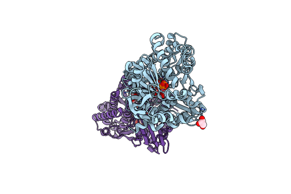 |
Co-Complex Structure Of Achromobactin Synthetase Protein D (Acsd) With Atp And N-Citryl-Ethylenediamine From Pectobacterium Chrysanthemi
Organism: Erwinia chrysanthemi
Method: X-RAY DIFFRACTION Resolution:2.00 Å Release Date: 2011-01-19 Classification: LIGASE Ligands: ATP, MG, GOL, X3J |
Organism: Erwinia chrysanthemi
Method: X-RAY DIFFRACTION
Release Date: 2011-01-19
Ligands: ATP, MG, GOL, X3J
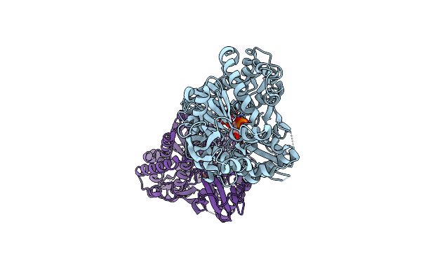 |
Co-Complex Structure Of Achromobactin Synthetase Protein D (Acsd) With Amp And Sulfate From Pectobacterium Chrysanthemi
Organism: Erwinia chrysanthemi
Method: X-RAY DIFFRACTION Resolution:2.50 Å Release Date: 2010-11-10 Classification: LIGASE Ligands: AMP, SO4 |
Organism: Erwinia chrysanthemi
Method: X-RAY DIFFRACTION
Release Date: 2010-11-10
Ligands: AMP, SO4
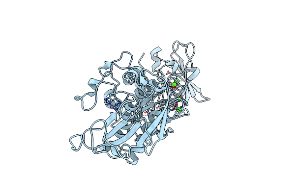 |
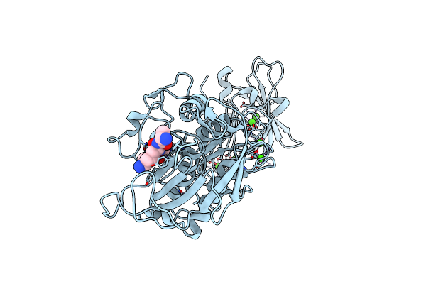
Prtc Methionine Mutants: M226I
Organism: Erwinia chrysanthemi
Method: X-RAY DIFFRACTION Resolution:1.75 Å Release Date: 2009-08-04 Classification: HYDROLASE Ligands: CA, ZN, CL |
Organism: Erwinia chrysanthemi
Method: X-RAY DIFFRACTION
Release Date: 2009-08-04
Ligands: CA, ZN, CL
 |
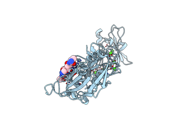
Prtc Methionine Mutants: M226H Desy
Organism: Erwinia chrysanthemi, Synthetic construct
Method: X-RAY DIFFRACTION Resolution:1.77 Å Release Date: 2009-07-21 Classification: HYDROLASE Ligands: CA, ZN, CL |
Organism: Erwinia chrysanthemi, Synthetic construct
Method: X-RAY DIFFRACTION
Release Date: 2009-07-21
Ligands: CA, ZN, CL
 |
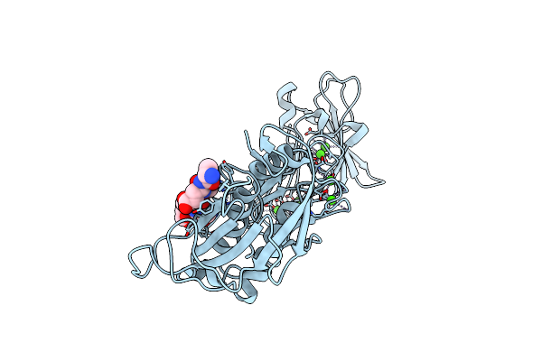
Prtc Methionine Mutants: M226A In-House
Organism: Erwinia chrysanthemi, Unidentified
Method: X-RAY DIFFRACTION Resolution:1.95 Å Release Date: 2009-06-30 Classification: HYDROLASE Ligands: CA, ZN, CL |
Organism: Erwinia chrysanthemi, Unidentified
Method: X-RAY DIFFRACTION
Release Date: 2009-06-30
Ligands: CA, ZN, CL
 |
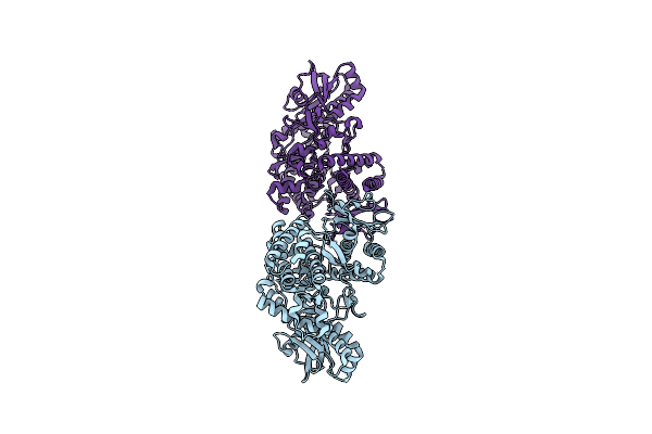
Prtc Methionine Mutants: M226A_Desy
Organism: Erwinia chrysanthemi, Synthetic construct
Method: X-RAY DIFFRACTION Resolution:2.13 Å Release Date: 2009-06-30 Classification: HYDROLASE Ligands: CA, CL |
Organism: Erwinia chrysanthemi, Synthetic construct
Method: X-RAY DIFFRACTION
Release Date: 2009-06-30
Ligands: CA, CL
 |
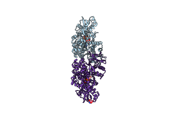
Structure Of Achromobactin Synthetase Protein D, (Acsd)
Organism: Erwinia chrysanthemi
Method: X-RAY DIFFRACTION Resolution:2.25 Å Release Date: 2009-02-03 Classification: BIOSYNTHETIC PROTEIN |
Organism: Erwinia chrysanthemi
Method: X-RAY DIFFRACTION
Release Date: 2009-02-03
 |
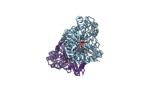
Co-Complex Structure Of Achromobactin Synthetase Protein D (Acsd) With Atp From Pectobacterium Chrysanthemi
Organism: Erwinia chrysanthemi
Method: X-RAY DIFFRACTION Resolution:2.20 Å Release Date: 2009-01-13 Classification: METAL TRANSPORT Ligands: ATP, MG, SER |
Organism: Erwinia chrysanthemi
Method: X-RAY DIFFRACTION
Release Date: 2009-01-13
Ligands: ATP, MG, SER
 |
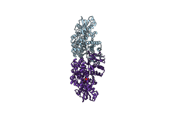
Co-Complex Structure Of Achromobactin Synthetase Protein D (Acsd) With Adenosine, Sulfate And Citrate From Pectobacterium Chrysanthemi
Organism: Erwinia chrysanthemi
Method: X-RAY DIFFRACTION Resolution:2.95 Å Release Date: 2009-01-13 Classification: METAL TRANSPORT Ligands: ADN, SO4, CIT |
Organism: Erwinia chrysanthemi
Method: X-RAY DIFFRACTION
Release Date: 2009-01-13
Ligands: ADN, SO4, CIT
 |
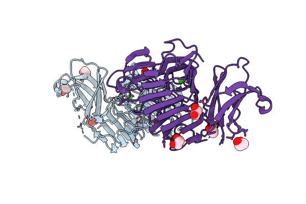
Co-Complex Structure Of Achromobactin Synthetase Protein D (Acsd) With Citrate In Atp Binding Site From Pectobacterium Chrysanthemi
Organism: Erwinia chrysanthemi
Method: X-RAY DIFFRACTION Resolution:2.80 Å Release Date: 2009-01-13 Classification: METAL TRANSPORT Ligands: FLC |
Organism: Erwinia chrysanthemi
Method: X-RAY DIFFRACTION
Release Date: 2009-01-13
Ligands: FLC
 |
Crystal Structure Analysis Of Pectate Lyase Peli From Erwinia Chrysanthemi
Organism: Erwinia chrysanthemi
Method: X-RAY DIFFRACTION Resolution:1.45 Å Release Date: 2008-04-29 Classification: LYASE Ligands: CA, ZN, EDO, SO4 |
Organism: Erwinia chrysanthemi
Method: X-RAY DIFFRACTION
Release Date: 2008-04-29
Ligands: CA, ZN, EDO, SO4