

Search Count: 41
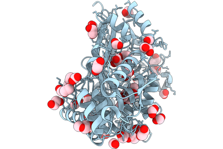 |
X-Ray Crystal Structure Of The Oligopeptide-Binding Protein Oppa3 From Borrelia Burgdorferi
Organism: Borreliella burgdorferi
Method: X-RAY DIFFRACTION Resolution:1.95 Å Release Date: 2026-08-05 Classification: PEPTIDE BINDING PROTEIN Ligands: EDO, FMT, ACT, ETX, PG0, CL |
Organism: Borreliella burgdorferi
Method: X-RAY DIFFRACTION
Release Date: 2026-08-05
Ligands: EDO, FMT, ACT, ETX, PG0, CL
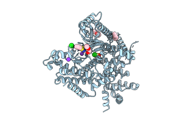 |
The Structure Of Human Vacuolar Protein Sorting 34 Catalytic Domain Bound To Rd-I-53
Organism: Homo sapiens
Method: X-RAY DIFFRACTION Resolution:2.16 Å Release Date: 2025-09-17 Classification: TRANSFERASE Ligands: ETZ, DMS, A1A6F, ETX, K, CL |
Organism: Homo sapiens
Method: X-RAY DIFFRACTION
Release Date: 2025-09-17
Ligands: ETZ, DMS, A1A6F, ETX, K, CL
 |
Non-Crystalline Proximal Tubulopathy And Crystalline Cast Nephropathy-Causing Bence-Jones Protein Pt-Cn: An Entire Immunoglobulin Kappa Light Chain Dimer
Organism: Homo sapiens
Method: X-RAY DIFFRACTION Resolution:2.30 Å Release Date: 2025-01-22 Classification: IMMUNE SYSTEM Ligands: PO4, ETX, PGE, GOL, EDO, PEG |
Organism: Homo sapiens
Method: X-RAY DIFFRACTION
Release Date: 2025-01-22
Ligands: PO4, ETX, PGE, GOL, EDO, PEG
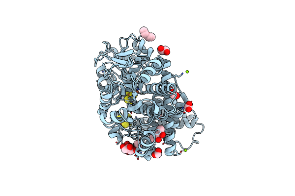 |
Hybrid Cluster Protein From The Thermophilic Methanogen Methanothermococcus Thermolithotrophicus As Isolated In A Reduced State At 1.45-A Resolution
Organism: Methanothermococcus thermolithotrophicus dsm 2095
Method: X-RAY DIFFRACTION Resolution:1.45 Å Release Date: 2023-04-19 Classification: OXIDOREDUCTASE Ligands: MG, ACT, SF4, ETX, FMT, EDO, GOL, SF3, MRD |
Organism: Methanothermococcus thermolithotrophicus dsm 2095
Method: X-RAY DIFFRACTION
Release Date: 2023-04-19
Ligands: MG, ACT, SF4, ETX, FMT, EDO, GOL, SF3, MRD
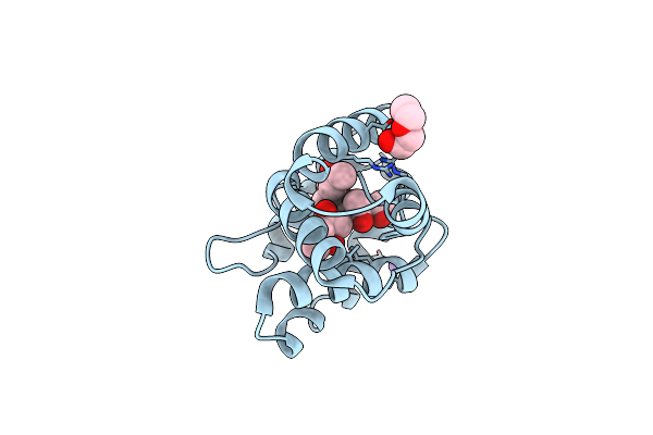 |
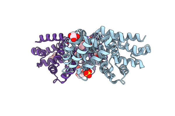
Crystal Structure Of Odorant Binding Protein 5 From Anopheles Gambiae (Agamobp5) With Mpd (2-Methyl-2,4-Pentanediol)
Organism: Anopheles gambiae
Method: X-RAY DIFFRACTION Resolution:1.35 Å Release Date: 2023-03-22 Classification: TRANSPORT PROTEIN Ligands: MPD, NA, ETX |
Organism: Anopheles gambiae
Method: X-RAY DIFFRACTION
Release Date: 2023-03-22
Ligands: MPD, NA, ETX
 |
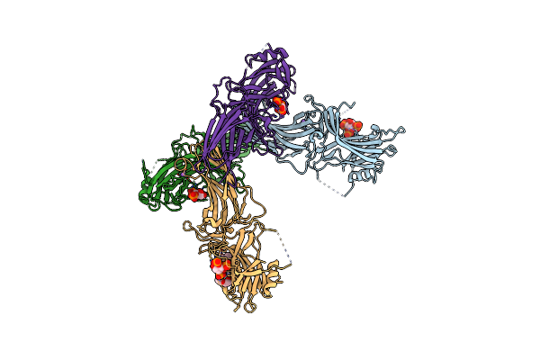
Crystal Structure Of The Anopheles Darlingi Ad-118 Long Form D7 Salivary Protein
Organism: Anopheles darlingi
Method: X-RAY DIFFRACTION Resolution:2.40 Å Release Date: 2022-05-25 Classification: BLOOD CLOTTING Ligands: ETX, SO4, PG0, GOL, PEG, EDO |
Organism: Anopheles darlingi
Method: X-RAY DIFFRACTION
Release Date: 2022-05-25
Ligands: ETX, SO4, PG0, GOL, PEG, EDO
 |
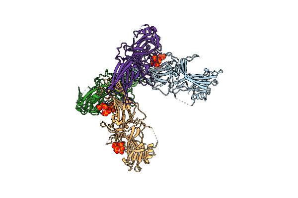
Crystal Structure Of Native Bovine Arrestin 1 In Complex With Inositol 1,4,5-Triphosphate
Organism: Bos taurus
Method: X-RAY DIFFRACTION Resolution:2.40 Å Release Date: 2021-10-13 Classification: PROTEIN BINDING Ligands: I3P, NA, ETX |
Organism: Bos taurus
Method: X-RAY DIFFRACTION
Release Date: 2021-10-13
Ligands: I3P, NA, ETX
 |
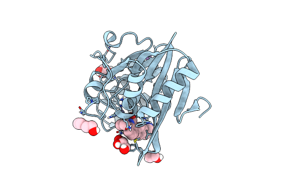
Crystal Structure Of Native Bovine Arrestin 1 In Complex With 1D-Myo-Inositol 5-Diphosphate Pentakisphosphate (5-Pp Ip5)
Organism: Bos taurus
Method: X-RAY DIFFRACTION Resolution:2.60 Å Release Date: 2021-10-13 Classification: PROTEIN BINDING Ligands: ETX, I7P |
Organism: Bos taurus
Method: X-RAY DIFFRACTION
Release Date: 2021-10-13
Ligands: ETX, I7P
 |
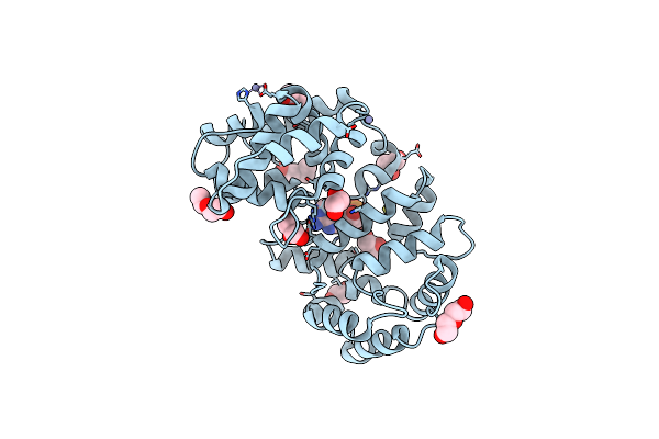
Crystal Structure Of The Metallo-Beta-Lactamase Ndm-7 With 407
Organism: Klebsiella pneumoniae
Method: X-RAY DIFFRACTION Resolution:1.02 Å Release Date: 2021-10-06 Classification: HYDROLASE Ligands: R8W, ZN, GOL, EDO, NA, ETX |
Organism: Klebsiella pneumoniae
Method: X-RAY DIFFRACTION
Release Date: 2021-10-06
Ligands: R8W, ZN, GOL, EDO, NA, ETX
 |
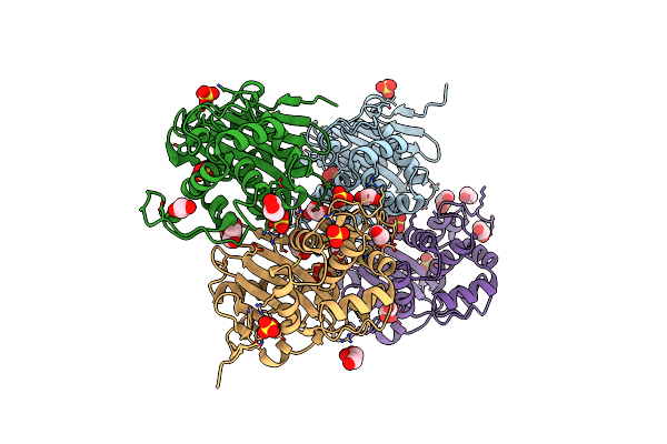
Culex Quinquefasciatus D7 Long Form 1- Cxd7L1 In Complex With Adp
Organism: Culex quinquefasciatus
Method: X-RAY DIFFRACTION Resolution:1.97 Å Release Date: 2020-06-24 Classification: PROTEIN BINDING Ligands: ADP, GOL, ETX, PEG, EDO, ZN |
Organism: Culex quinquefasciatus
Method: X-RAY DIFFRACTION
Release Date: 2020-06-24
Ligands: ADP, GOL, ETX, PEG, EDO, ZN
 |
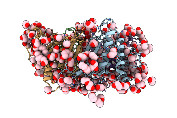
Crystal Structure Of Oxa-517 Beta-Lactamase
Organism: Klebsiella pneumoniae
Method: X-RAY DIFFRACTION Resolution:1.86 Å Release Date: 2019-08-28 Classification: HYDROLASE Ligands: SO4, ETX, GOL, CL, EDO |
Organism: Klebsiella pneumoniae
Method: X-RAY DIFFRACTION
Release Date: 2019-08-28
Ligands: SO4, ETX, GOL, CL, EDO
 |
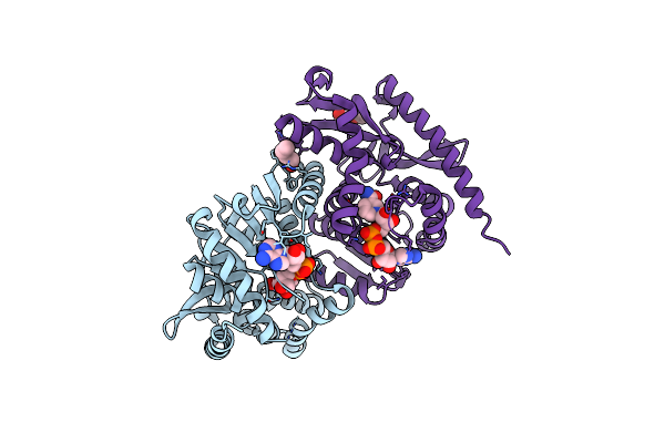
Structure Of The Poxvirus Protein F9
Organism: Vaccinia virus (strain western reserve)
Method: X-RAY DIFFRACTION Resolution:2.10 Å Release Date: 2019-03-06 Classification: VIRAL PROTEIN Ligands: PG4, PG0, PEG, EDO, GOL, PDO, EOH, MOH, PGO, ETX, ETE, PGE, P33, P6G |
 |
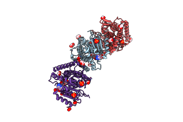
Crystal Structure Of Malate Dehydrogenase From Metallosphaera Sedula
Organism: Metallosphaera sedula
Method: X-RAY DIFFRACTION Resolution:2.30 Å Release Date: 2019-02-06 Classification: OXIDOREDUCTASE Ligands: NAD, ETX |
Organism: Metallosphaera sedula
Method: X-RAY DIFFRACTION
Release Date: 2019-02-06
Ligands: NAD, ETX
 |
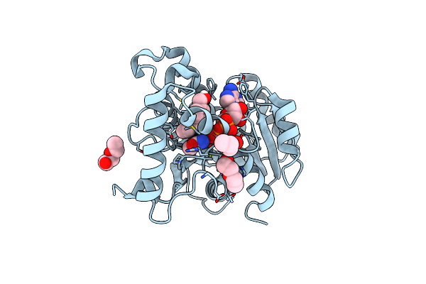
Cytosolic Malate Dehydrogenase 1 (Peroxide-Treated)
Organism: Arabidopsis thaliana
Method: X-RAY DIFFRACTION Resolution:1.35 Å Release Date: 2018-02-28 Classification: OXIDOREDUCTASE Ligands: NAD, SO4, GOL, PEO, FMT, TRS, ETX, ACT, EDO |
Organism: Arabidopsis thaliana
Method: X-RAY DIFFRACTION
Release Date: 2018-02-28
Ligands: NAD, SO4, GOL, PEO, FMT, TRS, ETX, ACT, EDO
 |
Inha (T2A Mutant) Complexed With (4-((1H-Pyrazol-1-Yl)Methyl)Phenyl)Methanol
Organism: Mycobacterium tuberculosis
Method: X-RAY DIFFRACTION Resolution:1.87 Å Release Date: 2018-02-14 Classification: OXIDOREDUCTASE Ligands: NAD, 9VQ, ETX |
Organism: Mycobacterium tuberculosis
Method: X-RAY DIFFRACTION
Release Date: 2018-02-14
Ligands: NAD, 9VQ, ETX
 |
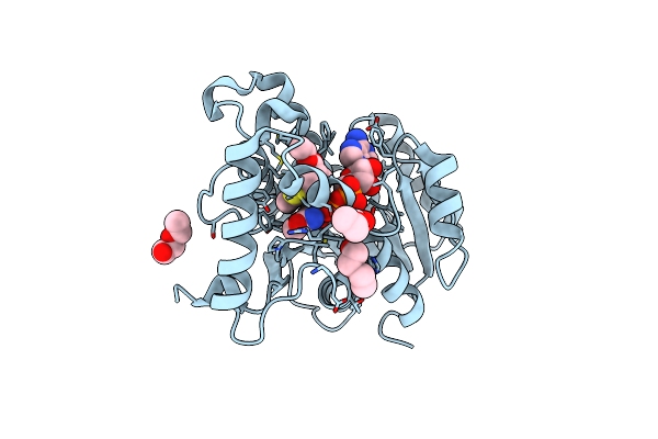
Inha (T2A Mutant) Complexed With 1-Cyclohexyl-3-(Pyridin-3-Ylmethyl)Urea
Organism: Mycobacterium tuberculosis (strain atcc 25618 / h37rv)
Method: X-RAY DIFFRACTION Resolution:2.76 Å Release Date: 2018-02-14 Classification: OXIDOREDUCTASE Ligands: NAD, 9VW, ETX |
Organism: Mycobacterium tuberculosis (strain atcc 25618 / h37rv)
Method: X-RAY DIFFRACTION
Release Date: 2018-02-14
Ligands: NAD, 9VW, ETX
 |
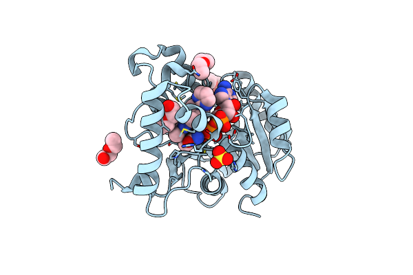
Inha (T2A Mutant) Complexed With Ethyl 2-Methyl-4,5,6,7-Tetrahydrobenzo[D]Thiazole-6-Carboxylate
Organism: Mycobacterium tuberculosis (strain atcc 25618 / h37rv)
Method: X-RAY DIFFRACTION Resolution:1.91 Å Release Date: 2018-02-14 Classification: OXIDOREDUCTASE Ligands: 9VZ, NAD, ETX |
Organism: Mycobacterium tuberculosis (strain atcc 25618 / h37rv)
Method: X-RAY DIFFRACTION
Release Date: 2018-02-14
Ligands: 9VZ, NAD, ETX
 |
Inha (T2A Mutant) Complexed With 2,6-Dimethyl-3-(1-(Pyrimidin-2-Yl)Piperidin-4-Yl)Pyridin-4(1H)-One
Organism: Mycobacterium tuberculosis
Method: X-RAY DIFFRACTION Resolution:1.97 Å Release Date: 2018-02-14 Classification: OXIDOREDUCTASE Ligands: NAD, 9W8, ETX, SO4 |
Organism: Mycobacterium tuberculosis
Method: X-RAY DIFFRACTION
Release Date: 2018-02-14
Ligands: NAD, 9W8, ETX, SO4
 |
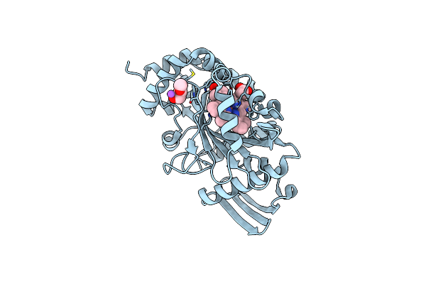
Crystal Structure Of Acetylcholinesterase In Complex With Crystal Violet
Organism: Mus musculus
Method: X-RAY DIFFRACTION Resolution:2.40 Å Release Date: 2017-09-06 Classification: HYDROLASE Ligands: NAG, PG0, ETX, EDO, CVI, PEG, PGE, AE3, PE4 |
Organism: Mus musculus
Method: X-RAY DIFFRACTION
Release Date: 2017-09-06
Ligands: NAG, PG0, ETX, EDO, CVI, PEG, PGE, AE3, PE4
 |
Crystal Structure Of The Alpha Subunit Of Heme Dependent Oxidative N-Demethylase (Hodm)
Organism: Pseudomonas mendocina (strain ymp)
Method: X-RAY DIFFRACTION Resolution:1.65 Å Release Date: 2016-11-09 Classification: OXIDOREDUCTASE Ligands: HEM, ETX, NA |
Organism: Pseudomonas mendocina (strain ymp)
Method: X-RAY DIFFRACTION
Release Date: 2016-11-09
Ligands: HEM, ETX, NA