
Search Count: 252
All
Selected
 |
Organism: Escherichia coli bl21(de3)
Method: X-RAY DIFFRACTION Resolution:2.50 Å Release Date: 2026-05-06 Classification: OXIDOREDUCTASE Ligands: HEM |
 |
Organism: Escherichia coli bw25113, Escherichia coli bl21(de3)
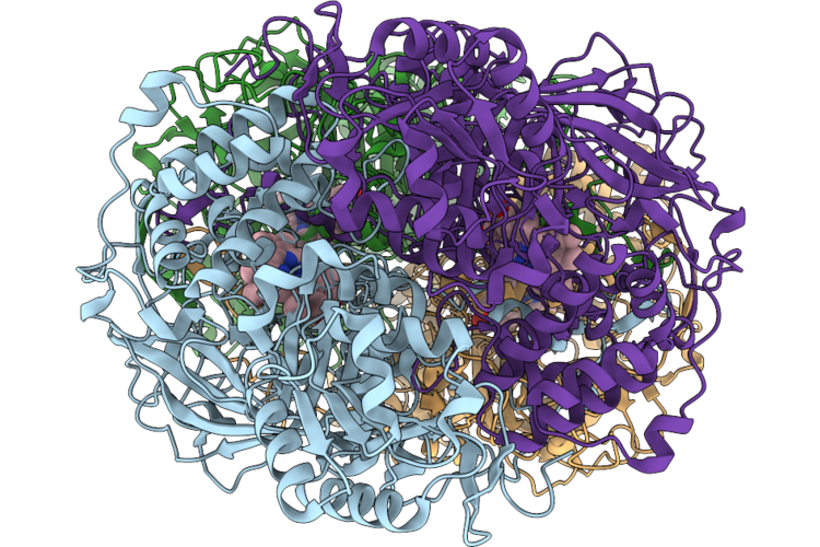
Method: ELECTRON MICROSCOPY Release Date: 2026-05-06 Classification: PROTON TRANSPORT Ligands: FES, CA, FMN, SF4, 3PE, LFA, CDL, TRD, UQ8 |
 |
Organism: Escherichia coli bl21(de3), Escherichia coli bw25113
Method: ELECTRON MICROSCOPY Release Date: 2026-05-06 Classification: PROTON TRANSPORT Ligands: FES, SF4, FMN, CA, 3PE, 7PH, UQ8 |
 |
Organism: Chloroflexi bacterium 13_1_20cm_2_59_7, Escherichia coli bl21(de3), Synthetic construct
Method: ELECTRON MICROSCOPY Release Date: 2026-04-29 Classification: RNA BINDING PROTEIN Ligands: MG |
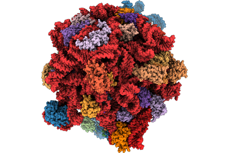 |
Doxycycline Bound E. Coli Ribosome With Rearranged Peptidyl Transferase Centre
Organism: Escherichia coli bl21(de3)
Method: ELECTRON MICROSCOPY Release Date: 2026-04-22 Classification: RIBOSOME Ligands: ZN, MG, K, DXT |
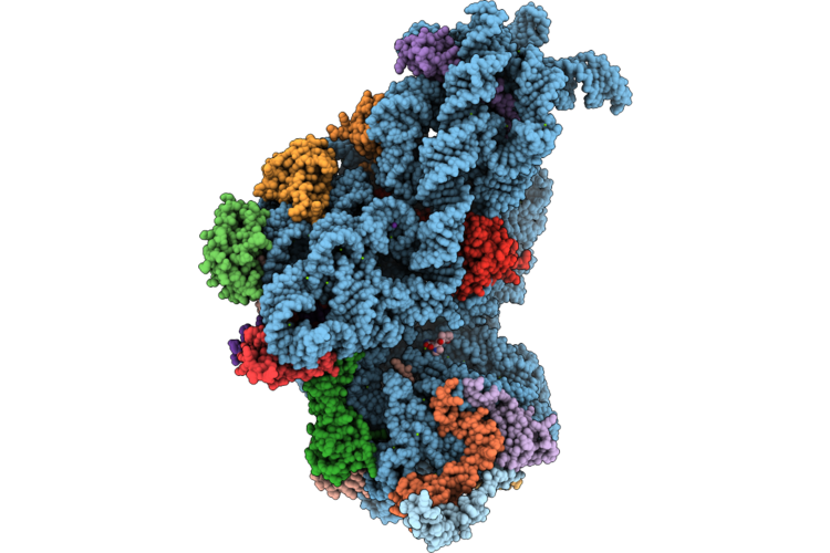 |
Organism: Escherichia coli bl21(de3)
Method: ELECTRON MICROSCOPY Release Date: 2026-04-22 Classification: RIBOSOME Ligands: ZN, DXT, MG, K |
 |
Organism: Escherichia coli bl21(de3)
Method: ELECTRON MICROSCOPY Release Date: 2026-04-01 Classification: STRUCTURAL PROTEIN |
 |
Organism: Escherichia coli bl21(de3)
Method: ELECTRON MICROSCOPY Release Date: 2026-04-01 Classification: STRUCTURAL PROTEIN Ligands: AMP |
 |
Organism: Escherichia coli bl21(de3)
Method: ELECTRON MICROSCOPY Release Date: 2026-04-01 Classification: STRUCTURAL PROTEIN Ligands: AMP |
 |
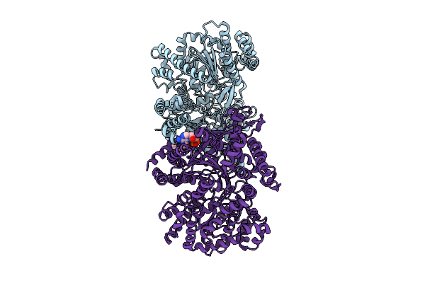
Crystal Structure Of Native Cytochrome B562 In Complex With The Synthetic Anti-Bril Antibody Bag2.
Organism: Synthetic construct, Escherichia coli bl21
Method: X-RAY DIFFRACTION Resolution:2.65 Å Release Date: 2026-03-25 Classification: PROTEIN BINDING Ligands: HEM |
 |
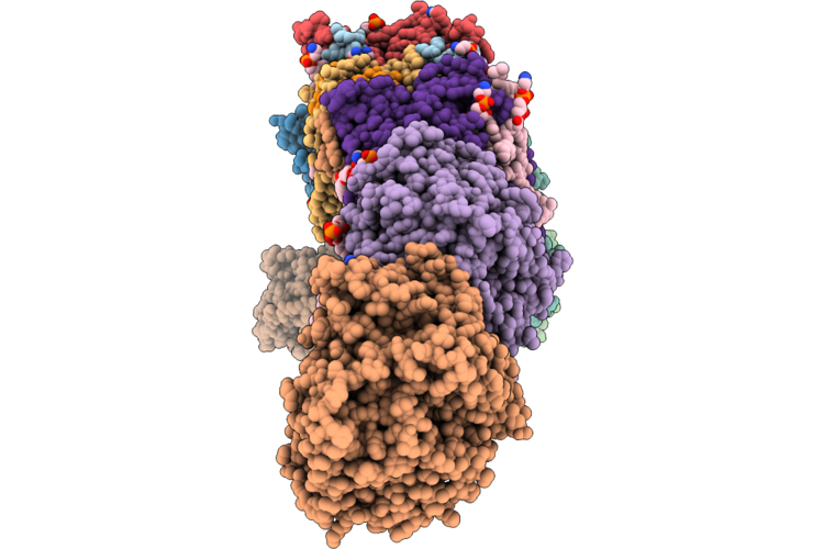
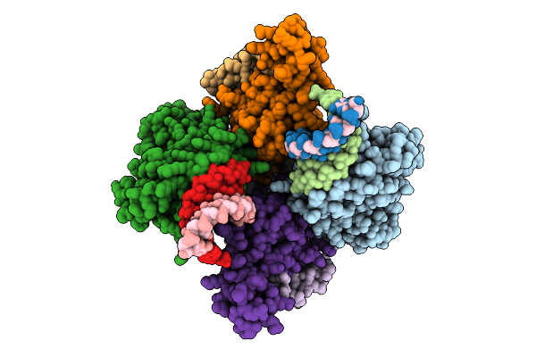
Heptamer Msp1 From S.Cerevisiae (With A Catalytic Dead Mutation) In Complex With An Unknown Peptide Substrate
Organism: Saccharomyces cerevisiae s288c, Escherichia coli bl21(de3)
Method: ELECTRON MICROSCOPY Release Date: 2026-03-18 Classification: MEMBRANE PROTEIN Ligands: ATP, MG |
 |
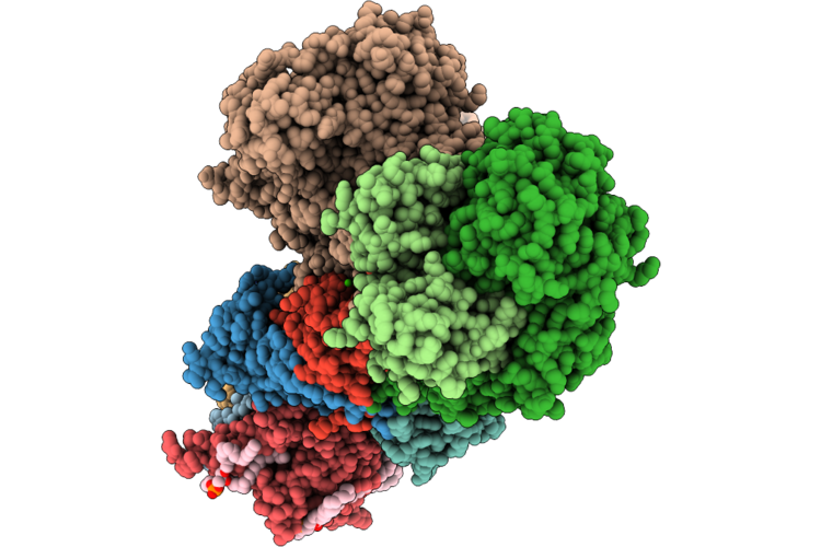
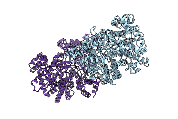
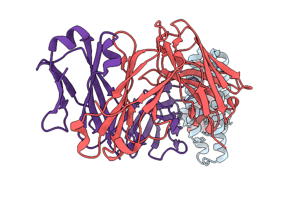
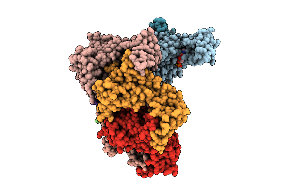
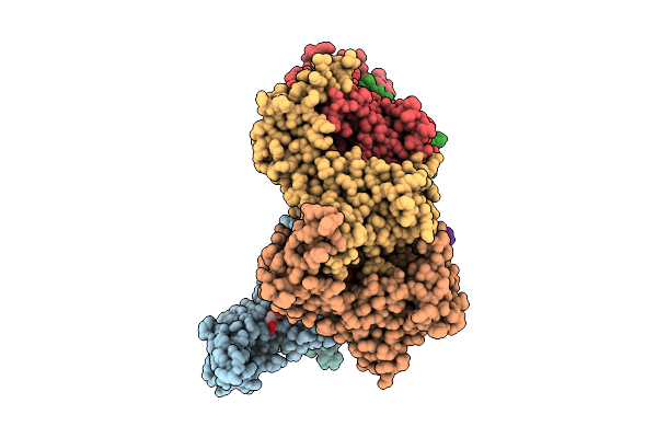
Structural Insights Into Hiv-1 Vif-Mediated Ubiquitination And Degradation Of Apobec3H
Organism: Pan troglodytes, Homo sapiens, Human immunodeficiency virus 1, Escherichia coli bl21(de3)
Method: ELECTRON MICROSCOPY Release Date: 2026-03-11 Classification: VIRAL PROTEIN/RNA Ligands: ZN |
 |
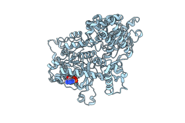
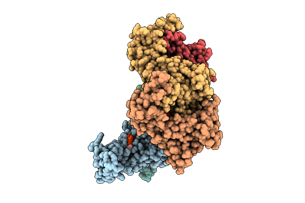
Structural Insights Into Hiv-1 Vif-Mediated Ubiquitination And Degradation Of Apobec3H
Organism: Pan troglodytes, Homo sapiens, Human immunodeficiency virus 1, Escherichia coli bl21(de3)
Method: ELECTRON MICROSCOPY Release Date: 2026-03-11 Classification: VIRAL PROTEIN/RNA Ligands: ZN |
 |
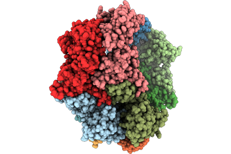
Hexamer Msp1 From S.Cerevisiae (With A Catalytic Dead Mutation) In Complex With An Unknown Peptide Substrate
Organism: Saccharomyces cerevisiae s288c, Escherichia coli bl21(de3)
Method: ELECTRON MICROSCOPY Release Date: 2026-03-11 Classification: MEMBRANE PROTEIN Ligands: ATP, MG |
 |
Hexamer Msp1 From S.Cerevisiae (With A Catalytic Dead Mutation) In Complex With An Unknown Peptide Substrate
Organism: Saccharomyces cerevisiae s288c, Escherichia coli bl21(de3)
Method: ELECTRON MICROSCOPY Release Date: 2026-03-11 Classification: MEMBRANE PROTEIN Ligands: ATP, MG |
 |
Hexamer Msp1 From S.Cerevisiae (With A Catalytic Dead Mutation) In Complex With An Unknown Peptide Substrate
Organism: Saccharomyces cerevisiae s288c, Escherichia coli bl21(de3)
Method: ELECTRON MICROSCOPY Release Date: 2026-03-11 Classification: MEMBRANE PROTEIN Ligands: ATP, MG |
 |
Octamer Msp1 From S.Cerevisiae (With A Catalytic Dead Mutation) In Complex With An Unknown Peptide Substrate
Organism: Saccharomyces cerevisiae s288c, Escherichia coli bl21(de3)
Method: ELECTRON MICROSCOPY Release Date: 2026-03-04 Classification: MEMBRANE PROTEIN Ligands: ATP, MG |
 |
Nonamer Msp1 From S.Cerevisiae (With A Catalytic Dead Mutation) In Complex With An Unknown Peptide Substrate
Organism: Saccharomyces cerevisiae s288c, Escherichia coli bl21(de3)
Method: ELECTRON MICROSCOPY Release Date: 2026-03-04 Classification: MEMBRANE PROTEIN Ligands: ATP, MG |
 |
Decamer Msp1 From S.Cerevisiae(With A Catalytic Dead Mutation) In Complex With An Unknown Peptide Substrate
Organism: Saccharomyces cerevisiae s288c, Escherichia coli bl21(de3)
Method: ELECTRON MICROSCOPY Release Date: 2026-03-04 Classification: MEMBRANE PROTEIN Ligands: ATP, MG |
 |
Organism: Escherichia coli bl21(de3)
Method: ELECTRON MICROSCOPY Release Date: 2026-02-18 Classification: STRUCTURAL PROTEIN |