
Search Count: 20,921
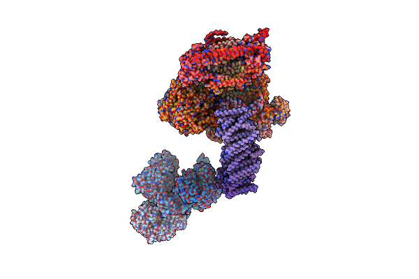 |
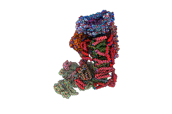
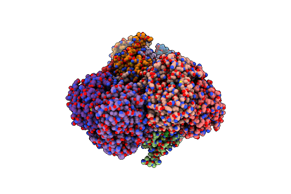
Fitted Model For Bovine Mitochondrial Supercomplex I1Iii2Iv1 By Single Particle Cryo-Em (Emd-1876)
Organism: Thermus thermophilus, Bos taurus, Escherichia coli
Method: ELECTRON MICROSCOPY Resolution:19.00 Å Release Date: 2011-10-19 Classification: OXIDOREDUCTASE Ligands: SF4, FMN, NAI, MG, FES, CA, HEM, SMA, UQ1, HEC, CDL, HEA, CU, ZN |
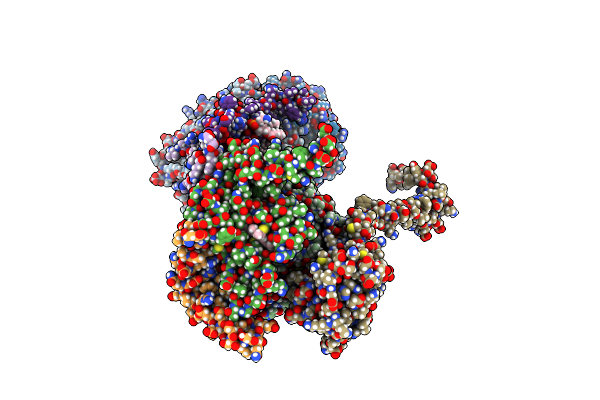 |
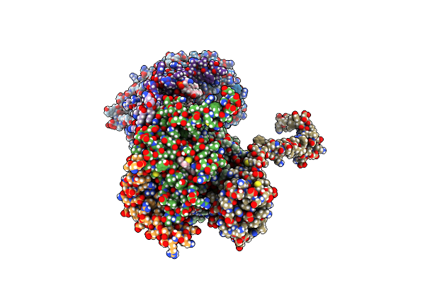
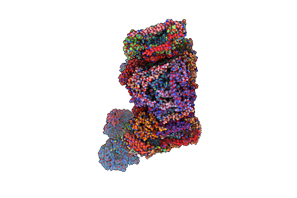
Rhodobacter Sphaeroides Mitochondrial Respiratory Chain Complex
Organism: Bos taurus
Method: X-RAY DIFFRACTION Resolution:2.80 Å Release Date: 2019-06-19 Classification: OXIDOREDUCTASE Ligands: 6PE, CDL, HEM, AZO, 8PE, HEC, FES, MC3 |
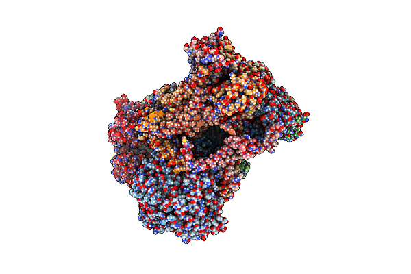 |
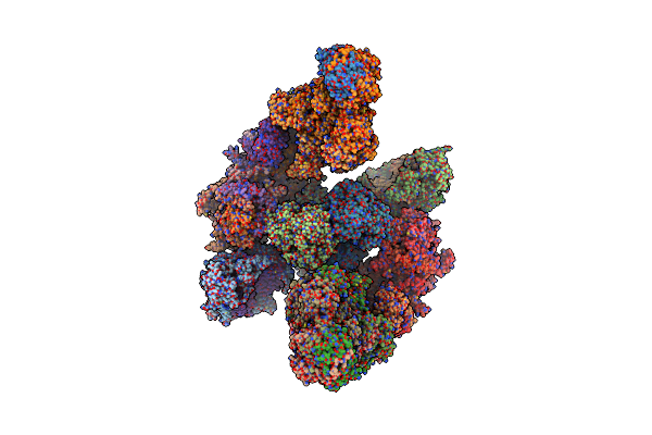
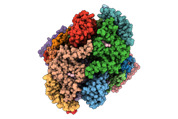
Structure Of Mitochondrial Bc1 In Complex With Ck-2-68
Organism: Bos taurus
Method: ELECTRON MICROSCOPY Release Date: 2023-02-22 Classification: OXIDOREDUCTASE Ligands: HEM, JHB, HEC, FES |
 |
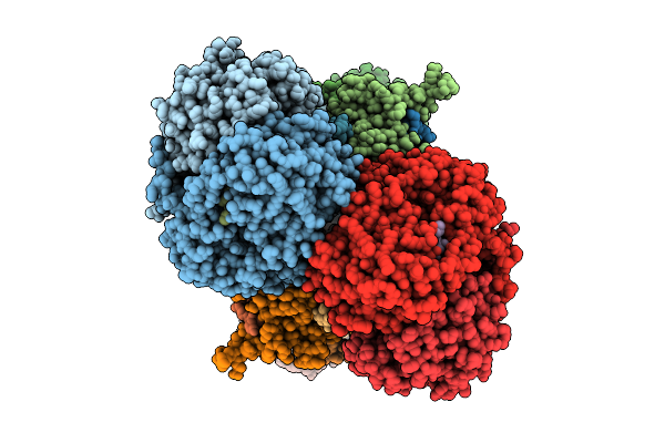
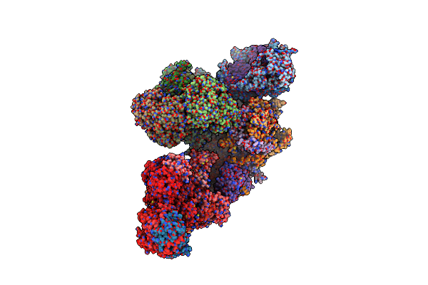
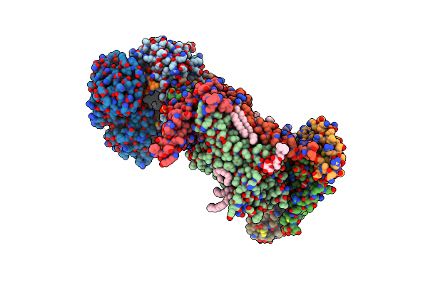
Bos Taurus Mitochondrial Bc1 In Complex With Pyramoxadone
Organism: Bos taurus
Method: X-RAY DIFFRACTION Resolution:2.95 Å Release Date: 2023-01-18 Classification: OXIDOREDUCTASE Ligands: 6PE, CDL, HEM, PQU, 8PE, PEF, CL, HEC, FES, PX4 |
 |
Cryo-Em Structure Of Complex Iii On The Bovine Heart Submitochondrial Particles, Iii-2
Organism: Bos taurus
Method: ELECTRON MICROSCOPY Release Date: 2026-04-01 Classification: OXIDOREDUCTASE Ligands: HEM, HEC, FES |
 |
Cryo-Em Structure Of Complex Iii On The Bovine Heart Submitochondrial Particles, Iii-1
Organism: Bos taurus
Method: ELECTRON MICROSCOPY Resolution:2.20 Å Release Date: 2026-04-01 Classification: MOTOR PROTEIN Ligands: HEM, HEC, FES |
 |
Cytochrome Bc1 Complex (Bos Taurus)
Organism: Bos taurus
Method: ELECTRON MICROSCOPY Release Date: 2024-07-03 Classification: MEMBRANE PROTEIN Ligands: HEM, HEC, FES |
 |
Iii2-Iv2 Mitochondrial Respiratory Supercomplex From S. Cerevisiae
Organism: Saccharomyces cerevisiae (strain atcc 204508 / s288c)
Method: ELECTRON MICROSCOPY Release Date: 2018-12-26 Classification: OXIDOREDUCTASE/ELECTRON TRANSPORT Ligands: PEF, HEM, UQ6, CDL, PCF, HEC, FES, CU, HEA, CA, MG, CUA, ZN |
 |
The Iii2-Iv(5B)2 Respiratory Supercomplex From S. Cerevisiae
Organism: Saccharomyces cerevisiae s288c
Method: ELECTRON MICROSCOPY Release Date: 2020-04-22 Classification: OXIDOREDUCTASE Ligands: CDL, PEF, HEM, PCF, HEC, FES, CU, HEA, CA, MG, CUA, ZN |
 |
Cytochrome Bc1 Bound To The 4(1H)-Pyridone Gw844520
Organism: Bos taurus
Method: X-RAY DIFFRACTION Resolution:3.57 Å Release Date: 2015-01-14 Classification: ELECTRON TRANSPORT Ligands: HEM, 4X9, PO4, PEE, HEC, CDL, FES, GOL |
 |
High Resolution Cryo-Em Structure Of Human Complex Iii In Mitochondria
Organism: Homo sapiens
Method: ELECTRON MICROSCOPY Release Date: 2025-11-12 Classification: MEMBRANE PROTEIN Ligands: CDL, 3PE, FES, PLX, HEC, HEM, PEE, U10, PC1 |
 |
Cryo-Em Of Bovine Respirasome
Organism: Bos taurus
Method: ELECTRON MICROSCOPY Release Date: 2016-11-30 Classification: OXIDOREDUCTASE Ligands: HEM, HEC, FES, CU, MG, HEA, ZN, SF4 |
 |
Structure Of Bos Taurus Cytochrome Bc1 With Fenamidone Inhibited
Organism: Bos taurus
Method: X-RAY DIFFRACTION Resolution:2.65 Å Release Date: 2016-10-12 Classification: OXIDOREDUCTASE Ligands: 6PE, CDL, GOL, HEM, FNM, 8PE, CL, HEC, PEF, FES, PX4 |
 |
Cryo-Em Architecture Of Human Respiratory Chain Megacomplex-I2Iii2Iv2
Organism: Homo sapiens, Bos taurus
Method: ELECTRON MICROSCOPY Release Date: 2017-09-13 Classification: OXIDOREDUCTASE/ELECTRON TRANSPORT Ligands: SF4, FMN, PLX, 8Q1, NDP, FES, CDL, PEE, CU, MG, HEA, ZN, HEC, HEM |
 |
Cryo-Em Structure Of Human Respiratory Supercomplex I1Iii2Iv1
Organism: Homo sapiens, Bos taurus
Method: ELECTRON MICROSCOPY Release Date: 2017-09-13 Classification: OXIDOREDUCTASE/ELECTRON TRANSPORT Ligands: SF4, FMN, PLX, 8Q1, NDP, FES, CDL, PEE, CU, MG, HEA, ZN, HEC, HEM |
 |
Cryo-Em Structure Of Human Respiratory Complex Iii (Cytochrome Bc1 Complex)
Organism: Homo sapiens
Method: ELECTRON MICROSCOPY Release Date: 2017-08-30 Classification: OXIDOREDUCTASE/ELECTRON TRANSPORT Ligands: CDL, FES, PEE, HEC, HEM, PLX |
 |
Activity Optimized Supercomplex State4
Organism: Bos taurus
Method: ELECTRON MICROSCOPY Release Date: 2022-05-18 Classification: OXIDOREDUCTASE Ligands: HEM, HEC, FES, 3PE, CDL, FMN, SF4, ZN, NAP, PC1, HEA, CU, MG |
 |
Human Kidney Respiratory Complex Iii
Organism: Homo sapiens
Method: ELECTRON MICROSCOPY Release Date: 2025-08-06 Classification: MEMBRANE PROTEIN Ligands: FES, HEC, HEM |
 |
Crystal Structure Of Bovine Cytochrome Bc1 In Complex With Inhibitor F8
Organism: Bos taurus
Method: X-RAY DIFFRACTION Resolution:3.52 Å Release Date: 2025-01-22 Classification: ELECTRON TRANSPORT Ligands: LOP, CDL, HEM, A1IKG, LMT, HEC, FES, PSC, SO4 |
 |
Activity Optimized Supercomplex State3
Organism: Bos taurus
Method: ELECTRON MICROSCOPY Release Date: 2022-05-18 Classification: OXIDOREDUCTASE Ligands: FES, CDL, 3PE, PC1, FMN, SF4, ZN, NAP, HEM, HEC, HEA, CU, MG |