

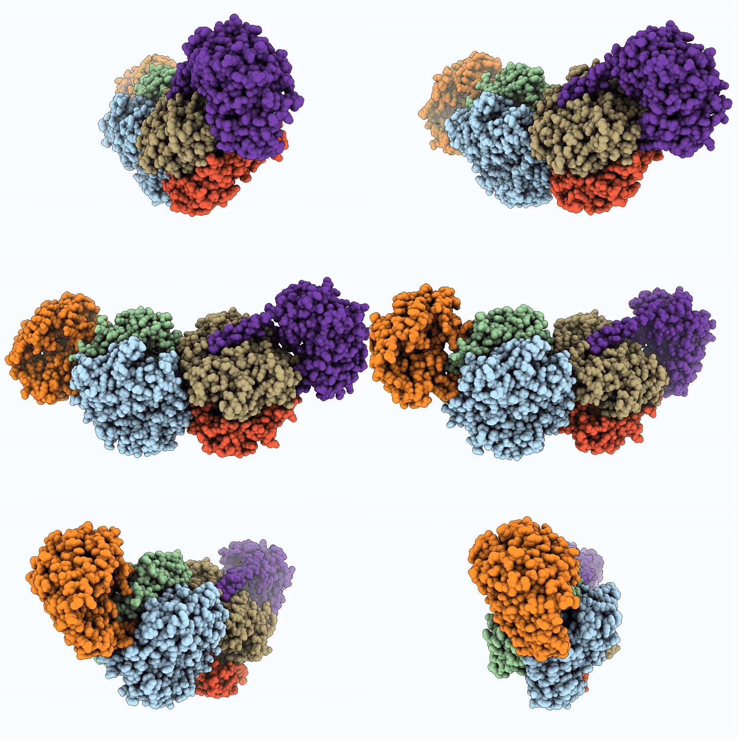
30GD image
Deposition Date 2026-04-23
Release Date 2026-05-13
Last Version Date 2026-07-15
Entry Detail
PDB ID:
30GD
Keywords:
Title:
CryoEM structure of human MATa2 in complex with MAT2B isoform v1 at 2.6 A resolution
Biological Source:
Source Organism(s):
Homo sapiens (Taxon ID: 9606)
Expression System(s):
Method Details:
Experimental Method:
Resolution:
2.90 Å
Aggregation State:
PARTICLE
Reconstruction Method:
SINGLE PARTICLE
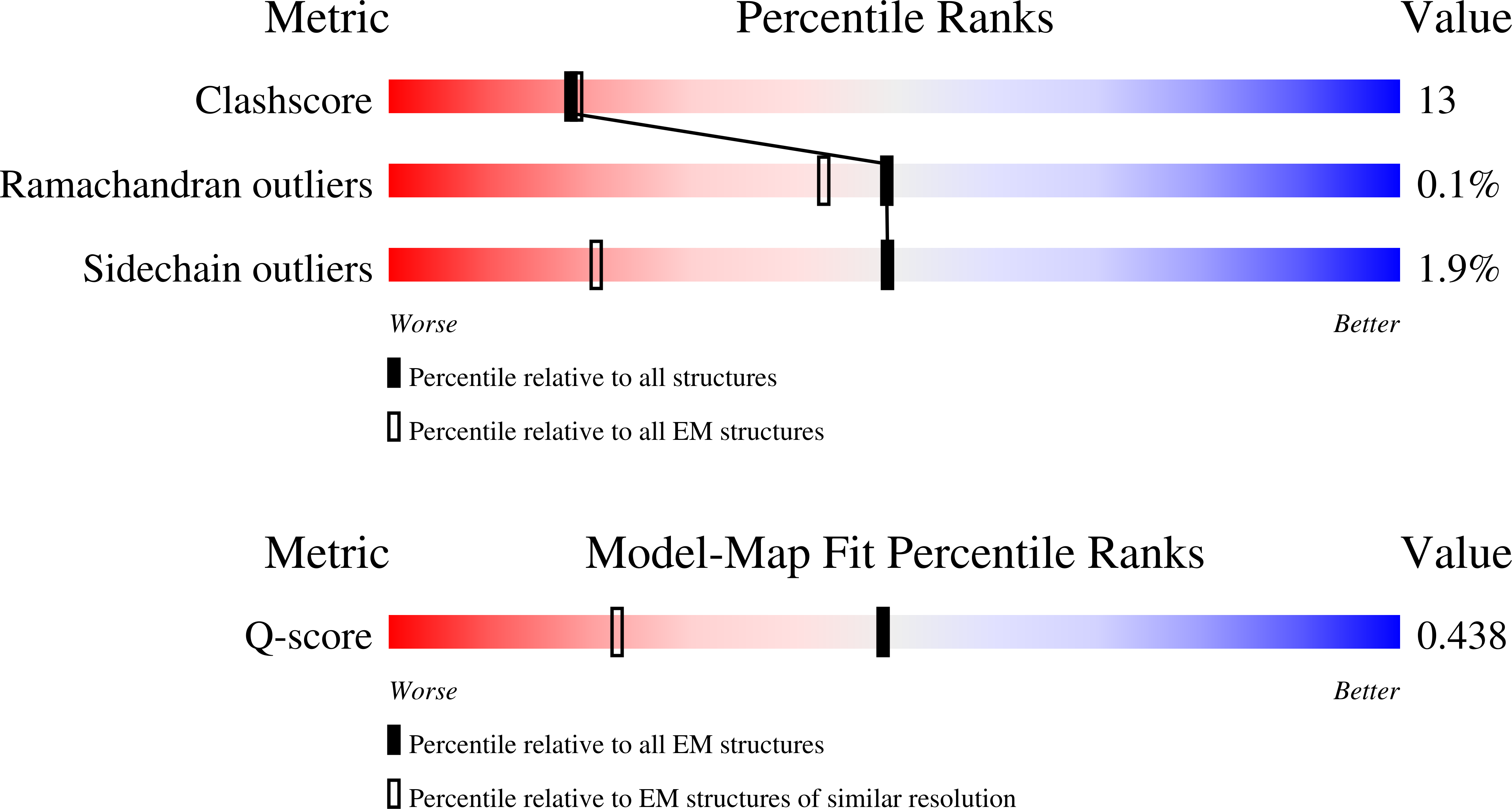
Macromolecular Entities
Polymer Type:polypeptide(L)
Molecule:S-adenosylmethionine synthase
Gene (Uniprot):MAT2A
Chain IDs:C (auth: B), D (auth: A), E (auth: C), F (auth: D)
Chain Length:395
Number of Molecules:4
Biological Source:Homo sapiens
Polymer Type:polypeptide(L)
Molecule:Isoform 1 of Methionine adeno
Gene (Uniprot):MAT2B
Chain IDs:A (auth: E), B (auth: F)
Chain Length:334
Number of Molecules:2
Biological Source:Homo sapiens
Ligand Molecules
Primary Citation
CryoEM structures reveal allosteric regulation of the catalytic activity of the multi-protein human MAT enzyme complexes.
Iucrj 13 373 384 (2026)
PMID: 42329745 DOI: 10.1107/S2052252526005075

Abstact

S-Adenosyl methionine (SAMe), the biological methyl donor essential for sustaining the life of most complex organisms, is the second most widely used cofactor, after ATP, in biochemical reactions and is synthesized by the enzyme methionine adenosyl transferase (MAT) from ATP and methionine. MAT, also known as S-adenosylmethionine synthetase, is found in almost every organism. SAMe is employed universally by different methyltransferases that catalyze the methylation of biomolecules such as nucleic acids, proteins and lipids. In plant cells SAMe produced by MAT enzymes controls the level of critical metabolites such as ethylene, polyamines and biotin, and regulates essential cellular processes such as cell division and synthesis of cell wall, chlorophyll and membrane. MAT enzyme complex MATalpha2beta, comprising the catalytic unit MATalpha2 and the regulatory protein MATbeta, is found in nearly all human tissues and is essential for providing the necessary SAMe flux for methylation of DNA and various proteins including histones. The enzymatic activity of MATalpha2 is enhanced by several fold upon complexation with both variants of MATbeta (betaV1 and betaV2). Using cryogenic electron microscopy, we determined the high-resolution resting-state structures of the MATalpha2(4)betaV1(2) and MATalpha2(4)betaV2(2) complexes, providing insights into the allosteric regulation of MAT catalytic activity, revealing how MATbetaV association could facilitate substrate binding, stabilize the transition state and promote product release to drive the catalytic cycle, and opening new possibilities for inhibitor binding.

Legend

Protein

Chemical

Disease

Primary Citation of related structures
Feedback Form
Name
Email
Institute
Feedback