

Search Count: 16
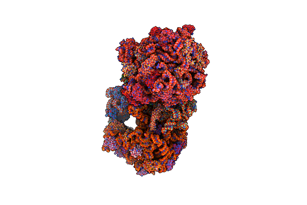 |
High-Resolution Structure Of The Escherichia Coli Ribosome
Organism: Escherichia coli (strain k12)
Method: X-RAY DIFFRACTION Resolution:3.12 Å Release Date: 2016-07-27 Classification: RIBOSOME Ligands: MG, PG4, MPD, PUT, ZN, PEG, EDO, PGE, SPD, 1PE, ACY, GUN, TRS Modified Residue(s): PSU, G7M, 2MG, 5MC, 4OC, UR3, MA6, D2T, 1MG, 5MU, 6MZ, 3TD, OMG, OMC, 2MA, OMU, MEQ, H2U |
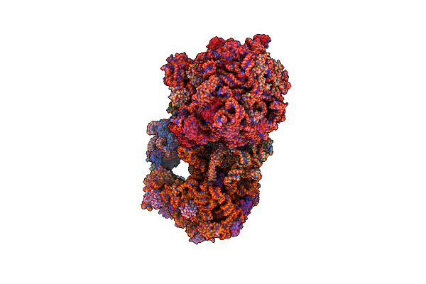 |
Structure Of The Wt E Coli Ribosome Bound To Tetracycline
Organism: Escherichia coli (strain k12)
Method: X-RAY DIFFRACTION Resolution:2.80 Å Release Date: 2016-07-27 Classification: RIBOSOME/ANTIBIOTIC Ligands: MG, PG4, MPD, PUT, TAC, ZN, PEG, EDO, PGE, SPD, 1PE, ACY, GUN, TRS Modified Residue(s): PSU, G7M, 2MG, 5MC, 4OC, UR3, MA6, D2T, MEQ, 1MG, 5MU, 6MZ, 3TD, OMG, OMC, 2MA, OMU, H2U |
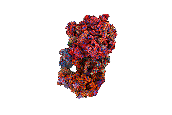 |
Structure Of The 70S E Coli Ribosome With The U1052G Mutation In The 16S Rrna Bound To Tetracycline
Organism: Escherichia coli (strain k12)
Method: X-RAY DIFFRACTION Resolution:3.00 Å Release Date: 2016-07-27 Classification: RIBOSOME/ANTIBIOTIC Ligands: MG, PG4, MPD, PUT, TAC, ZN, PEG, EDO, PGE, SPD, 1PE, ACY, GUN, TRS Modified Residue(s): PSU, G7M, 2MG, 5MC, 4OC, UR3, MA6, D2T, 1MG, 5MU, 6MZ, 3TD, OMG, OMC, 2MA, OMU, 4D4, H2U |
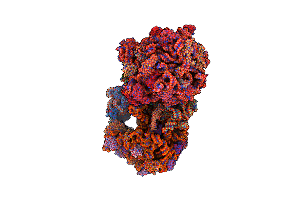 |
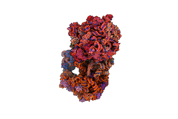
Structure Of The E Coli 70S Ribosome With The U1060A Mutation In 16S Rrna
Organism: Escherichia coli, Escherichia coli (strain k12)
Method: X-RAY DIFFRACTION Resolution:3.32 Å Release Date: 2016-07-06 Classification: ribosome/antibiotic Ligands: MG, PG4, MPD, PUT, ZN, PEG, EDO, PGE, SPD, 1PE, ACY, GUN, TRS Modified Residue(s): PSU, 7MG, 2MG, 5MC, 4OC, UR3, MA6, D2T, 1MG, 5MU, 6MZ, 3TD, OMG, OMC, 2MA, OMU, MEQ, H2U |
Organism: Escherichia coli, Escherichia coli (strain k12)
Method: X-RAY DIFFRACTION
Release Date: 2016-07-06
Ligands: MG, PG4, MPD, PUT, ZN, PEG, EDO, PGE, SPD, 1PE, ACY, GUN, TRS
 |
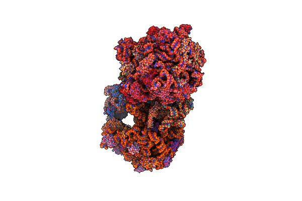
Structure Of The E Coli 70S Ribosome With The U1052G Mutation In 16S Rrna Bound To Tigecycline
Organism: Escherichia coli, Escherichia coli (strain k12), Escherichia coli o139:h28 (strain e24377a / etec)
Method: X-RAY DIFFRACTION Resolution:3.10 Å Release Date: 2016-07-06 Classification: ribosome/antibiotic Ligands: MG, PG4, MPD, PUT, T1C, ZN, PEG, EDO, PGE, SPD, 1PE, ACY, GUN, TRS Modified Residue(s): PSU, 7MG, 2MG, 5MC, 4OC, UR3, MA6, D2T, 1MG, 5MU, 6MZ, 3TD, OMG, OMC, 2MA, OMU, MEQ, H2U |
Organism: Escherichia coli, Escherichia coli (strain k12), Escherichia coli o139:h28 (strain e24377a / etec)
Method: X-RAY DIFFRACTION
Release Date: 2016-07-06
Ligands: MG, PG4, MPD, PUT, T1C, ZN, PEG, EDO, PGE, SPD, 1PE, ACY, GUN, TRS
 |
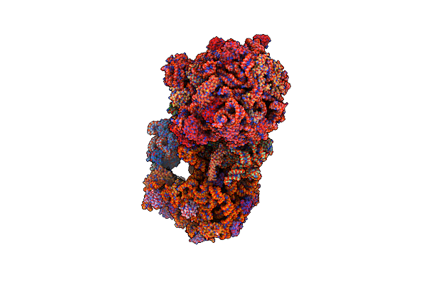
Structure Of The Wild-Type 70S E Coli Ribosome Bound To Tigecycline
Organism: Escherichia coli, Escherichia coli k-12, Escherichia coli (strain k12)
Method: X-RAY DIFFRACTION Resolution:2.96 Å Release Date: 2016-07-06 Classification: ribosome/antibiotic Ligands: MG, PG4, MPD, PUT, T1C, ZN, PEG, EDO, PGE, SPD, 1PE, ACY, GUN, TRS Modified Residue(s): PSU, G7M, 2MG, 5MC, 4OC, UR3, MA6, D2T, 1MG, 5MU, 6MZ, 3TD, OMG, OMC, 2MA, OMU, MEQ, H2U |
Organism: Escherichia coli, Escherichia coli k-12, Escherichia coli (strain k12)
Method: X-RAY DIFFRACTION
Release Date: 2016-07-06
Ligands: MG, PG4, MPD, PUT, T1C, ZN, PEG, EDO, PGE, SPD, 1PE, ACY, GUN, TRS
 |
Structure Of The Escherichia Coli Ribosome With The U1052G Mutation In The 16S Rrna
Organism: Escherichia coli k-12
Method: X-RAY DIFFRACTION Resolution:3.03 Å Release Date: 2016-07-06 Classification: RIBOSOME Ligands: MG, PG4, MPD, PUT, ZN, PEG, EDO, PGE, SPD, 1PE, ACY, GUN, TRS Modified Residue(s): PSU, G7M, 2MG, 5MC, 4OC, UR3, MA6, D2T, 1MG, 5MU, 6MZ, 3TD, OMG, OMC, 2MA, OMU, MEQ, H2U |
 |
Hin Glmu Bound To Wg188
Organism: Haemophilus influenzae
Method: X-RAY DIFFRACTION Resolution:2.10 Å Release Date: 2014-10-15 Classification: TRANSFERASE/TRANSFERASE INHIBITOR Ligands: 1S8, PG4, MG, SO4 |
Organism: Haemophilus influenzae
Method: X-RAY DIFFRACTION
Release Date: 2014-10-15
Ligands: 1S8, PG4, MG, SO4
 |
Hin Glmu Bound To Wg176
Organism: Haemophilus influenzae
Method: X-RAY DIFFRACTION Resolution:1.90 Å Release Date: 2014-10-15 Classification: TRANSFERASE/TRANSFERASE INHIBITOR Ligands: 1S9, PG4, MG, SO4 |
Organism: Haemophilus influenzae
Method: X-RAY DIFFRACTION
Release Date: 2014-10-15
Ligands: 1S9, PG4, MG, SO4
 |
Hin Glmu Bound To Wg766
Organism: Haemophilus influenzae
Method: X-RAY DIFFRACTION Resolution:2.21 Å Release Date: 2014-10-15 Classification: TRANSFERASE/TRANSFERASE INHIBITOR Ligands: SO4, MG, PG4, 1SE |
Organism: Haemophilus influenzae
Method: X-RAY DIFFRACTION
Release Date: 2014-10-15
Ligands: SO4, MG, PG4, 1SE
 |
Hin Glmu Bound To A Small Molecule Fragment
Organism: Haemophilus influenzae
Method: X-RAY DIFFRACTION Resolution:2.09 Å Release Date: 2014-10-15 Classification: TRANSFERASE Ligands: 1SF, SO4, MG, PG4 |
Organism: Haemophilus influenzae
Method: X-RAY DIFFRACTION
Release Date: 2014-10-15
Ligands: 1SF, SO4, MG, PG4
 |
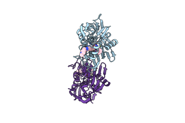
Hin Glmu Bound To Wg578
Organism: Haemophilus influenzae
Method: X-RAY DIFFRACTION Resolution:2.31 Å Release Date: 2014-10-15 Classification: TRANSFERASE Ligands: SO4, MG, PG4, 1SG |
Organism: Haemophilus influenzae
Method: X-RAY DIFFRACTION
Release Date: 2014-10-15
Ligands: SO4, MG, PG4, 1SG
 |
Lpxc From P.Aeruginosa With The Inhibitor 6-(Benzimidazol-1-Yl)-5-[4-[2-[6-[(4-Methylpiperazin-1-Yl)Methyl]-3-Pyridyl]Ethynyl]Phenyl]Pyridine-3-Carbohydroxamic Acid
Organism: Pseudomonas aeruginosa
Method: X-RAY DIFFRACTION Resolution:2.06 Å Release Date: 2014-09-24 Classification: HYDROLASE/HYDROLASE INHIBITOR Ligands: ZN, 2SZ |
Organism: Pseudomonas aeruginosa
Method: X-RAY DIFFRACTION
Release Date: 2014-09-24
Ligands: ZN, 2SZ
 |
Glmu In Complex With A Quinazoline Compound
Organism: Haemophilus influenzae
Method: X-RAY DIFFRACTION Resolution:2.00 Å Release Date: 2013-05-29 Classification: TRANSFERASE/TRANSFERASE INHIBITOR Ligands: SO4, PG4, 0N5 |
Organism: Haemophilus influenzae
Method: X-RAY DIFFRACTION
Release Date: 2013-05-29
Ligands: SO4, PG4, 0N5
 |
Thymidylate Kinase (Tmk) From S. Aureus In Complex With Tk-666
Organism: Staphylococcus aureus subsp. aureus
Method: X-RAY DIFFRACTION Resolution:1.80 Å Release Date: 2012-10-24 Classification: TRANSFERASE/TRANSFERASE INHIBITOR Ligands: 0YB |
Organism: Staphylococcus aureus subsp. aureus
Method: X-RAY DIFFRACTION
Release Date: 2012-10-24
Ligands: 0YB
 |
Glutamate Racemase (Muri) From E. Faecalis In Complex With A 9-Benzyl Purine Inhibitor
Organism: Enterococcus faecalis
Method: X-RAY DIFFRACTION Resolution:1.65 Å Release Date: 2008-06-24 Classification: ISOMERASE Ligands: I24, DGL |
Organism: Enterococcus faecalis
Method: X-RAY DIFFRACTION
Release Date: 2008-06-24
Ligands: I24, DGL