

Search Count: 21
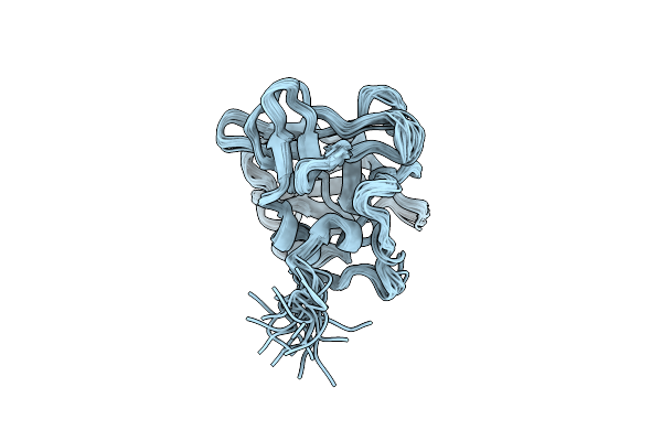 |
Structure Of The Endothelial Monocyte Activating Polypeptide Ii (Emap Ii) In Solution
Organism: Homo sapiens
Method: SOLUTION NMR, SOLUTION SCATTERING Release Date: 2024-04-10 Classification: CYTOKINE |
Organism: Homo sapiens
Method: SOLUTION NMR, SOLUTION SCATTERING
Release Date: 2024-04-10
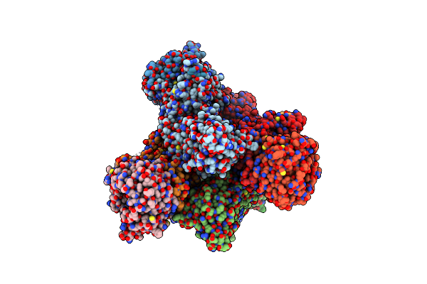 |
Structure Of The Methylosome-Lsm10/11 Complex
Organism: Homo sapiens, Mus musculus
Method: ELECTRON MICROSCOPY Release Date: 2023-08-23 Classification: TRANSFERASE Ligands: ADN |
Organism: Homo sapiens, Mus musculus
Method: ELECTRON MICROSCOPY
Release Date: 2023-08-23
Ligands: ADN
 |
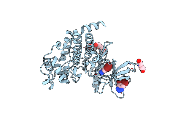
H. Sapiens Ck2 Kinase Alpha Subunit In Complex With The Atp-Competitive Inhibitor 5,6-Dibromobenzotriazole At Ph 6.5
Organism: Homo sapiens
Method: X-RAY DIFFRACTION Resolution:2.58 Å Release Date: 2022-10-19 Classification: TRANSFERASE Ligands: 7M0 |
Organism: Homo sapiens
Method: X-RAY DIFFRACTION
Release Date: 2022-10-19
Ligands: 7M0
 |
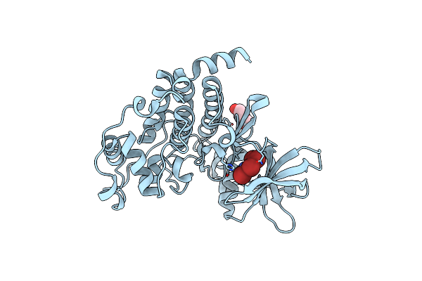
H. Sapiens Ck2 Kinase Alpha Subunit In Complex With The Atp-Competitive Inhibitor 5,6-Dibromobenzotriazole At Ph 8.5
Organism: Homo sapiens
Method: X-RAY DIFFRACTION Resolution:2.30 Å Release Date: 2022-10-19 Classification: TRANSFERASE Ligands: 7M0, CL, PEG, PG4, PGE |
Organism: Homo sapiens
Method: X-RAY DIFFRACTION
Release Date: 2022-10-19
Ligands: 7M0, CL, PEG, PG4, PGE
 |
H. Sapiens Ck2 Kinase Alpha Subunit In Complex With The Atp-Competitive Inhibitor 5,6,7,8-Tetrabromobenzotriazole (Tbbt) At Ph 8.5
Organism: Homo sapiens
Method: X-RAY DIFFRACTION Resolution:2.27 Å Release Date: 2022-10-19 Classification: TRANSFERASE Ligands: TBS, CL, PEG |
Organism: Homo sapiens
Method: X-RAY DIFFRACTION
Release Date: 2022-10-19
Ligands: TBS, CL, PEG
 |
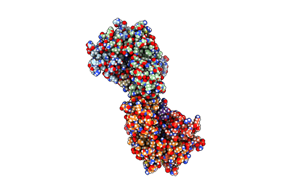
H. Sapiens Ck2 Kinase Alpha Subunit In Complex With The Atp-Competitive Inhibitor 5,6-Dibromobenzotriazole At Ph 5.5
Organism: Homo sapiens
Method: X-RAY DIFFRACTION Resolution:2.55 Å Release Date: 2022-09-28 Classification: TRANSFERASE Ligands: 7M0, FLC, PEG |
Organism: Homo sapiens
Method: X-RAY DIFFRACTION
Release Date: 2022-09-28
Ligands: 7M0, FLC, PEG
 |
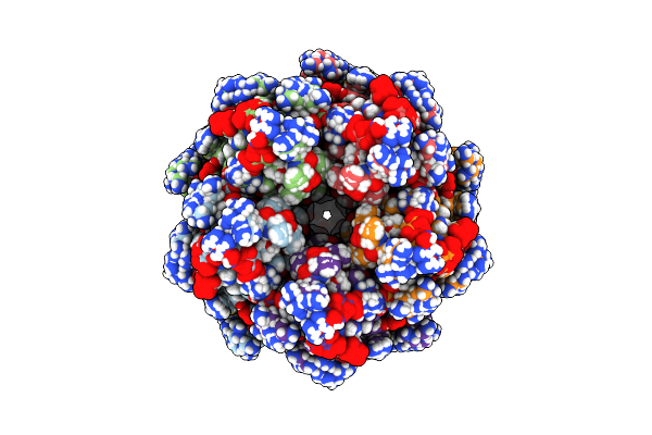
Crystal Structure Of Ctf19-Mcm21 Kinetochore Assembly Bound With Ctf19-Mcm21 Binding Motif Of Central Kinetochore Subunit Okp1
Organism: Kluyveromyces lactis nrrl y-1140
Method: X-RAY DIFFRACTION Resolution:2.10 Å Release Date: 2017-11-01 Classification: CELL CYCLE |
Organism: Kluyveromyces lactis nrrl y-1140
Method: X-RAY DIFFRACTION
Release Date: 2017-11-01
 |
Structure Of The Nucleoplasmin-Like N-Terminal Domain Of Drosophila Fkbp39
Organism: Drosophila melanogaster
Method: SOLUTION NMR Release Date: 2014-10-29 Classification: ISOMERASE |
Organism: Drosophila melanogaster
Method: SOLUTION NMR
Release Date: 2014-10-29
 |
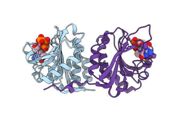
Restriction Endonuclease Dpni In Complex With Two Dna Molecules
Organism: Streptococcus pneumoniae
Method: X-RAY DIFFRACTION Resolution:2.35 Å Release Date: 2014-06-04 Classification: HYDROLASE/DNA Ligands: ZN, CA, NA |
Organism: Streptococcus pneumoniae
Method: X-RAY DIFFRACTION
Release Date: 2014-06-04
Ligands: ZN, CA, NA
 |
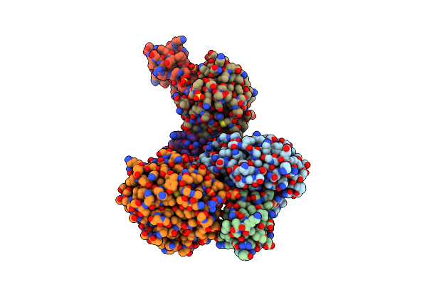
Cocrystal Structure Of The Messenger Rna 5' Cap-Binding Protein (Eif4E) Bound To 7-Methylgpppg
Organism: Mus musculus
Method: X-RAY DIFFRACTION Resolution:1.80 Å Release Date: 2002-06-12 Classification: RNA BINDING PROTEIN Ligands: MGP |
Organism: Mus musculus
Method: X-RAY DIFFRACTION
Release Date: 2002-06-12
Ligands: MGP
 |
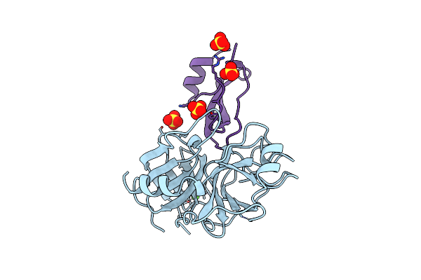
Crystal Structure Of The Bpti Ala16Leu Mutant In Complex With Bovine Trypsin
Organism: Bos taurus
Method: X-RAY DIFFRACTION Resolution:1.85 Å Release Date: 2001-03-03 Classification: HYDROLASE/INHIBITOR Ligands: SO4 |
Organism: Bos taurus
Method: X-RAY DIFFRACTION
Release Date: 2001-03-03
Ligands: SO4
 |
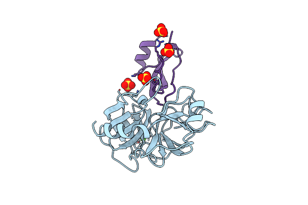
The Crystal Structures Of The Complexes Between Bovine Beta-Trypsin And Ten P1 Variants Of Bpti.
Organism: Bos taurus
Method: X-RAY DIFFRACTION Resolution:1.85 Å Release Date: 2000-03-15 Classification: HYDROLASE Ligands: CA, SO4 |
Organism: Bos taurus
Method: X-RAY DIFFRACTION
Release Date: 2000-03-15
Ligands: CA, SO4
 |
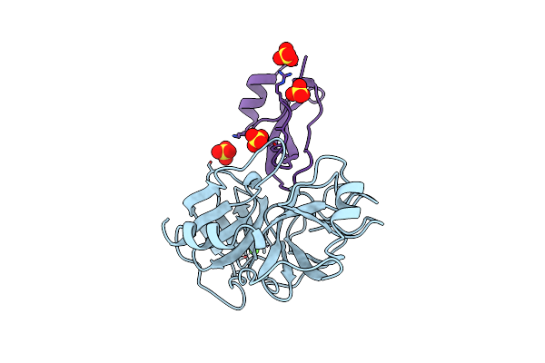
The Crystal Structures Of The Complexes Between Bovine Beta-Trypsin And Ten P1 Variants Of Bpti
Organism: Bos taurus
Method: X-RAY DIFFRACTION Resolution:1.90 Å Release Date: 2000-03-15 Classification: HYDROLASE/HYDROLASE INHIBITOR Ligands: CA, SO4 |
Organism: Bos taurus
Method: X-RAY DIFFRACTION
Release Date: 2000-03-15
Ligands: CA, SO4
 |
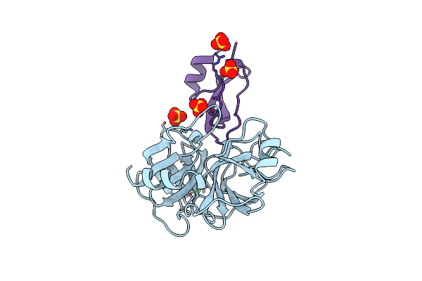
The Crystal Structures Of The Complexes Between The Bovine Beta-Trypsin And Ten P1 Variants Of Bpti.
Organism: Bos taurus
Method: X-RAY DIFFRACTION Resolution:1.90 Å Release Date: 2000-03-13 Classification: HYDROLASE/HYDROLASE INHIBITOR Ligands: CA, SO4 |
Organism: Bos taurus
Method: X-RAY DIFFRACTION
Release Date: 2000-03-13
Ligands: CA, SO4
 |
The Crystal Structures Of The Complexes Between Bovine Beta-Trypsin And Ten P1 Variants Of Bpti.
Organism: Bos taurus
Method: X-RAY DIFFRACTION Resolution:1.80 Å Release Date: 2000-03-13 Classification: HYDROLASE/HYDROLASE INHIBITOR Ligands: CA, SO4 |
Organism: Bos taurus
Method: X-RAY DIFFRACTION
Release Date: 2000-03-13
Ligands: CA, SO4
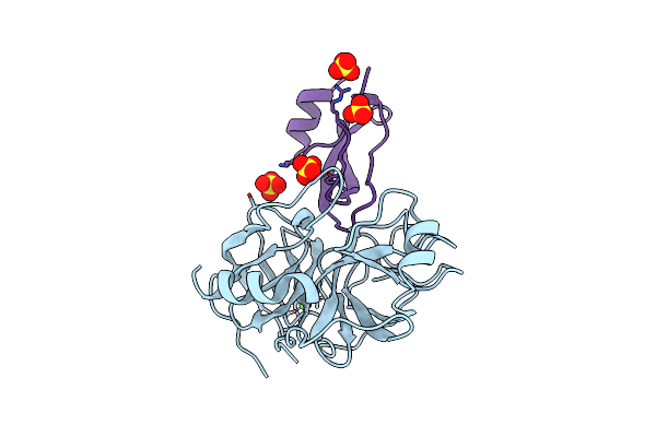 |
The Crystal Structures Of The Complexes Between Bovine Beta-Trypsin And Ten P1 Variants Of Bpti
Organism: Bos taurus
Method: X-RAY DIFFRACTION Resolution:1.90 Å Release Date: 2000-03-13 Classification: HYDROLASE/HYDROLASE INHIBITOR Ligands: CA, SO4 |
Organism: Bos taurus
Method: X-RAY DIFFRACTION
Release Date: 2000-03-13
Ligands: CA, SO4
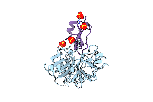 |
The Crystal Structures Of The Complexes Between Bovine Beta-Trypsin And Ten P1 Variants Of Bpti
Organism: Bos taurus
Method: X-RAY DIFFRACTION Resolution:1.75 Å Release Date: 2000-03-13 Classification: HYDROLASE/HYDROLASE INHIBITOR Ligands: CA, SO4 |
Organism: Bos taurus
Method: X-RAY DIFFRACTION
Release Date: 2000-03-13
Ligands: CA, SO4
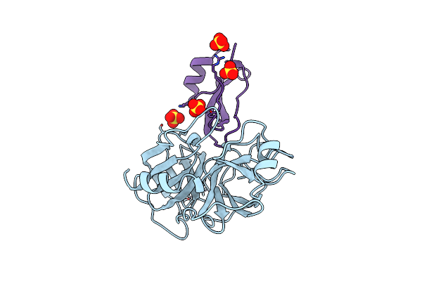 |
The Crystal Structures Of The Complexes Between Bovine Beta-Trypsin And Ten P1 Variants Of Bpti
Organism: Bos taurus
Method: X-RAY DIFFRACTION Resolution:1.85 Å Release Date: 2000-03-13 Classification: HYDROLASE/HYDROLASE INHIBITOR Ligands: CA, SO4 |
Organism: Bos taurus
Method: X-RAY DIFFRACTION
Release Date: 2000-03-13
Ligands: CA, SO4
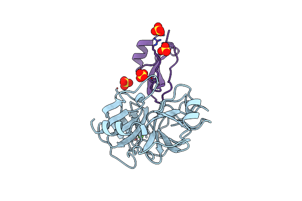 |
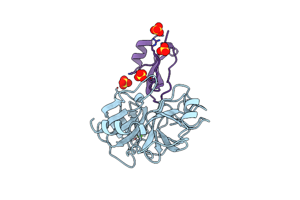
The Crystal Structures Of The Complexes Between Bovine Beta-Trypsin And Ten P1 Variants Of Bpti
Organism: Bos taurus
Method: X-RAY DIFFRACTION Resolution:1.80 Å Release Date: 2000-03-13 Classification: HYDROLASE/HYDROLASE INHIBITOR Ligands: CA, SO4 |
Organism: Bos taurus
Method: X-RAY DIFFRACTION
Release Date: 2000-03-13
Ligands: CA, SO4
 |
The Crystal Structures Of The Complexes Between Bovine Beta-Trypsin And Ten P1 Variants Of Bpti
Organism: Bos taurus
Method: X-RAY DIFFRACTION Resolution:1.90 Å Release Date: 2000-03-13 Classification: HYDROLASE/HYDROLASE INHIBITOR Ligands: CA, SO4 |
Organism: Bos taurus
Method: X-RAY DIFFRACTION
Release Date: 2000-03-13
Ligands: CA, SO4