
Search Count: 33,713
All
Selected
 |
Organism: Homo sapiens
Method: X-RAY DIFFRACTION Resolution:2.00 Å Release Date: 2026-05-06 Classification: IMMUNE SYSTEM |
 |
Organism: Homo sapiens
Method: X-RAY DIFFRACTION Resolution:2.40 Å Release Date: 2026-05-06 Classification: IMMUNE SYSTEM |
 |
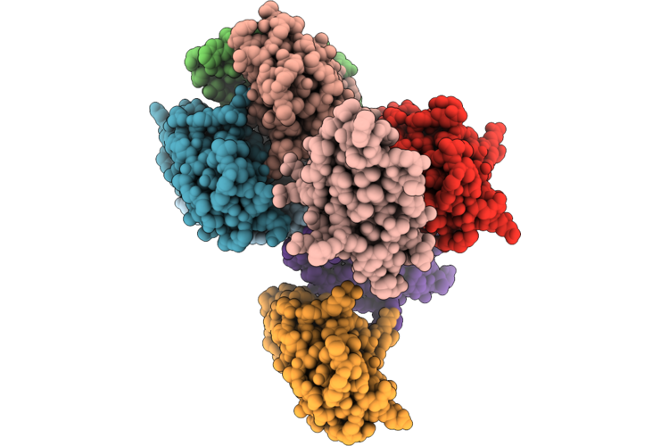
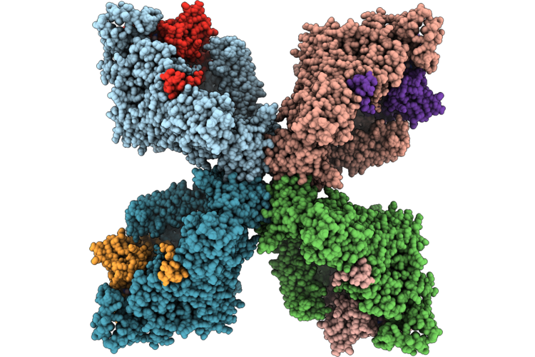
Organism: Homo sapiens, Mus musculus, Bos taurus, Escherichia coli, Synthetic construct
Method: ELECTRON MICROSCOPY Release Date: 2026-05-06 Classification: SIGNALING PROTEIN |
 |
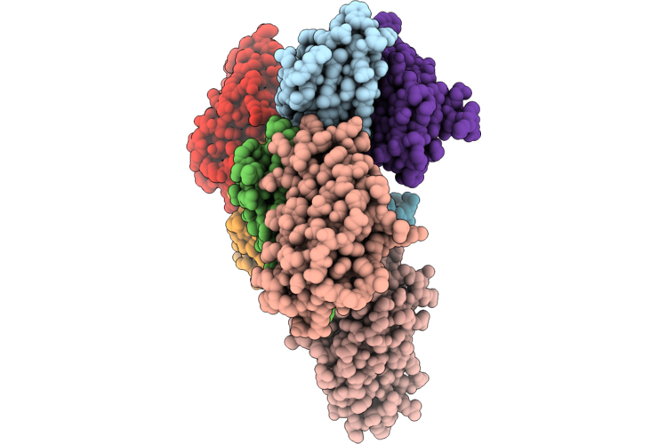
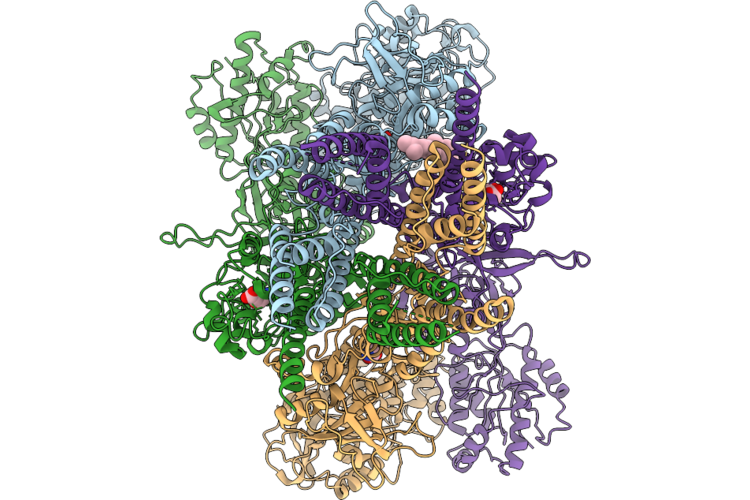
Organism: Rattus norvegicus, Bos taurus, Homo sapiens, Synthetic construct
Method: ELECTRON MICROSCOPY Release Date: 2026-05-06 Classification: SIGNALING PROTEIN/IMMUNE SYSTEM Ligands: CLR |
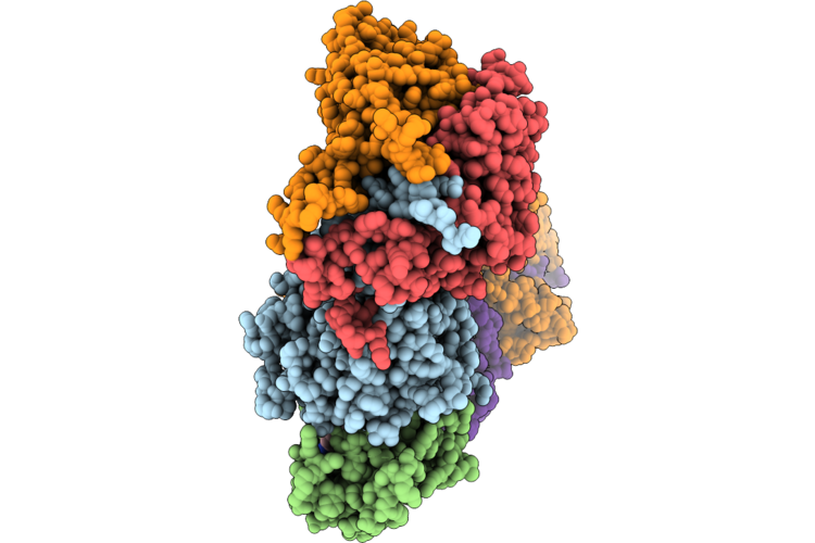 |
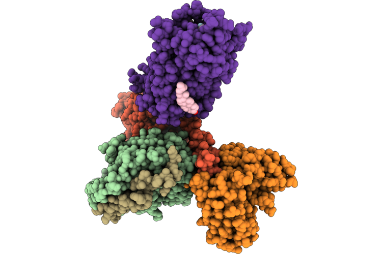
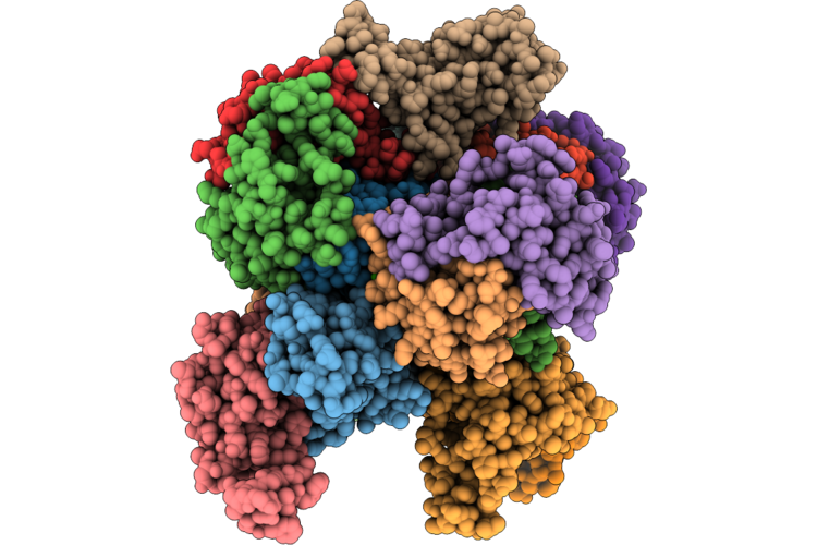
Cryo-Em Structure Of The Cul1-Rbx1-Skp1-Fbxo22 Scf Ubiquition Ligase In Complex With Nsd2 Via Unc10088
Organism: Homo sapiens
Method: ELECTRON MICROSCOPY Release Date: 2026-05-06 Classification: LIGASE Ligands: A1J20 |
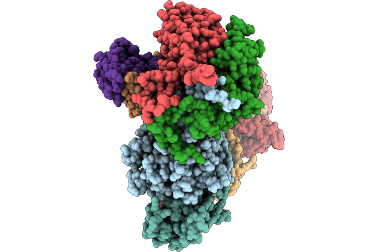 |
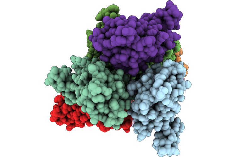
Cryo-Em Structure Of The Cul1-Rbx1-Skp1-Fbxo22 Scf Ubiquition Ligase In Complex With Nsd2, Unc10088 And Bach1
Organism: Homo sapiens
Method: ELECTRON MICROSCOPY Release Date: 2026-05-06 Classification: LIGASE Ligands: A1J20 |
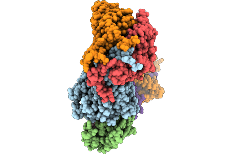 |
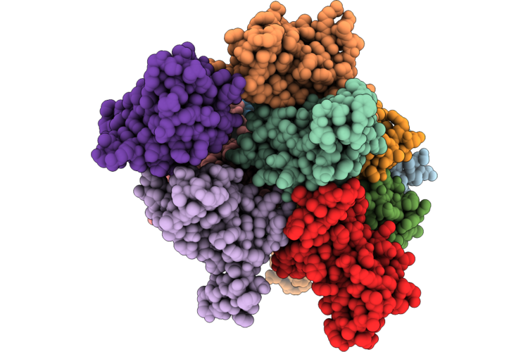
Cryo-Em Structure Of The Cul1-Rbx1-Skp1-Fbxo22 Scf Ubiquition Ligase In Complex With Nsd2 Via Unc10415667
Organism: Homo sapiens
Method: ELECTRON MICROSCOPY Release Date: 2026-05-06 Classification: LIGASE Ligands: A1J21 |
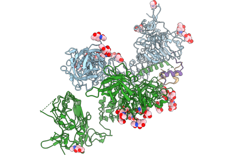 |
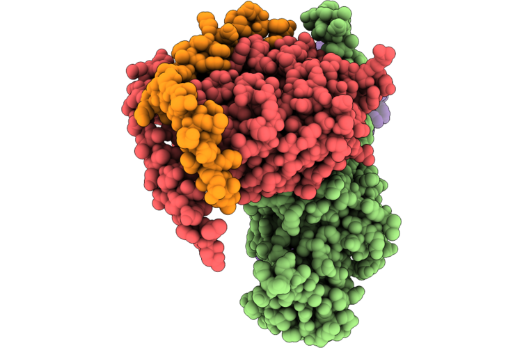
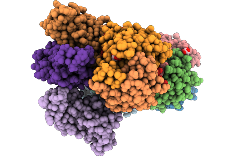
Drosophila Melanogaster Insulin Receptor Ectodomain In Complex With One Dilp2 Molecule
Organism: Drosophila melanogaster
Method: ELECTRON MICROSCOPY Resolution:3.70 Å Release Date: 2026-05-06 Classification: SIGNALING PROTEIN Ligands: NAG |
 |
Drosophila Melanogaster Insulin Receptor Ectodomain In Complex With Two Dilp2 Molecules
Organism: Drosophila melanogaster
Method: ELECTRON MICROSCOPY Release Date: 2026-05-06 Classification: SIGNALING PROTEIN Ligands: NAG |
 |
Organism: Homo sapiens
Method: X-RAY DIFFRACTION Resolution:1.54 Å Release Date: 2026-05-06 Classification: CYTOKINE Ligands: CA |
 |
Organism: Homo sapiens
Method: X-RAY DIFFRACTION Resolution:1.64 Å Release Date: 2026-05-06 Classification: CYTOKINE |
 |
Organism: Homo sapiens
Method: X-RAY DIFFRACTION Resolution:1.53 Å Release Date: 2026-05-06 Classification: CYTOKINE Ligands: TRS |
 |
Organism: Homo sapiens
Method: X-RAY DIFFRACTION Resolution:1.49 Å Release Date: 2026-05-06 Classification: CYTOKINE Ligands: MLI |
 |
Organism: Escherichia coli, Escherichia phage phiv-1
Method: ELECTRON MICROSCOPY Resolution:2.33 Å Release Date: 2026-05-06 Classification: ANTIVIRAL PROTEIN Ligands: ATP, MG |
 |
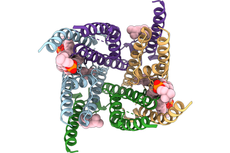
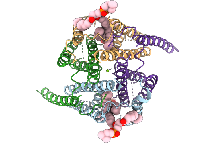
Organism: Rattus norvegicus
Method: ELECTRON MICROSCOPY Resolution:3.18 Å Release Date: 2026-05-06 Classification: MEMBRANE PROTEIN Ligands: GLY, GLU, CLR |
 |
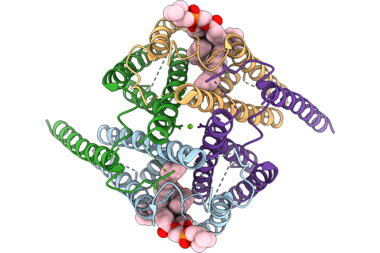
Organism: Rattus norvegicus
Method: ELECTRON MICROSCOPY Resolution:3.01 Å Release Date: 2026-05-06 Classification: MEMBRANE PROTEIN Ligands: POV, CLR |
 |
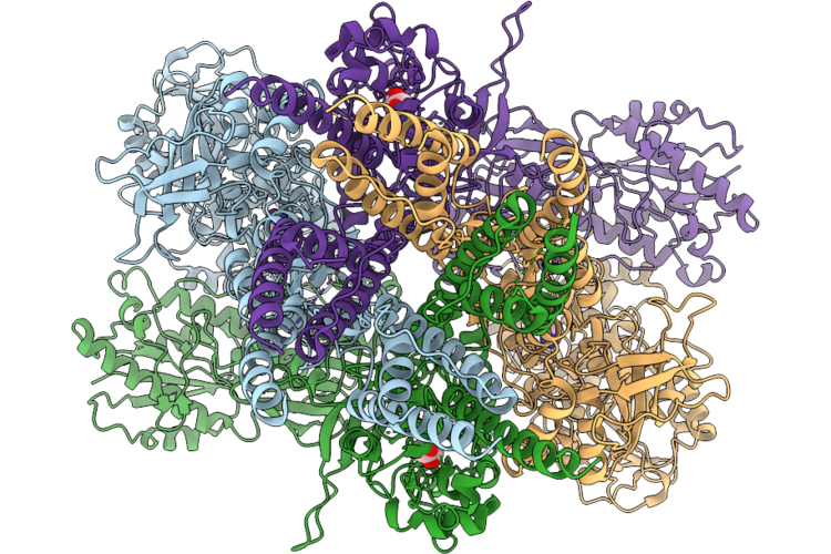
Organism: Rattus norvegicus
Method: ELECTRON MICROSCOPY Release Date: 2026-05-06 Classification: MEMBRANE PROTEIN |
 |
Organism: Rattus norvegicus
Method: ELECTRON MICROSCOPY Resolution:3.21 Å Release Date: 2026-05-06 Classification: MEMBRANE PROTEIN Ligands: POV, MG |
 |
Organism: Rattus norvegicus
Method: ELECTRON MICROSCOPY Resolution:3.15 Å Release Date: 2026-05-06 Classification: MEMBRANE PROTEIN Ligands: POV, MG |
 |
Organism: Rattus norvegicus
Method: ELECTRON MICROSCOPY Resolution:2.59 Å Release Date: 2026-05-06 Classification: MEMBRANE PROTEIN Ligands: GLY, GLU |