

Search Count: 78
 |
Hpfcold Bound Hibernating C. Burnetii 30S Ribosome
Organism: Coxiella burnetii
Method: ELECTRON MICROSCOPY Resolution:2.87 Å Release Date: 2026-04-22 Classification: RIBOSOME Ligands: ZN, K, MG |
Organism: Coxiella burnetii
Method: ELECTRON MICROSCOPY
Release Date: 2026-04-22
Ligands: ZN, K, MG
 |
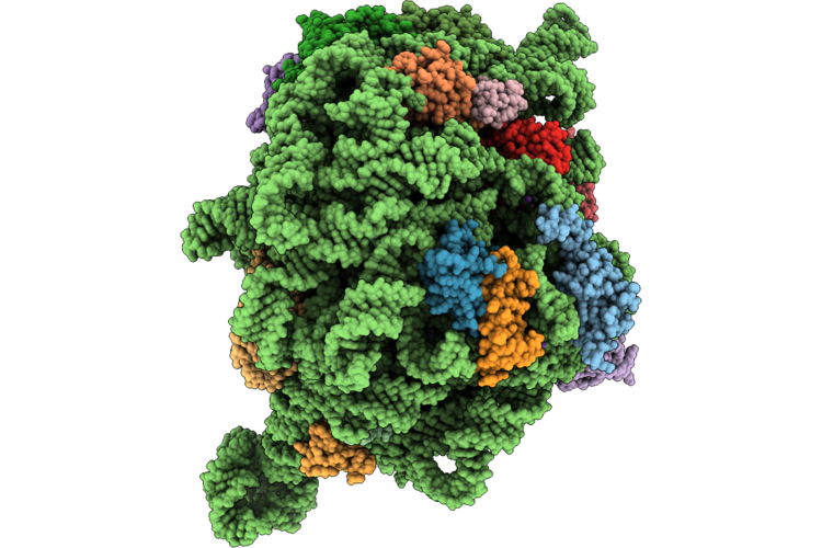
Doxycycline Bound C. Burnetii 30S Ribosome
Organism: Coxiella burnetii
Method: ELECTRON MICROSCOPY Release Date: 2026-04-22 Classification: RIBOSOME Ligands: DXT, K, MG, ZN |
Organism: Coxiella burnetii
Method: ELECTRON MICROSCOPY
Release Date: 2026-04-22
Ligands: DXT, K, MG, ZN
 |
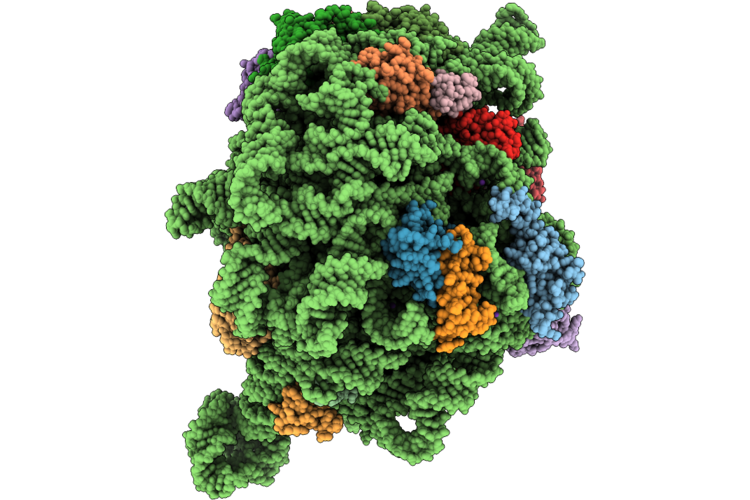
Triple Stack Doxycycline Bound 50S Subunit Of The Coxiella Burnetii Ribosome
Organism: Coxiella burnetii
Method: ELECTRON MICROSCOPY Release Date: 2026-04-22 Classification: ANTIBIOTIC Ligands: ZN, MG, K, DXT |
Organism: Coxiella burnetii
Method: ELECTRON MICROSCOPY
Release Date: 2026-04-22
Ligands: ZN, MG, K, DXT
 |
Empty 50S Subunit Of The Coxiella Burnetii Ribosome
Organism: Coxiella burnetii
Method: ELECTRON MICROSCOPY Release Date: 2026-04-22 Classification: RIBOSOME Ligands: ZN, MG, K |
Organism: Coxiella burnetii
Method: ELECTRON MICROSCOPY
Release Date: 2026-04-22
Ligands: ZN, MG, K
 |
70S Coxiella Burnetii Ribosome With Doxycycline And Hpfcold
Organism: Coxiella burnetii
Method: ELECTRON MICROSCOPY Release Date: 2026-04-22 Classification: RIBOSOME Ligands: MG, K, ZN, DXT |
Organism: Coxiella burnetii
Method: ELECTRON MICROSCOPY
Release Date: 2026-04-22
Ligands: MG, K, ZN, DXT
 |
Crystal Structure Of The Coxiella Burnetii 2-Methylisocitrate Lyase
Organism: Coxiella burnetii
Method: X-RAY DIFFRACTION Resolution:1.60 Å Release Date: 2025-08-13 Classification: CYTOSOLIC PROTEIN Ligands: EPE, EDO, PEG, CL |
Organism: Coxiella burnetii
Method: X-RAY DIFFRACTION
Release Date: 2025-08-13
Ligands: EPE, EDO, PEG, CL
 |
Crystal Structure Of The Coxiella Burnetii E110Q Mutant 2-Methylisocitrate Lyase
Organism: Coxiella burnetii
Method: X-RAY DIFFRACTION Resolution:1.82 Å Release Date: 2025-08-13 Classification: CYTOSOLIC PROTEIN Ligands: EDO, CL |
Organism: Coxiella burnetii
Method: X-RAY DIFFRACTION
Release Date: 2025-08-13
Ligands: EDO, CL
 |
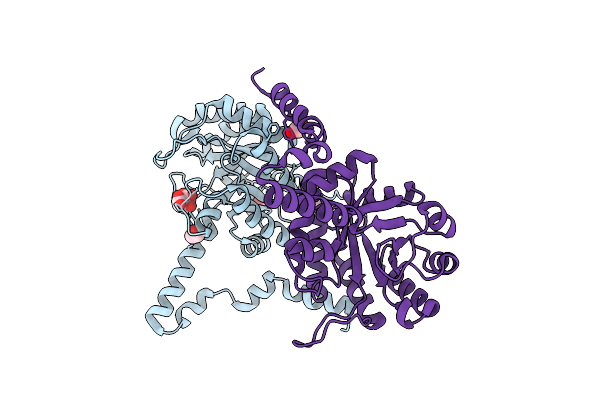
Crystal Structure Of The Coxiella Burnetii 2-Methylisocitrate Lyase Bound To Substrate 2-Mic
Organism: Coxiella burnetii
Method: X-RAY DIFFRACTION Resolution:1.90 Å Release Date: 2025-08-13 Classification: CYTOSOLIC PROTEIN Ligands: MIC, EOH, EDO, MG, CL |
Organism: Coxiella burnetii
Method: X-RAY DIFFRACTION
Release Date: 2025-08-13
Ligands: MIC, EOH, EDO, MG, CL
 |
Crystal Structure Of The Coxiella Burnetii R152Q Mutant 2-Methylisocitrate Lyase
Organism: Coxiella burnetii
Method: X-RAY DIFFRACTION Resolution:1.70 Å Release Date: 2025-08-13 Classification: CYTOSOLIC PROTEIN Ligands: CIT, EDO, CL |
Organism: Coxiella burnetii
Method: X-RAY DIFFRACTION
Release Date: 2025-08-13
Ligands: CIT, EDO, CL
 |
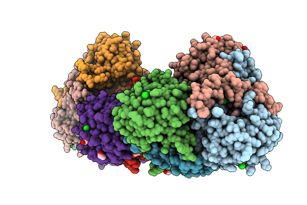
Crystal Structure Of The Coxiella Burnetii 2-Methylisocitrate Lyase Bound To Inhibitor Isocitric Acid
Organism: Coxiella burnetii
Method: X-RAY DIFFRACTION Resolution:2.33 Å Release Date: 2025-08-13 Classification: CYTOSOLIC PROTEIN Ligands: ICT, EDO, MG, CL, PEG, PGE |
Organism: Coxiella burnetii
Method: X-RAY DIFFRACTION
Release Date: 2025-08-13
Ligands: ICT, EDO, MG, CL, PEG, PGE
 |
Crystal Structure Of The Coxiella Burnetii 2-Methylisocitrate Lyase Bound To Products Succinic And Pyruvic Acid
Organism: Coxiella burnetii
Method: X-RAY DIFFRACTION Resolution:2.48 Å Release Date: 2025-08-13 Classification: CYTOSOLIC PROTEIN Ligands: SIN, PYR, EDO, MG, CL, DMS, PGE, PEG |
Organism: Coxiella burnetii
Method: X-RAY DIFFRACTION
Release Date: 2025-08-13
Ligands: SIN, PYR, EDO, MG, CL, DMS, PGE, PEG
 |
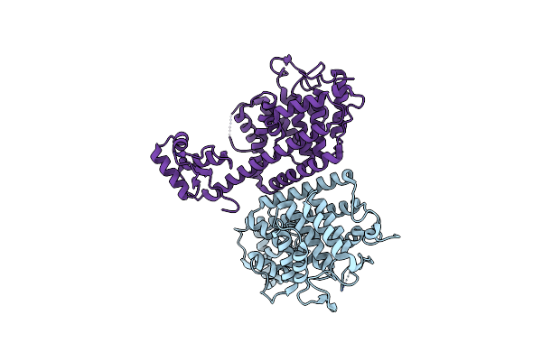
Crystal Structure Of Acetyltransferase Cbu0801 From Coxiella Burnetii
Organism: Coxiella burnetii
Method: X-RAY DIFFRACTION Resolution:1.69 Å Release Date: 2025-01-22 Classification: TRANSFERASE Ligands: COA |
Organism: Coxiella burnetii
Method: X-RAY DIFFRACTION
Release Date: 2025-01-22
Ligands: COA
 |
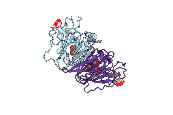
Crystal Structure Of Coxiella Burnetii Fic Protein 2
Organism: Coxiella burnetii
Method: X-RAY DIFFRACTION Resolution:1.98 Å Release Date: 2023-11-15 Classification: LIGASE |
Organism: Coxiella burnetii
Method: X-RAY DIFFRACTION
Release Date: 2023-11-15
 |
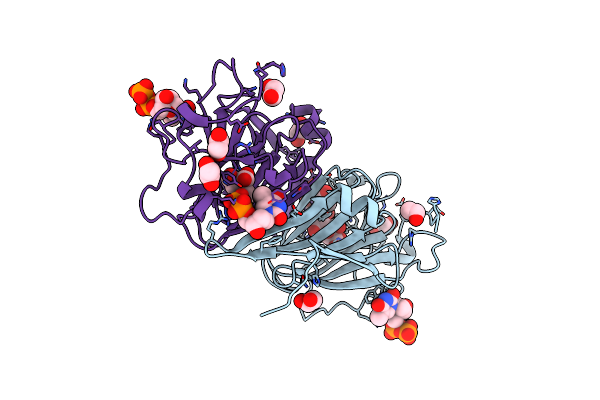
Dtdp-Sugar Epimerase
Organism: Coxiella burnetii (strain rsa 493 / nine mile phase i)
Method: X-RAY DIFFRACTION Resolution:1.43 Å Release Date: 2022-04-20 Classification: SUGAR BINDING PROTEIN Ligands: XYP, XYS, NA, FLC |
Organism: Coxiella burnetii (strain rsa 493 / nine mile phase i)
Method: X-RAY DIFFRACTION
Release Date: 2022-04-20
Ligands: XYP, XYS, NA, FLC
 |
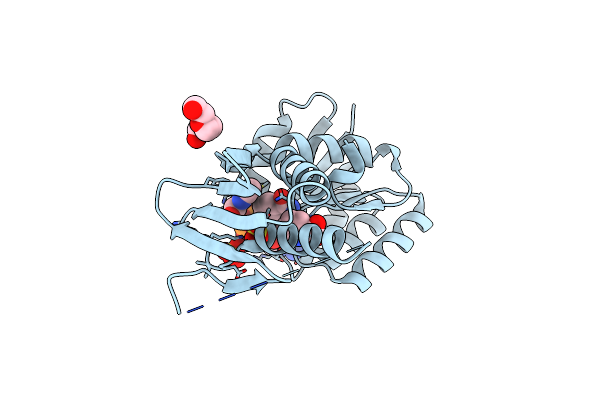
Dtdp-Sugar Epimerase From Coxiella Burnetii In Complex With Dtdp
Organism: Coxiella burnetii (strain rsa 493 / nine mile phase i)
Method: X-RAY DIFFRACTION Resolution:1.87 Å Release Date: 2022-04-20 Classification: SUGAR BINDING PROTEIN Ligands: TYD, CIT, EDO |
Organism: Coxiella burnetii (strain rsa 493 / nine mile phase i)
Method: X-RAY DIFFRACTION
Release Date: 2022-04-20
Ligands: TYD, CIT, EDO
 |
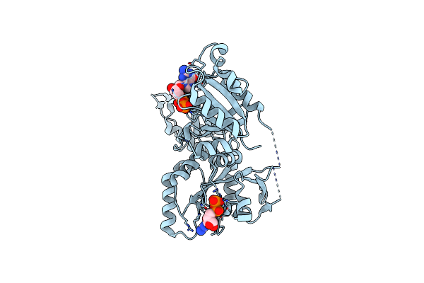
Crystal Structure Of Dihydrodipicolinate Reductase Dapb From Coxiella Burnetii
Organism: Coxiella burnetii (strain rsa 493 / nine mile phase i)
Method: X-RAY DIFFRACTION Resolution:1.70 Å Release Date: 2017-08-16 Classification: OXIDOREDUCTASE Ligands: NAP, SO4, 6PC, PE3 |
Organism: Coxiella burnetii (strain rsa 493 / nine mile phase i)
Method: X-RAY DIFFRACTION
Release Date: 2017-08-16
Ligands: NAP, SO4, 6PC, PE3
 |
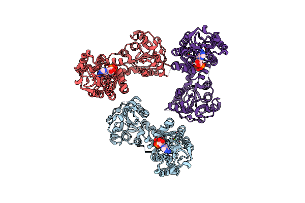
1.76 Angstrom Crystal Structure Of Gtp-Binding Protein Der From Coxiella Burnetii In Complex With Gdp.
Organism: Coxiella burnetii (strain rsa 493 / nine mile phase i)
Method: X-RAY DIFFRACTION Resolution:1.76 Å Release Date: 2015-10-14 Classification: GTP-binding protein Ligands: GDP |
Organism: Coxiella burnetii (strain rsa 493 / nine mile phase i)
Method: X-RAY DIFFRACTION
Release Date: 2015-10-14
Ligands: GDP
 |
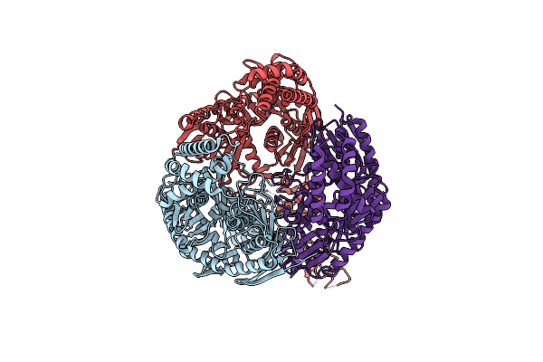
Structure Of Phosphoglycerate Kinase (Cbu_1782) From Coxiella Burnetii
Organism: Coxiella burnetii
Method: X-RAY DIFFRACTION Resolution:2.78 Å Release Date: 2015-06-24 Classification: TRANSFERASE Ligands: MG, ADP |
Organism: Coxiella burnetii
Method: X-RAY DIFFRACTION
Release Date: 2015-06-24
Ligands: MG, ADP
 |
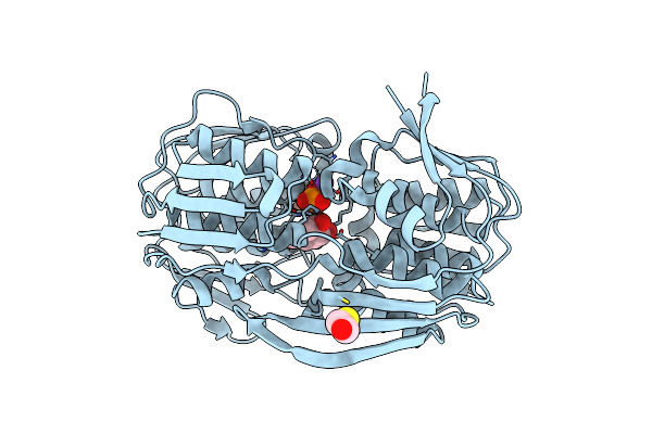
Structure Of The Polynucleotide Phosphorylase (Cbu_0852) From Coxiella Burnetii
Organism: Coxiella burnetii
Method: X-RAY DIFFRACTION Resolution:2.91 Å Release Date: 2015-06-17 Classification: TRANSFERASE Ligands: SO4 |
Organism: Coxiella burnetii
Method: X-RAY DIFFRACTION
Release Date: 2015-06-17
Ligands: SO4
 |
2.55 Angstrom Resolution Structure Of 3-Phosphoshikimate 1-Carboxyvinyltransferase (Aroa) From Coxiella Burnetii In Complex With Shikimate-3-Phosphate, Phosphate, And Potassium
Organism: Coxiella burnetii (strain rsa 493 / nine mile phase i)
Method: X-RAY DIFFRACTION Resolution:2.55 Å Release Date: 2015-05-13 Classification: TRANSFERASE Ligands: S3P, PO4, K, BME |
Organism: Coxiella burnetii (strain rsa 493 / nine mile phase i)
Method: X-RAY DIFFRACTION
Release Date: 2015-05-13
Ligands: S3P, PO4, K, BME