

Search Count: 23
 |
Crystal Structure Of Beta-Trcp Bound By Monophosphorylated Nrf2 Degron Peptide.
Organism: Homo sapiens
Method: X-RAY DIFFRACTION Resolution:1.80 Å Release Date: 2026-04-08 Classification: LIGASE |
Organism: Homo sapiens
Method: X-RAY DIFFRACTION
Release Date: 2026-04-08
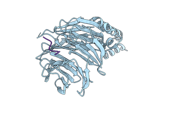 |
Crystal Structure Of Beta-Trcp Bound By Diphosphorylated Nrf2 Degron Peptide.
Organism: Homo sapiens
Method: X-RAY DIFFRACTION Resolution:2.15 Å Release Date: 2026-04-08 Classification: LIGASE |
Organism: Homo sapiens
Method: X-RAY DIFFRACTION
Release Date: 2026-04-08
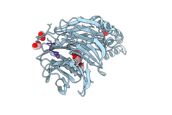 |
Crystal Structure Of Beta-Trcp Bound By Diphosphorylated I-Kappa-B-Alpha Degron Peptide
Organism: Homo sapiens
Method: X-RAY DIFFRACTION Resolution:1.16 Å Release Date: 2026-04-08 Classification: TRANSFERASE Ligands: EDO |
Organism: Homo sapiens
Method: X-RAY DIFFRACTION
Release Date: 2026-04-08
Ligands: EDO
 |
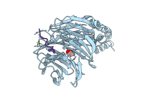
Crystal Structure Of Beta-Trcp Bound By Diphosphorylated Claspin Degron Peptide
Organism: Homo sapiens
Method: X-RAY DIFFRACTION Resolution:1.35 Å Release Date: 2026-04-08 Classification: TRANSFERASE Ligands: EDO, MG |
Organism: Homo sapiens
Method: X-RAY DIFFRACTION
Release Date: 2026-04-08
Ligands: EDO, MG
 |
Crystal Structure Of Beta-Trcp Bound By Diphosphorylated Pdcd4 Degron Peptide
Organism: Homo sapiens
Method: X-RAY DIFFRACTION Resolution:1.22 Å Release Date: 2026-04-08 Classification: TRANSFERASE Ligands: MG, EDO |
Organism: Homo sapiens
Method: X-RAY DIFFRACTION
Release Date: 2026-04-08
Ligands: MG, EDO
 |
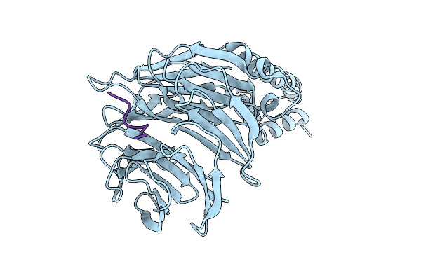
Crystal Structure Of Beta-Trcp Bound By Monophosphorylated Atf4 Degron Peptide
Organism: Homo sapiens
Method: X-RAY DIFFRACTION Resolution:2.00 Å Release Date: 2026-04-08 Classification: TRANSFERASE |
Organism: Homo sapiens
Method: X-RAY DIFFRACTION
Release Date: 2026-04-08
 |
Crystal Structure Of Beta-Trcp Bound By Monophosphorylated Wee1 Degron Peptide
Organism: Homo sapiens
Method: X-RAY DIFFRACTION Resolution:1.68 Å Release Date: 2026-04-08 Classification: TRANSFERASE |
Organism: Homo sapiens
Method: X-RAY DIFFRACTION
Release Date: 2026-04-08
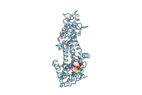 |
Structure Of Sos1 In Complex With Compound 3
Organism: Homo sapiens
Method: X-RAY DIFFRACTION Resolution:1.88 Å Release Date: 2025-09-17 Classification: CYTOSOLIC PROTEIN Ligands: A1I6H |
Organism: Homo sapiens
Method: X-RAY DIFFRACTION
Release Date: 2025-09-17
Ligands: A1I6H
 |
Polysaccharide Lyase Btpl33Ha (Bt4410) Apo Form 2
Organism: Bacteroides thetaiotaomicron vpi-5482
Method: X-RAY DIFFRACTION Resolution:2.03 Å Release Date: 2024-12-04 Classification: LYASE Ligands: ZN |
Organism: Bacteroides thetaiotaomicron vpi-5482
Method: X-RAY DIFFRACTION
Release Date: 2024-12-04
Ligands: ZN
 |
Polysaccharide Lyase Btpl33Ha (Bt4410) Y291A With Ha Dp4
Organism: Bacteroides thetaiotaomicron vpi-5482
Method: X-RAY DIFFRACTION Resolution:1.80 Å Release Date: 2024-12-04 Classification: LYASE Ligands: P4K, MES, ZN |
Organism: Bacteroides thetaiotaomicron vpi-5482
Method: X-RAY DIFFRACTION
Release Date: 2024-12-04
Ligands: P4K, MES, ZN
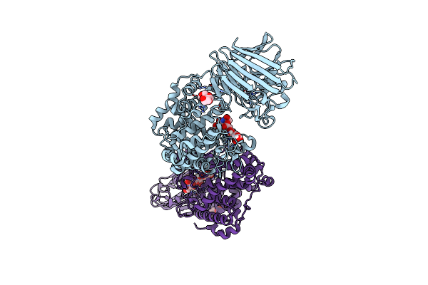 |
Polysaccharide Lyase Btpl33Ha (Bt4410) Y291A With Hadp4 Collected At 1.22 A
Organism: Bacteroides thetaiotaomicron vpi-5482
Method: X-RAY DIFFRACTION Resolution:1.92 Å Release Date: 2024-12-04 Classification: LYASE Ligands: P4K, ZN |
Organism: Bacteroides thetaiotaomicron vpi-5482
Method: X-RAY DIFFRACTION
Release Date: 2024-12-04
Ligands: P4K, ZN
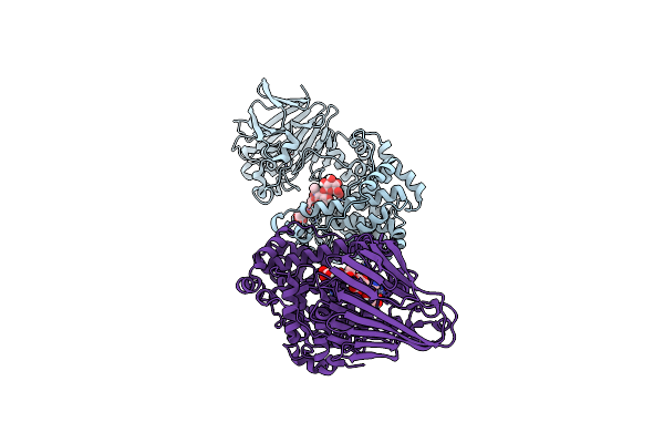 |
Polysaccharide Lyase Btpl33Ha (Bt4410) Y291A With Ha Dp4 Collected At 1.33 A
Organism: Bacteroides thetaiotaomicron vpi-5482
Method: X-RAY DIFFRACTION Resolution:2.58 Å Release Date: 2024-12-04 Classification: LYASE Ligands: ZN |
Organism: Bacteroides thetaiotaomicron vpi-5482
Method: X-RAY DIFFRACTION
Release Date: 2024-12-04
Ligands: ZN
 |
Polysaccharide Lyase Bcpl33Ha (Bcellwh2_04512) Apo Form
Organism: Bacteroides cellulosilyticus wh2
Method: X-RAY DIFFRACTION Resolution:2.11 Å Release Date: 2024-12-04 Classification: LYASE Ligands: ZN |
Organism: Bacteroides cellulosilyticus wh2
Method: X-RAY DIFFRACTION
Release Date: 2024-12-04
Ligands: ZN
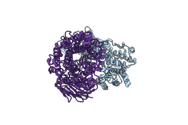 |
Polysaccharide Lyase Btpl33Ha (Bt4410) Apo Form 1
Organism: Bacteroides thetaiotaomicron vpi-5482
Method: X-RAY DIFFRACTION Resolution:2.70 Å Release Date: 2024-12-04 Classification: LYASE Ligands: ZN |
Organism: Bacteroides thetaiotaomicron vpi-5482
Method: X-RAY DIFFRACTION
Release Date: 2024-12-04
Ligands: ZN
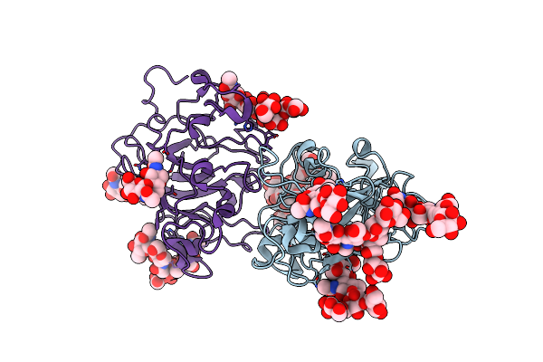 |
Crystal Structure Of A Xylan-Active Lytic Polysaccharide Monooxygenase From Pycnoporus Coccineus.
Organism: Pycnoporus cinnabarinus
Method: X-RAY DIFFRACTION Resolution:3.01 Å Release Date: 2018-01-17 Classification: OXIDOREDUCTASE Ligands: SO4 |
Organism: Pycnoporus cinnabarinus
Method: X-RAY DIFFRACTION
Release Date: 2018-01-17
Ligands: SO4
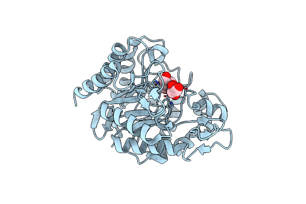 |
Crystal Structure Of Podospora Anserina Gh5 Beta-(1,4)-Mannanase
Organism: Podospora anserina
Method: X-RAY DIFFRACTION Resolution:1.40 Å Release Date: 2013-04-17 Classification: HYDROLASE Ligands: GOL, TRS |
Organism: Podospora anserina
Method: X-RAY DIFFRACTION
Release Date: 2013-04-17
Ligands: GOL, TRS
 |
Crystal Structure Of Podospora Anserina Gh26-Cbm35 Beta-(1,4)- Mannanase
Organism: Podospora anserina
Method: X-RAY DIFFRACTION Resolution:2.85 Å Release Date: 2013-04-17 Classification: HYDROLASE Ligands: CA, HG, TLA |
Organism: Podospora anserina
Method: X-RAY DIFFRACTION
Release Date: 2013-04-17
Ligands: CA, HG, TLA
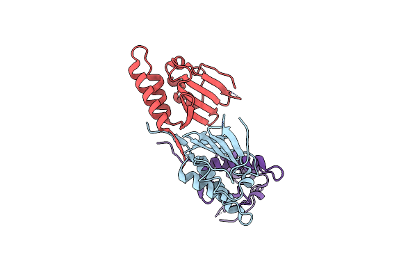 |
Crystal Structure Of Cagd From Helicobacter Pylori Pathogenicity Island
Organism: Helicobacter pylori
Method: X-RAY DIFFRACTION Resolution:2.30 Å Release Date: 2008-12-30 Classification: UNKNOWN FUNCTION |
Organism: Helicobacter pylori
Method: X-RAY DIFFRACTION
Release Date: 2008-12-30
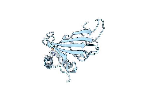 |
Structure Of Cagd From H. Pylori Pathogenicity Island Crystallized In The Presence Of Cu(Ii) Ions
Organism: Helicobacter pylori
Method: X-RAY DIFFRACTION Resolution:2.75 Å Release Date: 2008-12-30 Classification: UNKNOWN FUNCTION Ligands: CU |
Organism: Helicobacter pylori
Method: X-RAY DIFFRACTION
Release Date: 2008-12-30
Ligands: CU
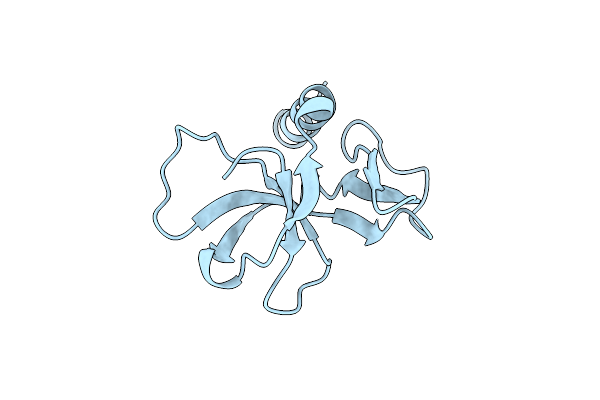 |
Ccdb, A Topoisomerase Poison From Escherichia Coli
Organism: Escherichia coli
Method: X-RAY DIFFRACTION Resolution:1.45 Å Release Date: 1998-10-14 Classification: PLASMID Ligands: CL |
Organism: Escherichia coli
Method: X-RAY DIFFRACTION
Release Date: 1998-10-14
Ligands: CL