

Search Count: 48
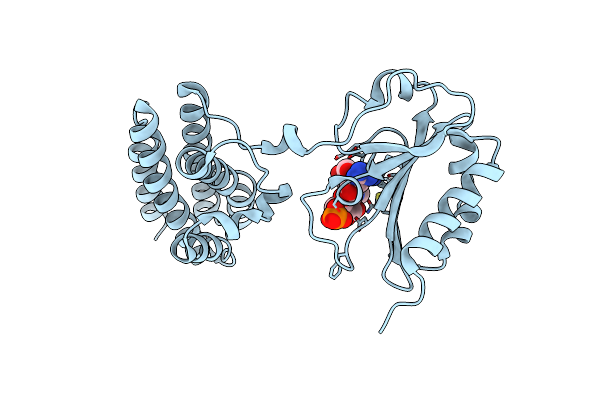 |
Structure Of Ant(6)-Ib From Campylobacter Fetus Subsp Fetus Complexed With Hydrated Streptomycin
Organism: Campylobacter fetus subsp. fetus
Method: X-RAY DIFFRACTION Resolution:1.85 Å Release Date: 2024-09-18 Classification: TRANSFERASE Ligands: 5I0, PO4 |
Organism: Campylobacter fetus subsp. fetus
Method: X-RAY DIFFRACTION
Release Date: 2024-09-18
Ligands: 5I0, PO4
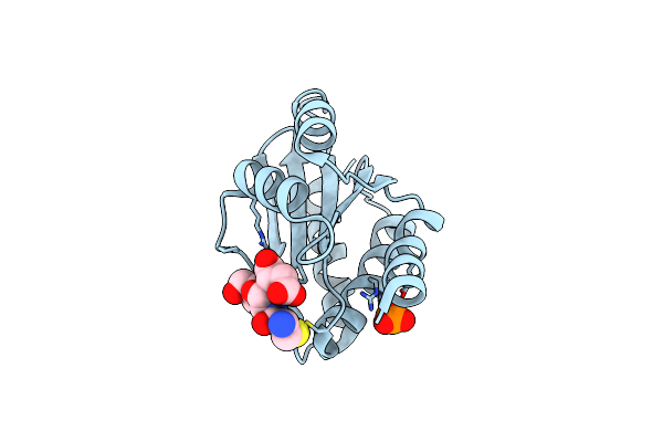 |
Brxa From Staphylococcus Aureus With Bacillithiol Mixed Disulfide
Organism: Staphylococcus aureus
Method: X-RAY DIFFRACTION Resolution:1.60 Å Release Date: 2022-04-13 Classification: OXIDOREDUCTASE Ligands: G5M, PO4 |
Organism: Staphylococcus aureus
Method: X-RAY DIFFRACTION
Release Date: 2022-04-13
Ligands: G5M, PO4
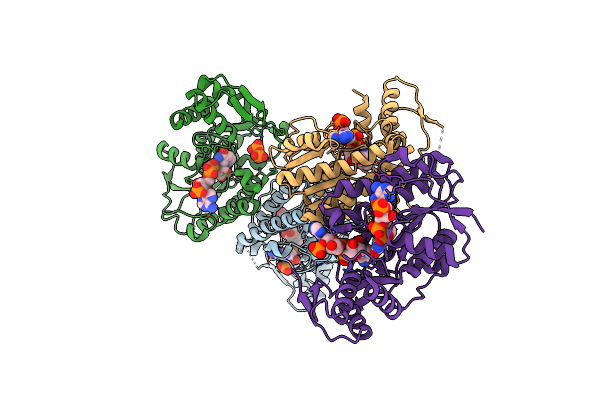 |
Structure Of Rmd From Pseudomonas Aeruginosa Complexed With Nadph
Organism: Pseudomonas aeruginosa
Method: X-RAY DIFFRACTION Resolution:1.91 Å Release Date: 2021-05-26 Classification: OXIDOREDUCTASE Ligands: NDP, NO3, POP |
Organism: Pseudomonas aeruginosa
Method: X-RAY DIFFRACTION
Release Date: 2021-05-26
Ligands: NDP, NO3, POP
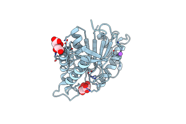 |
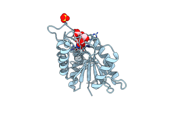
Bshb From Bacillus Subtilis Complexed With Citrate
Organism: Bacillus subtilis (strain 168)
Method: X-RAY DIFFRACTION Resolution:1.85 Å Release Date: 2020-01-08 Classification: HYDROLASE Ligands: ZN, CIT, NA |
Organism: Bacillus subtilis (strain 168)
Method: X-RAY DIFFRACTION
Release Date: 2020-01-08
Ligands: ZN, CIT, NA
 |
Bshb From Bacillus Subtilis Complexed With A Substrate Analogue
Organism: Bacillus subtilis (strain 168)
Method: X-RAY DIFFRACTION Resolution:1.45 Å Release Date: 2020-01-08 Classification: HYDROLASE Ligands: RWI, ZN, SO4 |
Organism: Bacillus subtilis (strain 168)
Method: X-RAY DIFFRACTION
Release Date: 2020-01-08
Ligands: RWI, ZN, SO4
 |
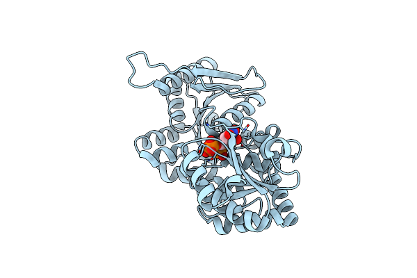
Bsha From Staphylococcus Aureus Complexed With Udp And N-Acetylglucosamine
Organism: Staphylococcus aureus subsp. aureus cn1
Method: X-RAY DIFFRACTION Resolution:2.35 Å Release Date: 2019-08-21 Classification: TRANSFERASE Ligands: UDP, NDG |
Organism: Staphylococcus aureus subsp. aureus cn1
Method: X-RAY DIFFRACTION
Release Date: 2019-08-21
Ligands: UDP, NDG
 |
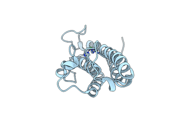
Bsha From Staphylococcus Aureus Complexed With Udp
Organism: Staphylococcus aureus
Method: X-RAY DIFFRACTION Resolution:2.00 Å Release Date: 2019-05-08 Classification: TRANSFERASE Ligands: UDP |
Organism: Staphylococcus aureus
Method: X-RAY DIFFRACTION
Release Date: 2019-05-08
Ligands: UDP
 |
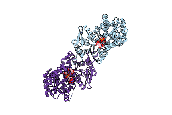
Crystal Structure Of The Bacillithiol Transferase Bsta From Staphylococcus Aureus.
Organism: Staphylococcus sp. hmsc055h04
Method: X-RAY DIFFRACTION Resolution:1.34 Å Release Date: 2018-02-21 Classification: UNKNOWN FUNCTION Ligands: NI |
Organism: Staphylococcus sp. hmsc055h04
Method: X-RAY DIFFRACTION
Release Date: 2018-02-21
Ligands: NI
 |
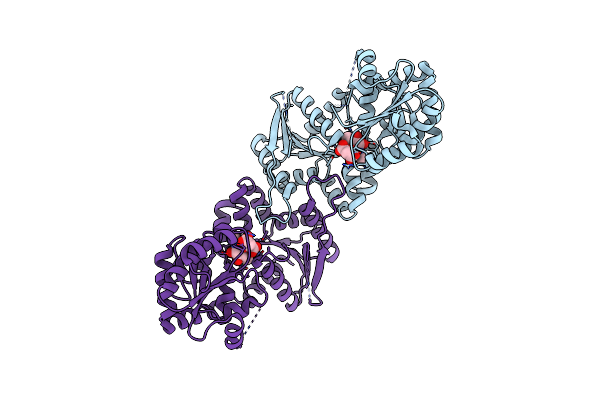
Crystal Structure Of Bsha From B. Subtilis Complexed With N-Acetylglucosaminyl-Malate And Ump
Organism: Bacillus subtilis (strain 168)
Method: X-RAY DIFFRACTION Resolution:2.15 Å Release Date: 2016-09-07 Classification: TRANSFERASE Ligands: GMT, PO4, U5P |
Organism: Bacillus subtilis (strain 168)
Method: X-RAY DIFFRACTION
Release Date: 2016-09-07
Ligands: GMT, PO4, U5P
 |
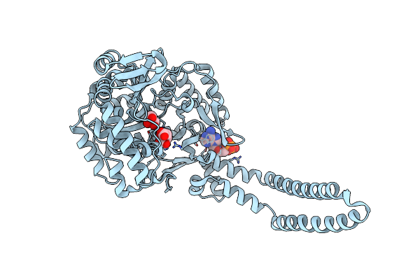
Crystal Structure Of Bsha From B. Subtilis Complexed With N-Acetylglucosaminyl-Malate
Organism: Bacillus subtilis (strain 168)
Method: X-RAY DIFFRACTION Resolution:2.02 Å Release Date: 2016-09-07 Classification: TRANSFERASE Ligands: GMT |
Organism: Bacillus subtilis (strain 168)
Method: X-RAY DIFFRACTION
Release Date: 2016-09-07
Ligands: GMT
 |
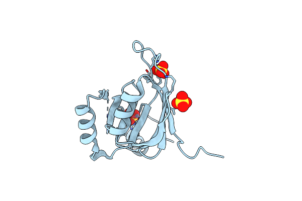
The Crystal Structure Of Bshc From Bacillus Subtilis Complexed With Citrate And Adp
Organism: Bacillus subtilis
Method: X-RAY DIFFRACTION Resolution:1.77 Å Release Date: 2014-12-31 Classification: LIGASE Ligands: ADP, CIT, GOL |
Organism: Bacillus subtilis
Method: X-RAY DIFFRACTION
Release Date: 2014-12-31
Ligands: ADP, CIT, GOL
 |
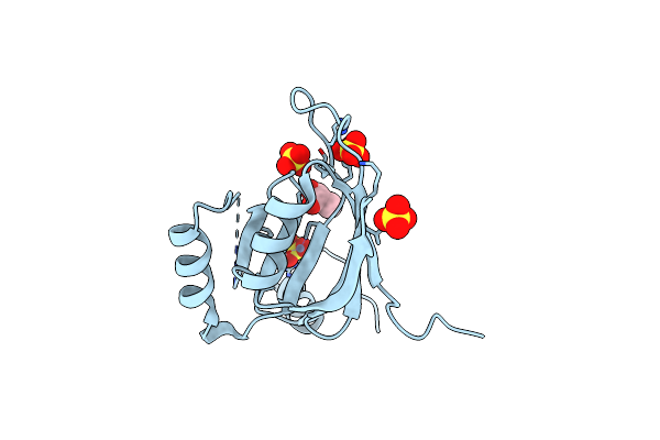
Crystal Structure Of Fosb From Staphylococcus Aureus With Zn And Sulfate At 1.42 Angstrom Resolution - Sad Phasing
Organism: Staphylococcus aureus subsp. aureus
Method: X-RAY DIFFRACTION Resolution:1.42 Å Release Date: 2014-02-26 Classification: TRANSFERASE Ligands: SO4, ZN |
Organism: Staphylococcus aureus subsp. aureus
Method: X-RAY DIFFRACTION
Release Date: 2014-02-26
Ligands: SO4, ZN
 |
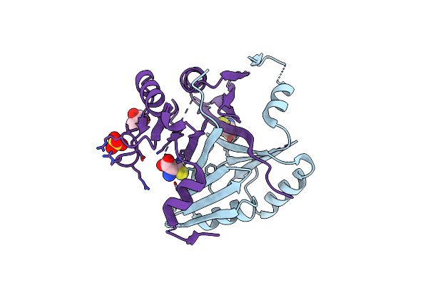
Crystal Structure Of Fosb From Staphylococcus Aureus With Zn And Sulfate At 1.15 Angstrom Resolution
Organism: Staphylococcus aureus subsp. aureus
Method: X-RAY DIFFRACTION Resolution:1.15 Å Release Date: 2014-02-26 Classification: TRANSFERASE Ligands: SO4, GOL, ZN |
Organism: Staphylococcus aureus subsp. aureus
Method: X-RAY DIFFRACTION
Release Date: 2014-02-26
Ligands: SO4, GOL, ZN
 |
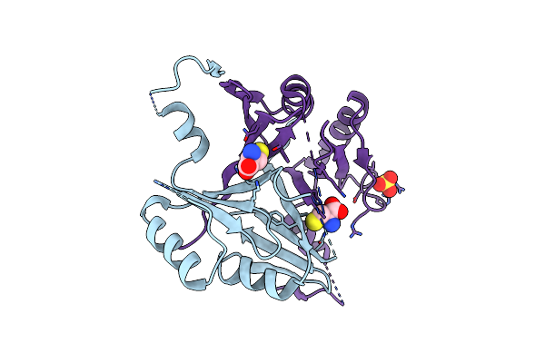
Crystal Structure Of Fosb From Staphylococcus Aureus With Bs-Cys9 Disulfide At 1.62 Angstrom Resolution
Organism: Staphylococcus aureus subsp. aureus
Method: X-RAY DIFFRACTION Resolution:1.62 Å Release Date: 2014-02-26 Classification: TRANSFERASE Ligands: SO4, CYS, GOL |
Organism: Staphylococcus aureus subsp. aureus
Method: X-RAY DIFFRACTION
Release Date: 2014-02-26
Ligands: SO4, CYS, GOL
 |
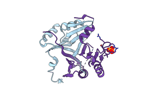
Crystal Structure Of Fosb From Staphylococcus Aureus At 1.80 Angstrom Resolution With L-Cysteine-Cys9 Disulfide
Organism: Staphylococcus aureus subsp. aureus
Method: X-RAY DIFFRACTION Resolution:1.80 Å Release Date: 2014-02-26 Classification: TRANSFERASE Ligands: CYS, SO4 |
Organism: Staphylococcus aureus subsp. aureus
Method: X-RAY DIFFRACTION
Release Date: 2014-02-26
Ligands: CYS, SO4
 |
Crystal Structure Of Fosb From Staphylococcus Aureus At 1.89 Angstrom Resolution - Apo Structure
Organism: Staphylococcus aureus
Method: X-RAY DIFFRACTION Resolution:1.89 Å Release Date: 2014-02-26 Classification: TRANSFERASE Ligands: SO4 |
Organism: Staphylococcus aureus
Method: X-RAY DIFFRACTION
Release Date: 2014-02-26
Ligands: SO4
 |
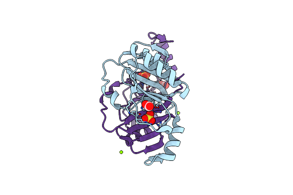
Crystal Structure Of Fosb From Bacillus Cereus With Zinc And Sulfate At 1.55 A Resolution -Sad Phasing
Organism: Bacillus cereus
Method: X-RAY DIFFRACTION Resolution:1.55 Å Release Date: 2013-10-02 Classification: TRANSFERASE Ligands: ZN, SO4, MG, GOL |
Organism: Bacillus cereus
Method: X-RAY DIFFRACTION
Release Date: 2013-10-02
Ligands: ZN, SO4, MG, GOL
 |
Crystal Structure Of Fosb From Bacillus Cereus With Zinc And Sulfate At 1.27 A Resolution
Organism: Bacillus cereus
Method: X-RAY DIFFRACTION Resolution:1.27 Å Release Date: 2013-10-02 Classification: TRANSFERASE Ligands: ZN, SO4, FMT, MG |
Organism: Bacillus cereus
Method: X-RAY DIFFRACTION
Release Date: 2013-10-02
Ligands: ZN, SO4, FMT, MG
 |
Crystal Structure Of Fosb From Bacillus Cereus With Zinc And Fosfomycin
Organism: Bacillus cereus
Method: X-RAY DIFFRACTION Resolution:1.50 Å Release Date: 2013-10-02 Classification: TRANSFERASE Ligands: ZN, MG, FCN, GOL, FMT |
Organism: Bacillus cereus
Method: X-RAY DIFFRACTION
Release Date: 2013-10-02
Ligands: ZN, MG, FCN, GOL, FMT
 |
Crystal Structure Of Fosb From Bacillus Cereus With Nickel And Fosfomycin
Organism: Bacillus cereus
Method: X-RAY DIFFRACTION Resolution:1.90 Å Release Date: 2013-10-02 Classification: TRANSFERASE Ligands: NI, FCN |
Organism: Bacillus cereus
Method: X-RAY DIFFRACTION
Release Date: 2013-10-02
Ligands: NI, FCN