

Search Count: 155
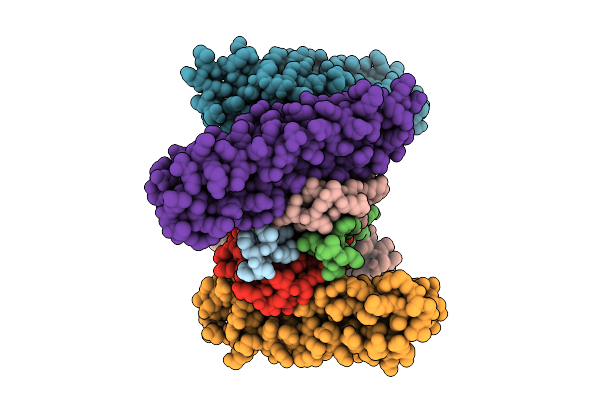 |
Crystal Structure Of Bak Bound To The Inhibitory Abak
Organism: Homo sapiens, Synthetic construct
Method: X-RAY DIFFRACTION Resolution:2.86 Å Release Date: 2025-07-16 Classification: APOPTOSIS |
Organism: Homo sapiens, Synthetic construct
Method: X-RAY DIFFRACTION
Release Date: 2025-07-16
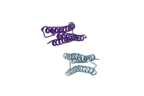 |
Computational Design Of Potent And Selective Inhibitors Of Bak And Bax
Organism: Synthetic construct
Method: X-RAY DIFFRACTION Resolution:2.81 Å Release Date: 2024-03-20 Classification: DE NOVO PROTEIN |
Organism: Synthetic construct
Method: X-RAY DIFFRACTION
Release Date: 2024-03-20
 |
Crystal Structure Of Bax Core Domain Bh3-Groove Dimer - Tetrameric Fraction P21
Organism: Homo sapiens
Method: X-RAY DIFFRACTION Resolution:2.09 Å Release Date: 2023-12-27 Classification: APOPTOSIS Ligands: EDO |
Organism: Homo sapiens
Method: X-RAY DIFFRACTION
Release Date: 2023-12-27
Ligands: EDO
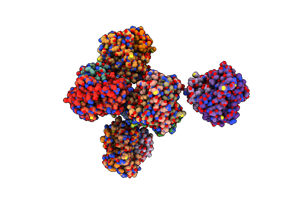 |
Crystal Structure Of Bax Core Domain Bh3-Groove Dimer - Tetrameric Fraction P31
Organism: Homo sapiens
Method: X-RAY DIFFRACTION Resolution:2.30 Å Release Date: 2023-12-27 Classification: APOPTOSIS Ligands: ZN, EDO, PEG |
Organism: Homo sapiens
Method: X-RAY DIFFRACTION
Release Date: 2023-12-27
Ligands: ZN, EDO, PEG
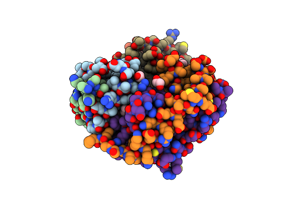 |
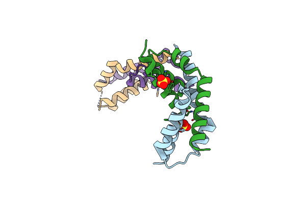
Crystal Structure Of Bax Core Domain Bh3-Groove Dimer - Hexameric Fraction With 2-Stearoyl Lysopc
Organism: Homo sapiens
Method: X-RAY DIFFRACTION Resolution:2.20 Å Release Date: 2023-12-27 Classification: APOPTOSIS Ligands: 1GP, EDO, OCT, D12, DD9 |
Organism: Homo sapiens
Method: X-RAY DIFFRACTION
Release Date: 2023-12-27
Ligands: 1GP, EDO, OCT, D12, DD9
 |
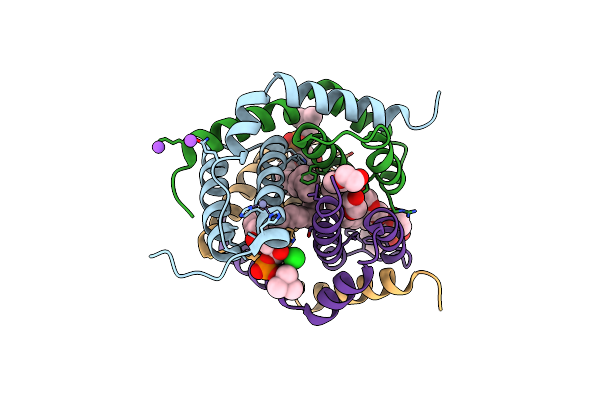
Crystal Structure Of Bax Core Domain Bh3-Groove Dimer - Hexameric Fraction With Dioctanoyl Phosphatidylserine
Organism: Homo sapiens
Method: X-RAY DIFFRACTION Resolution:2.40 Å Release Date: 2023-12-27 Classification: APOPTOSIS Ligands: SO4 |
Organism: Homo sapiens
Method: X-RAY DIFFRACTION
Release Date: 2023-12-27
Ligands: SO4
 |
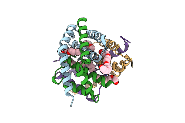
Crystal Structure Of Bak-Bax Heterodimer With Lysopc
Organism: Homo sapiens
Method: X-RAY DIFFRACTION Resolution:2.09 Å Release Date: 2023-12-27 Classification: APOPTOSIS Ligands: K6G, ZN, NA, PEG, EDO, CL |
Organism: Homo sapiens
Method: X-RAY DIFFRACTION
Release Date: 2023-12-27
Ligands: K6G, ZN, NA, PEG, EDO, CL
 |
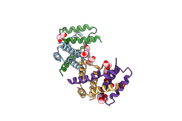
Crystal Structure Of Bak-Bax Heterodimer With C12E8
Organism: Homo sapiens
Method: X-RAY DIFFRACTION Resolution:2.40 Å Release Date: 2023-12-27 Classification: APOPTOSIS Ligands: PG0, PEG, N8E, P33, PG4 |
Organism: Homo sapiens
Method: X-RAY DIFFRACTION
Release Date: 2023-12-27
Ligands: PG0, PEG, N8E, P33, PG4
 |
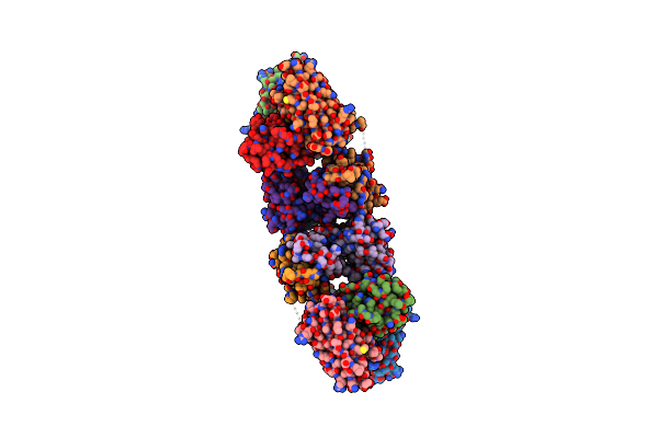
Crystal Structure Of Bax D71N Core Domain Bh3-Groove Dimer
Organism: Homo sapiens
Method: X-RAY DIFFRACTION Resolution:2.25 Å Release Date: 2023-12-27 Classification: APOPTOSIS Ligands: PEG, PGE, PG4, SO4 |
Organism: Homo sapiens
Method: X-RAY DIFFRACTION
Release Date: 2023-12-27
Ligands: PEG, PGE, PG4, SO4
 |
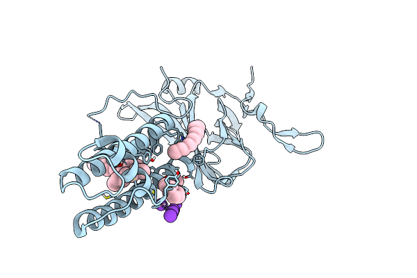
Crystal Structure Of Bak L100A In Complex With Activating Antibody Fragments
Organism: Homo sapiens, Rattus norvegicus
Method: X-RAY DIFFRACTION Resolution:3.10 Å Release Date: 2022-02-09 Classification: APOPTOSIS Ligands: MPD |
Organism: Homo sapiens, Rattus norvegicus
Method: X-RAY DIFFRACTION
Release Date: 2022-02-09
Ligands: MPD
 |
Kirbac3.1 L124M Mutant
Organism: Magnetospirillum magnetotacticum
Method: X-RAY DIFFRACTION Resolution:2.72 Å Release Date: 2021-12-29 Classification: MEMBRANE PROTEIN Ligands: PCW, TMO, OCT, K |
Organism: Magnetospirillum magnetotacticum
Method: X-RAY DIFFRACTION
Release Date: 2021-12-29
Ligands: PCW, TMO, OCT, K
 |
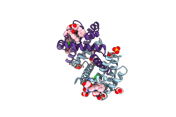
Kirbac3.1 C71S C262S
Organism: Magnetospirillum magnetotacticum
Method: X-RAY DIFFRACTION Resolution:2.40 Å Release Date: 2021-12-29 Classification: MEMBRANE PROTEIN Ligands: PCW, TMO, K |
Organism: Magnetospirillum magnetotacticum
Method: X-RAY DIFFRACTION
Release Date: 2021-12-29
Ligands: PCW, TMO, K
 |
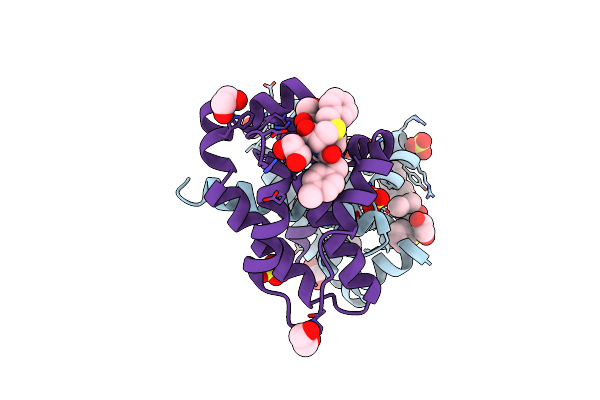
Crystal Structure Of Bcl-Xl Bound To Compound 8: (R)-3-(Benzylthio)-2-(3-(2-((4'-Chloro-[1,1'-Biphenyl]-2-Yl)Methyl)-1,2,3,4-Tetrahydroisoquinoline-6-Carbonyl)-3-(4-Methylbenzyl)Ureido)Propanoic Acid
Organism: Homo sapiens
Method: X-RAY DIFFRACTION Resolution:1.90 Å Release Date: 2021-05-05 Classification: Apoptosis/INHIBITOR Ligands: XOY, SO4, EDO |
Organism: Homo sapiens
Method: X-RAY DIFFRACTION
Release Date: 2021-05-05
Ligands: XOY, SO4, EDO
 |
Crystal Structure Of Bcl-Xl Bound To Compound 2: (2R)-3-(Benzylsulfanyl)-2-({[(4-Methylphenyl)Methyl] [(4 Phenylphenyl)Carbonyl] Carbamoyl}Amino) Propanoic Acid
Organism: Homo sapiens
Method: X-RAY DIFFRACTION Resolution:2.15 Å Release Date: 2021-05-05 Classification: Apoptosis/INHIBITOR Ligands: XOU, SO4, EDO |
Organism: Homo sapiens
Method: X-RAY DIFFRACTION
Release Date: 2021-05-05
Ligands: XOU, SO4, EDO
 |
Crystal Structure Of Bcl-Xl Bound To Compound 7: (R)-3-(Benzylthio)-2-(3-(4-Chloro-[1,1':2',1'':3'',1'''-Quaterphenyl]-4'''-Carbonyl)-3-(4-Methylbenzyl)Ureido)Propanoic Acid
Organism: Homo sapiens
Method: X-RAY DIFFRACTION Resolution:2.87 Å Release Date: 2021-05-05 Classification: Apoptosis/INHIBITOR Ligands: QHV, EDO |
Organism: Homo sapiens
Method: X-RAY DIFFRACTION
Release Date: 2021-05-05
Ligands: QHV, EDO
 |
Crystal Structure Of Bcl-Xl Bound To Compound 12: (R)-2-(3-([1,1'-Biphenyl]-4-Carbonyl)-3-(4-Methylbenzyl)Ureido)-3-((Cyclohexylmethyl)Sulfonyl)Propanoic Acid
Organism: Homo sapiens
Method: X-RAY DIFFRACTION Resolution:2.24 Å Release Date: 2021-05-05 Classification: Apoptosis/INHIBITOR Ligands: SO4, EDO, QHS |
Organism: Homo sapiens
Method: X-RAY DIFFRACTION
Release Date: 2021-05-05
Ligands: SO4, EDO, QHS
 |
Crystal Structure Of Bcl-Xl Bound To Compound 13: (R)-2-(3-([1,1'-Biphenyl]-4-Carbonyl)-3-(4-Methylbenzyl)Ureido)-3-(((3R,5R,7R)-Adamantan-1-Ylmethyl)Sulfonyl)Propanoic Acid
Organism: Homo sapiens
Method: X-RAY DIFFRACTION Resolution:2.10 Å Release Date: 2021-05-05 Classification: Apoptosis/INHIBITOR Ligands: QHP, SO4, EDO |
Organism: Homo sapiens
Method: X-RAY DIFFRACTION
Release Date: 2021-05-05
Ligands: QHP, SO4, EDO
 |
Crystal Structure Of Bcl-Xl Bound To Compound 15: (R)-2-(3-(2-((4'-Chloro-[1,1'-Biphenyl]-2-Yl)Methyl)-1,2,3,4-Tetrahydroisoquinoline-6-Carbonyl)-3-(4-Methylbenzyl)Ureido)-3-((Cyclohexylmethyl)Sulfonyl)Propanoic Acid
Organism: Homo sapiens
Method: X-RAY DIFFRACTION Resolution:2.19 Å Release Date: 2021-05-05 Classification: Apoptosis/INHIBITOR Ligands: EDO, SO4, QHJ |
Organism: Homo sapiens
Method: X-RAY DIFFRACTION
Release Date: 2021-05-05
Ligands: EDO, SO4, QHJ
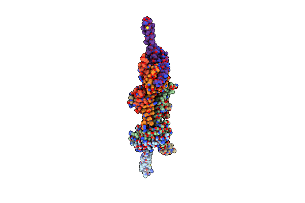 |
The Crystal Structure Of A Bak Dimer Activated By Detergent
Organism: Homo sapiens
Method: X-RAY DIFFRACTION Resolution:3.40 Å Release Date: 2021-03-31 Classification: APOPTOSIS |
Organism: Homo sapiens
Method: X-RAY DIFFRACTION
Release Date: 2021-03-31
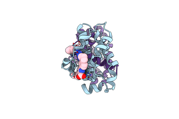 |
Crystal Structure Of Bcl-Xl In Complex With Compound 1620116, Crystal Form 2
Organism: Homo sapiens
Method: X-RAY DIFFRACTION Resolution:2.05 Å Release Date: 2021-02-24 Classification: APOPTOSIS Ligands: V9S |
Organism: Homo sapiens
Method: X-RAY DIFFRACTION
Release Date: 2021-02-24
Ligands: V9S