

Search Count: 89
 |
The Solution Structure Of The Cyanobacterial Calcium Binding Protein Cse At 293 K
Organism: Nostoc sp. pcc 7120 = fachb-418
Method: SOLUTION NMR Release Date: 2026-07-29 Classification: METAL BINDING PROTEIN Ligands: CA |
Organism: Nostoc sp. pcc 7120 = fachb-418
Method: SOLUTION NMR
Release Date: 2026-07-29
Ligands: CA
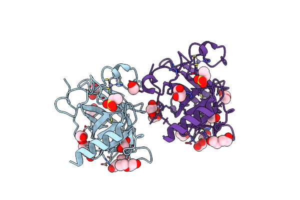 |
P53-Y220C Core Domain Covalently Bound To 2,5,6-Trifluoropyridine-3-Carbonitrile Soaked At 5 Mm
Organism: Homo sapiens
Method: X-RAY DIFFRACTION Resolution:1.57 Å Release Date: 2024-11-27 Classification: CELL CYCLE Ligands: YLW, PEG, EDO, EPE, ZN, GOL |
Organism: Homo sapiens
Method: X-RAY DIFFRACTION
Release Date: 2024-11-27
Ligands: YLW, PEG, EDO, EPE, ZN, GOL
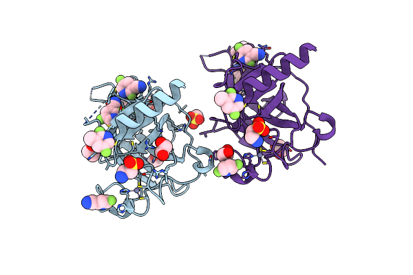 |
P53-Y220C Core Domain Covalently Bound To 2,5,6-Trifluoropyridine-3-Carbonitrile Soaked At 40 Mm
Organism: Homo sapiens
Method: X-RAY DIFFRACTION Resolution:1.69 Å Release Date: 2024-11-27 Classification: CELL CYCLE Ligands: YLW, GOL, EDO, EPE, ZN |
Organism: Homo sapiens
Method: X-RAY DIFFRACTION
Release Date: 2024-11-27
Ligands: YLW, GOL, EDO, EPE, ZN
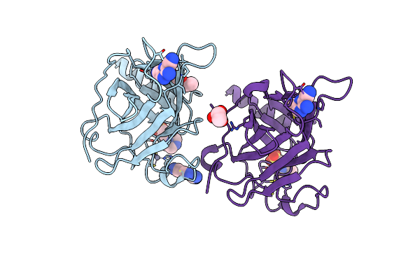 |
P53-Y220C Core Domain Covalently Bound To 3-Amino-5-Chloropyrazine-2,6-Dicarbonitrile Soaked At 5 Mm
Organism: Homo sapiens
Method: X-RAY DIFFRACTION Resolution:2.06 Å Release Date: 2024-11-27 Classification: CELL CYCLE Ligands: YM0, EPE, PEG, ZN |
Organism: Homo sapiens
Method: X-RAY DIFFRACTION
Release Date: 2024-11-27
Ligands: YM0, EPE, PEG, ZN
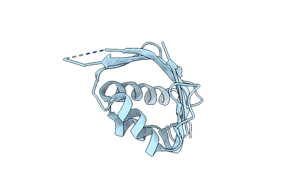 |
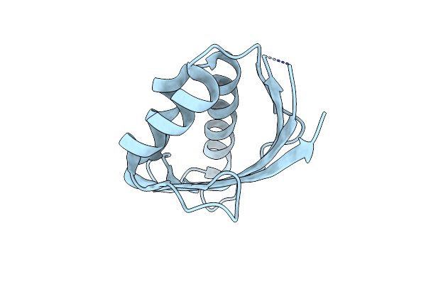
Crystal Structure Of Sam0.2
Organism: Synthetic construct
Method: X-RAY DIFFRACTION Resolution:2.65 Å Release Date: 2024-05-22 Classification: DE NOVO PROTEIN |
Organism: Synthetic construct
Method: X-RAY DIFFRACTION
Release Date: 2024-05-22
 |
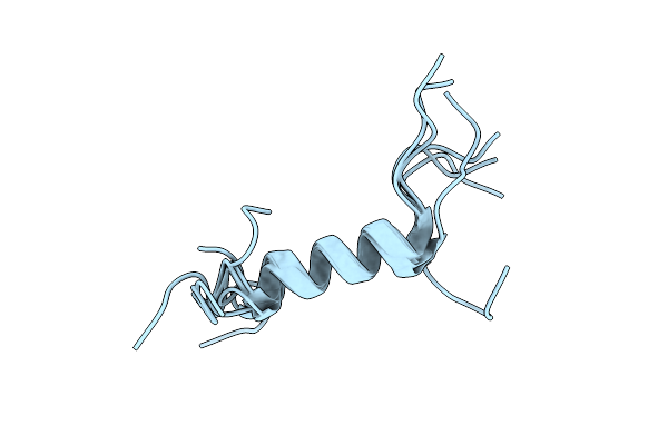
Crystal Structure Of Sam0.7
Organism: Synthetic construct
Method: X-RAY DIFFRACTION Resolution:1.80 Å Release Date: 2024-05-22 Classification: DE NOVO PROTEIN |
Organism: Synthetic construct
Method: X-RAY DIFFRACTION
Release Date: 2024-05-22
 |
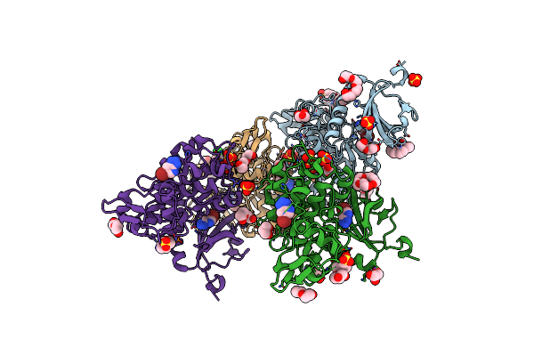
Solution Nmr Structure Of The Single Alpha Helix Peptide (P3-7)2
Organism: Unidentified
Method: SOLUTION NMR Release Date: 2023-01-25 Classification: DE NOVO PROTEIN |
Organism: Unidentified
Method: SOLUTION NMR
Release Date: 2023-01-25
 |
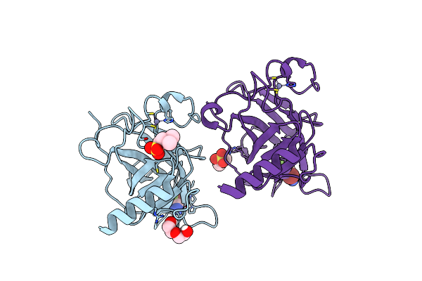
Dyrk1A In Complex With A Bromo-Triazolo-Pyridine
Organism: Homo sapiens
Method: X-RAY DIFFRACTION Resolution:2.30 Å Release Date: 2022-11-09 Classification: TRANSFERASE Ligands: IWU, PG4, PEG, PGE, GOL, EPE, 1PE, SO4, CL |
Organism: Homo sapiens
Method: X-RAY DIFFRACTION
Release Date: 2022-11-09
Ligands: IWU, PG4, PEG, PGE, GOL, EPE, 1PE, SO4, CL
 |
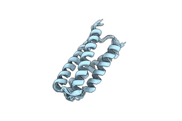
P53-Y220C Core Domain In Complex With A Bromo-Trifluoro-Pyrazole-Amine
Organism: Homo sapiens
Method: X-RAY DIFFRACTION Resolution:1.37 Å Release Date: 2022-11-09 Classification: CELL CYCLE Ligands: LE9, EPE, ZN, PEG |
Organism: Homo sapiens
Method: X-RAY DIFFRACTION
Release Date: 2022-11-09
Ligands: LE9, EPE, ZN, PEG
 |
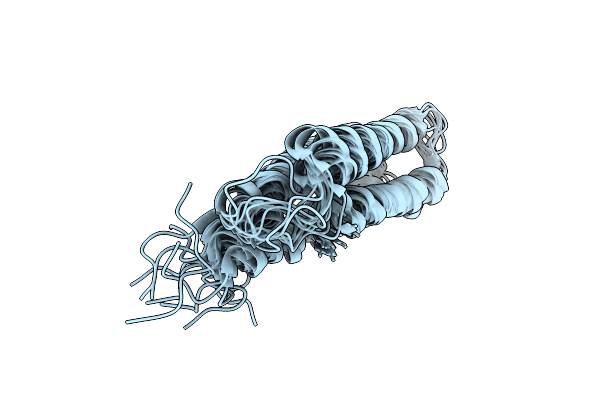
Solution Structure Of Boskar4; A De Novo Designed G-Csf Agonist
Organism: Synthetic construct
Method: SOLUTION NMR Release Date: 2021-10-06 Classification: DE NOVO PROTEIN |
Organism: Synthetic construct
Method: SOLUTION NMR
Release Date: 2021-10-06
 |
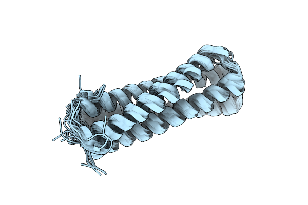
Moevan: A Designed Granulopoietic Protein By Topological Rescaffolding
Organism: Synthetic construct
Method: SOLUTION NMR Release Date: 2020-03-18 Classification: DE NOVO PROTEIN |
Organism: Synthetic construct
Method: SOLUTION NMR
Release Date: 2020-03-18
 |
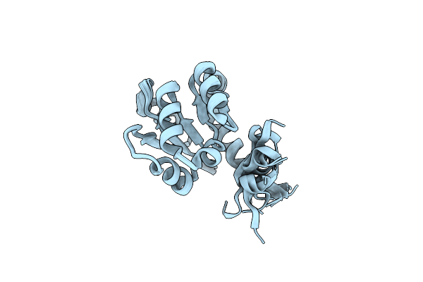
Designing A Granulopoietic Protein By Topological Rescaffolding 1: Sohair
Organism: Synthetic construct
Method: SOLUTION NMR Release Date: 2020-03-18 Classification: DE NOVO PROTEIN |
Organism: Synthetic construct
Method: SOLUTION NMR
Release Date: 2020-03-18
 |
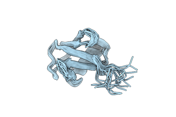
Reconstructing The Origins Of The Hemd-Like Fold
Organism: Thermus thermophilus
Method: SOLUTION NMR Release Date: 2019-11-27 Classification: FLAVOPROTEIN |
Organism: Thermus thermophilus
Method: SOLUTION NMR
Release Date: 2019-11-27
 |
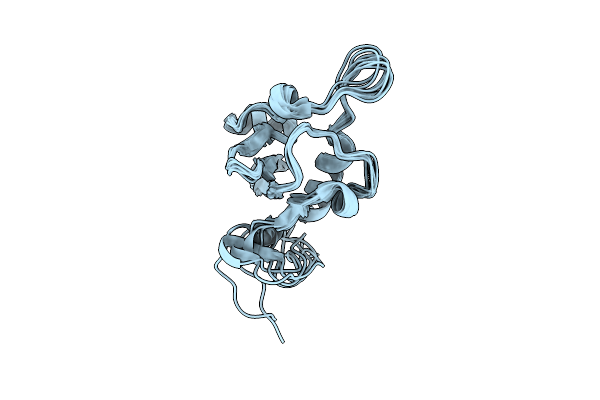
Solution Nmr Ensemble For Human Ubiquitin At 298K Compiled Using The Comand Method
Organism: Homo sapiens
Method: SOLUTION NMR Release Date: 2019-04-10 Classification: SIGNALING PROTEIN |
Organism: Homo sapiens
Method: SOLUTION NMR
Release Date: 2019-04-10
 |
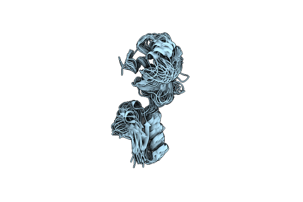
Solution Nmr Ensemble For Mlbq At 298K Compiled Using The Comand Method
Organism: Microbispora sp. atcc pta-5024
Method: SOLUTION NMR Release Date: 2019-04-10 Classification: SIGNALING PROTEIN |
Organism: Microbispora sp. atcc pta-5024
Method: SOLUTION NMR
Release Date: 2019-04-10
 |
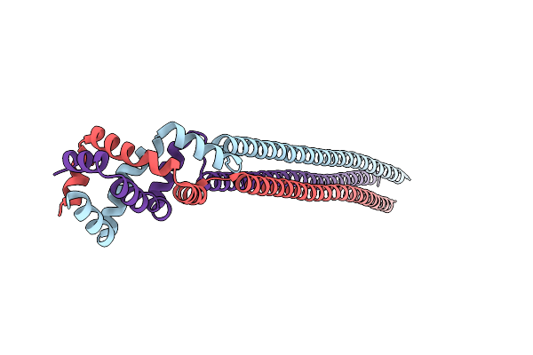
Solution Nmr Ensemble For A Chimeric Kh-S1 Domain Construct Of Exosomal Polynucleotide Phosphrylase At 298K Compiled Using The Comand Method
Organism: Escherichia coli (strain se11)
Method: SOLUTION NMR Release Date: 2019-04-10 Classification: SIGNALING PROTEIN |
Organism: Escherichia coli (strain se11)
Method: SOLUTION NMR
Release Date: 2019-04-10
 |
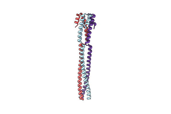
Kcr_0859 Delta Tm From Korarchaeum Cryptofilum
Organism: Korarchaeum cryptofilum (strain opf8)
Method: X-RAY DIFFRACTION Resolution:2.19 Å Release Date: 2019-02-13 Classification: MEMBRANE PROTEIN |
Organism: Korarchaeum cryptofilum (strain opf8)
Method: X-RAY DIFFRACTION
Release Date: 2019-02-13
 |
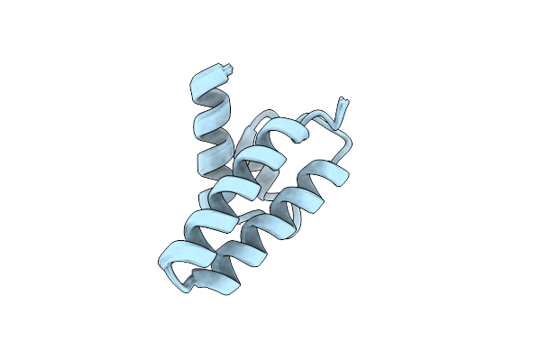
Coiled-Coil Domain-Containing Protein 90B Residues 43-125 From Homo Sapiens Fused To A Gcn4 Adaptor
Organism: Homo sapiens, Saccharomyces cerevisiae (strain atcc 204508 / s288c)
Method: X-RAY DIFFRACTION Resolution:2.10 Å Release Date: 2019-02-13 Classification: MEMBRANE PROTEIN |
Organism: Homo sapiens, Saccharomyces cerevisiae (strain atcc 204508 / s288c)
Method: X-RAY DIFFRACTION
Release Date: 2019-02-13
 |
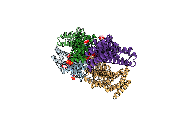
A Computationally Designed Drp Lyase Domain Reconstructed From Two Heterologous Fragments
Organism: Synthetic construct
Method: SOLUTION NMR Release Date: 2018-12-12 Classification: UNKNOWN FUNCTION |
Organism: Synthetic construct
Method: SOLUTION NMR
Release Date: 2018-12-12
 |
Crystal Structure Of Novel Repeat Protein Bric2 Fused To Darpin D12
Organism: Synthetic construct
Method: X-RAY DIFFRACTION Resolution:3.00 Å Release Date: 2018-09-05 Classification: DE NOVO PROTEIN Ligands: SO4, EDO |
Organism: Synthetic construct
Method: X-RAY DIFFRACTION
Release Date: 2018-09-05
Ligands: SO4, EDO