

Search Count: 20
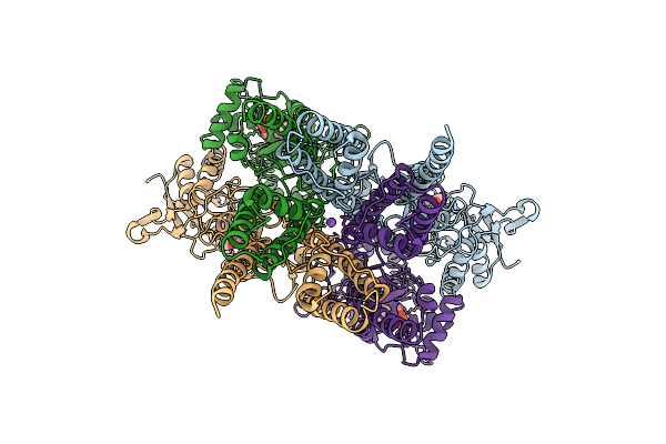 |
Heteromeric Glua1/A2 In The Inactive State, Composite Map Of Lbd-Tmd
Organism: Rattus norvegicus
Method: ELECTRON MICROSCOPY Release Date: 2026-04-08 Classification: MEMBRANE PROTEIN Ligands: ZK1, NA |
Organism: Rattus norvegicus
Method: ELECTRON MICROSCOPY
Release Date: 2026-04-08
Ligands: ZK1, NA
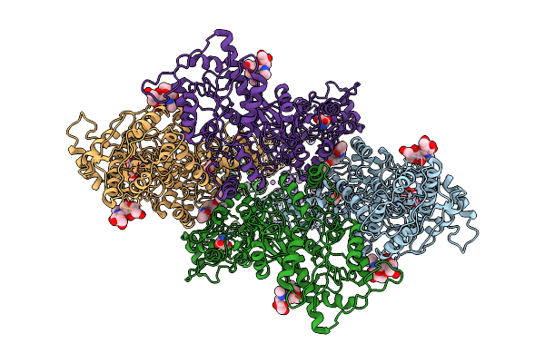 |
Composite Map Of Glua1/A2 In The Activated State, In Complex With Positive Allosteric Modulator (R,R)-2B And Agonist Glutamate (Atd-Lbd-Tmd)
Organism: Rattus norvegicus
Method: ELECTRON MICROSCOPY Release Date: 2026-04-08 Classification: MEMBRANE PROTEIN Ligands: GLU, NA, FWF |
Organism: Rattus norvegicus
Method: ELECTRON MICROSCOPY
Release Date: 2026-04-08
Ligands: GLU, NA, FWF
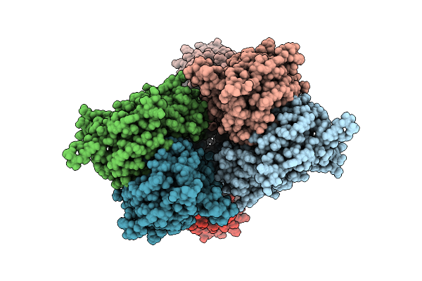 |
Heteromeric Glua1/A2-Cnih1 In The Activated State, Composite Map Of Lbd-Tmd
Organism: Rattus norvegicus, Homo sapiens
Method: ELECTRON MICROSCOPY Release Date: 2026-04-08 Classification: MEMBRANE PROTEIN Ligands: GLU, POV, FWF, NA |
Organism: Rattus norvegicus, Homo sapiens
Method: ELECTRON MICROSCOPY
Release Date: 2026-04-08
Ligands: GLU, POV, FWF, NA
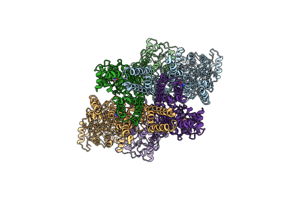 |
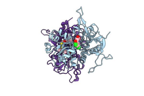
Heteromeric Glua1/A2 In The Desensitized State, Composite Map Of Atd-Lbd-Tmd
Organism: Rattus norvegicus
Method: ELECTRON MICROSCOPY Release Date: 2026-04-08 Classification: MEMBRANE PROTEIN Ligands: QUS |
Organism: Rattus norvegicus
Method: ELECTRON MICROSCOPY
Release Date: 2026-04-08
Ligands: QUS
 |
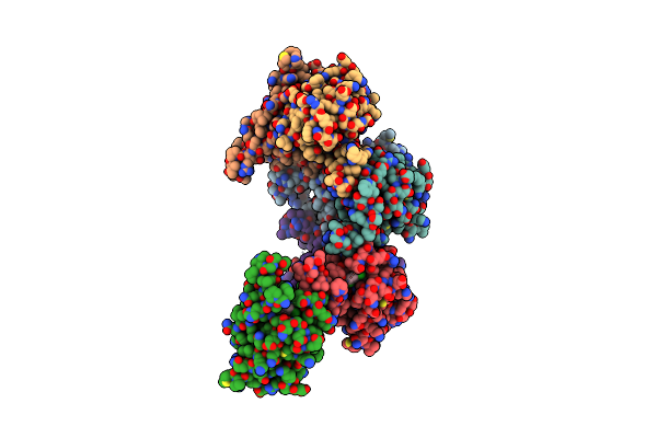
Crystal Structure Of Dcka/Glutamate-Bound Glun1/Glun2A Agonist Binding Domains With Ucm-101
Organism: Rattus norvegicus
Method: X-RAY DIFFRACTION Resolution:1.71 Å Release Date: 2025-10-29 Classification: MEMBRANE PROTEIN Ligands: DK1, A1B7E, GLU |
Organism: Rattus norvegicus
Method: X-RAY DIFFRACTION
Release Date: 2025-10-29
Ligands: DK1, A1B7E, GLU
 |
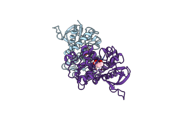
Structure Of Thr354Asn, Glu355Gln, Thr412Asn, Ile414Met, Ile464His, And Phe467Met Mutant Human Camkii Alpha Hub Bound To 5-Hdc
Organism: Homo sapiens
Method: X-RAY DIFFRACTION Resolution:2.20 Å Release Date: 2021-07-21 Classification: SIGNALING PROTEIN Ligands: 7ZV, NA |
Organism: Homo sapiens
Method: X-RAY DIFFRACTION
Release Date: 2021-07-21
Ligands: 7ZV, NA
 |
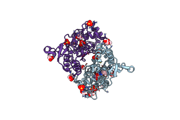
Crystal Structure Of Glun2A Agonist Binding Domain With 4-Butyl-(S)-Ccg-Iv
Organism: Rattus norvegicus
Method: X-RAY DIFFRACTION Resolution:2.30 Å Release Date: 2020-04-01 Classification: TRANSPORT PROTEIN Ligands: M7V |
Organism: Rattus norvegicus
Method: X-RAY DIFFRACTION
Release Date: 2020-04-01
Ligands: M7V
 |
Structure Of Glua2 Ligand-Binding Domain (S1S2J) In Complex With The Agonist (S)-2-Amino-3-(1-Ethyl-4-Hydroxy-1H-1,2,3-Triazol-5-Yl)Propanoic Acid At 1.4 A Resolution
Organism: Rattus norvegicus
Method: X-RAY DIFFRACTION Resolution:1.40 Å Release Date: 2019-04-17 Classification: MEMBRANE PROTEIN Ligands: SO4, HJ8, GOL, PGE, CL, CIT, LI, PEG |
Organism: Rattus norvegicus
Method: X-RAY DIFFRACTION
Release Date: 2019-04-17
Ligands: SO4, HJ8, GOL, PGE, CL, CIT, LI, PEG
 |
Structure Of Glua2 Ligand-Binding Domain (S1S2J) In Complex With The Agonist (S)-2-Amino-3-(2-Methyl-5-Hydroxy-2H-1,2,3-Triazol-4-Yl)Propanoic Acid At 1.55 A Resolution
Organism: Rattus norvegicus
Method: X-RAY DIFFRACTION Resolution:1.55 Å Release Date: 2019-04-17 Classification: MEMBRANE PROTEIN Ligands: HJH, SO4, GOL, CL, LI |
Organism: Rattus norvegicus
Method: X-RAY DIFFRACTION
Release Date: 2019-04-17
Ligands: HJH, SO4, GOL, CL, LI
 |
Crystal Structure Of The Glua2 Ligand-Binding Domain (S1S2J) In Complex With (S)-2-Amino-3-(5-(2-(3-(Aminomethyl)Benzyl)-2H-Tetrazol-5-Yl)-3-Hydroxyisoxazol-4-Yl)Propanoic Acid At Resolution 1.55 A Resolution
Organism: Rattus norvegicus
Method: X-RAY DIFFRACTION Resolution:1.55 Å Release Date: 2016-03-02 Classification: MEMBRANE PROTEIN Ligands: SO4, 5XP, GOL, ACT, EDO, LI |
Organism: Rattus norvegicus
Method: X-RAY DIFFRACTION
Release Date: 2016-03-02
Ligands: SO4, 5XP, GOL, ACT, EDO, LI
 |
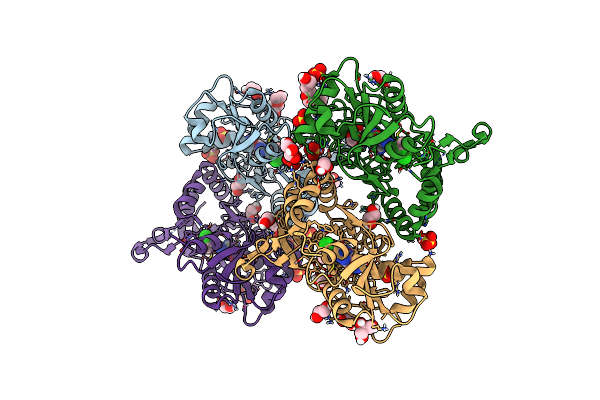
Crystal Structure Of The Glua2 Ligand-Binding Domain (S1S2J) In Complex With (S)-2-Amino-3-(5-(2-(3-Methylbenzyl)-2H-Tetrazol-5-Yl)-3-Hydroxyisoxazol-4-Yl)Propanoic Acid At 1.6 A Resolution
Organism: Rattus norvegicus
Method: X-RAY DIFFRACTION Resolution:1.60 Å Release Date: 2016-03-02 Classification: MEMBRANE PROTEIN Ligands: SO4, GOL, ACT, 5XO, EDO |
Organism: Rattus norvegicus
Method: X-RAY DIFFRACTION
Release Date: 2016-03-02
Ligands: SO4, GOL, ACT, 5XO, EDO
 |
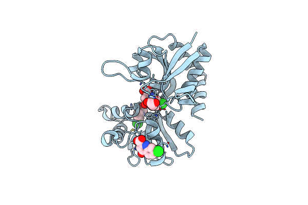
Crystal Structure Of The Glua2 Ligand-Binding Domain (S1S2J) In Complex With (S)-2-Amino-3-(5-(2-(3-Chlorobenzyl)-2H-Tetrazol-5-Yl)-3-Hydroxyisoxazol-4-Yl)Propanoic Acid At 2.3 A Resolution
Organism: Rattus norvegicus
Method: X-RAY DIFFRACTION Resolution:2.30 Å Release Date: 2016-03-02 Classification: MEMBRANE PROTEIN Ligands: 5XN, SO4, GOL, CL, EDO, PG4 |
Organism: Rattus norvegicus
Method: X-RAY DIFFRACTION
Release Date: 2016-03-02
Ligands: 5XN, SO4, GOL, CL, EDO, PG4
 |
Structure Of The Ligand-Binding Domain Of The Ionotropic Glutamate Receptor-Like Glud2 In Complex With 7-Cka
Organism: Rattus norvegicus
Method: X-RAY DIFFRACTION Resolution:2.50 Å Release Date: 2015-12-30 Classification: SIGNALING PROTEIN Ligands: CKA, GOL, CL |
Organism: Rattus norvegicus
Method: X-RAY DIFFRACTION
Release Date: 2015-12-30
Ligands: CKA, GOL, CL
 |
Glun2A Ligand-Binding Core In Complex With Propyl-Nhp5G
Organism: Rattus norvegicus
Method: X-RAY DIFFRACTION Resolution:1.50 Å Release Date: 2013-05-29 Classification: UNKNOWN FUNCTION Ligands: 1N4 |
Organism: Rattus norvegicus
Method: X-RAY DIFFRACTION
Release Date: 2013-05-29
Ligands: 1N4
 |
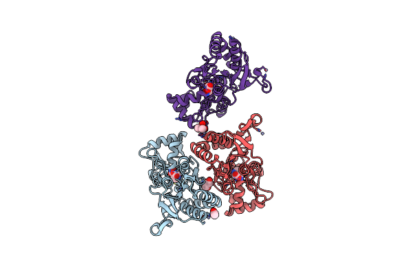
Glun2D Ligand-Binding Core In Complex With Propyl-Nhp5G
Organism: Rattus norvegicus
Method: X-RAY DIFFRACTION Resolution:2.00 Å Release Date: 2013-05-29 Classification: TRANSPORT PROTEIN Ligands: 1N4 |
Organism: Rattus norvegicus
Method: X-RAY DIFFRACTION
Release Date: 2013-05-29
Ligands: 1N4
 |
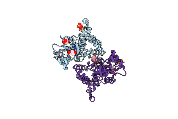
X-Ray Structure Of The Ligand-Binding Core Of Glua2 In Complex With (S)-7-Hpca At 2.5 A Resolution
Organism: Rattus norvegicus
Method: X-RAY DIFFRACTION Resolution:2.48 Å Release Date: 2010-12-29 Classification: MEMBRANE PROTEIN Ligands: ZN, HA7, GOL, ACY, CAC |
Organism: Rattus norvegicus
Method: X-RAY DIFFRACTION
Release Date: 2010-12-29
Ligands: ZN, HA7, GOL, ACY, CAC
 |
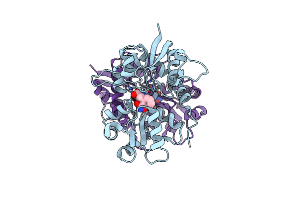
X-Ray Structure Of The Ligand-Binding Core Of Glua2 In Complex With (R)-5-Hpca At 2.1 A Resolution
Organism: Rattus norvegicus
Method: X-RAY DIFFRACTION Resolution:2.10 Å Release Date: 2010-12-29 Classification: MEMBRANE PROTEIN Ligands: HA5, SO4, GOL, CL |
Organism: Rattus norvegicus
Method: X-RAY DIFFRACTION
Release Date: 2010-12-29
Ligands: HA5, SO4, GOL, CL
 |
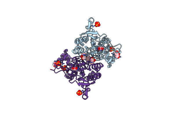
Crystal Structure Of The Ligand-Binding Core Of Glur5 In Complex With The Agonist 4-Ahcp
Organism: Rattus norvegicus
Method: X-RAY DIFFRACTION Resolution:2.20 Å Release Date: 2009-07-21 Classification: MEMBRANE PROTEIN Ligands: CL, IBC |
Organism: Rattus norvegicus
Method: X-RAY DIFFRACTION
Release Date: 2009-07-21
Ligands: CL, IBC
 |
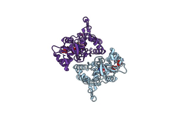
Crystal Structure Of The Iglur2 Ligand Binding Core (S1S2J-N775S) In Complex With A Dimeric Positive Modulator As Well As Glutamate At 2.25 A Resolution
Organism: Rattus norvegicus
Method: X-RAY DIFFRACTION Resolution:2.25 Å Release Date: 2007-12-04 Classification: MEMBRANE PROTEIN Ligands: SO4, CL, GLU, BHY, GOL |
Organism: Rattus norvegicus
Method: X-RAY DIFFRACTION
Release Date: 2007-12-04
Ligands: SO4, CL, GLU, BHY, GOL
 |
X-Ray Structure Of The Glur2 Ligand Binding Core (S1S2J) In Complex With 2-Bn-Tet-Ampa At 2.26A Resolution
Organism: Rattus norvegicus
Method: X-RAY DIFFRACTION Resolution:2.26 Å Release Date: 2007-06-19 Classification: MEMBRANE PROTEIN Ligands: SO4, GLU, MP9 |
Organism: Rattus norvegicus
Method: X-RAY DIFFRACTION
Release Date: 2007-06-19
Ligands: SO4, GLU, MP9