

Search Count: 73
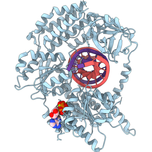 |
Cryoem Structure Of Human Mda5 With Dsrna (One Protein Subunit On Dsrna)
Organism: Homo sapiens, In vitro transcription vector pt7-fluc(deltai)
Method: ELECTRON MICROSCOPY Release Date: 2026-09-09 Classification: IMMUNE SYSTEM Ligands: ANP, MG |
Organism: Homo sapiens, In vitro transcription vector pt7-fluc(deltai)
Method: ELECTRON MICROSCOPY
Release Date: 2026-09-09
Ligands: ANP, MG
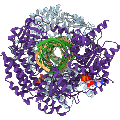 |
Cryoem Structure Of Human Mda5 In Complex With Dsrna (Two Protein Subunits On Dsrna)
Organism: Homo sapiens, In vitro transcription vector pt7-fluc(deltai)
Method: ELECTRON MICROSCOPY Release Date: 2026-09-09 Classification: IMMUNE SYSTEM Ligands: ANP |
Organism: Homo sapiens, In vitro transcription vector pt7-fluc(deltai)
Method: ELECTRON MICROSCOPY
Release Date: 2026-09-09
Ligands: ANP
 |
Structure Of Mc5Ar2 In Complex With Mc5A-Desarg (Monomer)
Organism: Mus musculus
Method: ELECTRON MICROSCOPY Resolution:3.42 Å Release Date: 2026-07-01 Classification: SIGNALING PROTEIN |
Organism: Mus musculus
Method: ELECTRON MICROSCOPY
Release Date: 2026-07-01
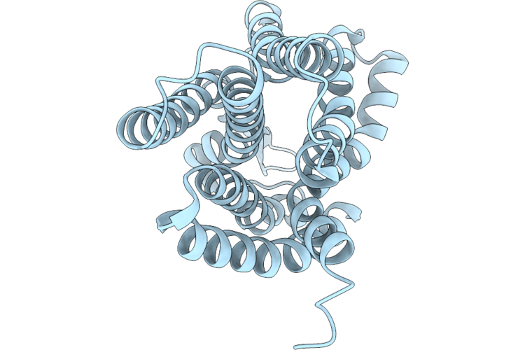 |
Structure Of C5A Anaphylatoxin Chemotactic Receptor 2, C5Ar2 In The Apo State
Organism: Homo sapiens
Method: ELECTRON MICROSCOPY Release Date: 2026-07-01 Classification: SIGNALING PROTEIN |
Organism: Homo sapiens
Method: ELECTRON MICROSCOPY
Release Date: 2026-07-01
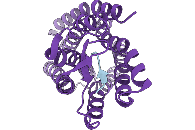 |
Structure Of C5A Anaphylatoxin Chemotactic Receptor 2, C5Ar2 Bound To Ep54
Organism: Homo sapiens
Method: ELECTRON MICROSCOPY Release Date: 2026-07-01 Classification: SIGNALING PROTEIN |
Organism: Homo sapiens
Method: ELECTRON MICROSCOPY
Release Date: 2026-07-01
 |
Structure Of C5A Anaphylatoxin Chemotactic Receptor 2, C5Ar2 Bound To C5A
Organism: Homo sapiens
Method: ELECTRON MICROSCOPY Release Date: 2026-07-01 Classification: SIGNALING PROTEIN |
Organism: Homo sapiens
Method: ELECTRON MICROSCOPY
Release Date: 2026-07-01
 |
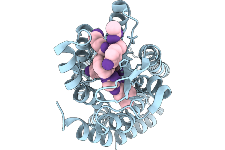
Structure Of C5A Anaphylatoxin Chemotactic Receptor 2, C5Ar2 Bound To C5A-Pep
Organism: Homo sapiens
Method: ELECTRON MICROSCOPY Release Date: 2026-07-01 Classification: SIGNALING PROTEIN |
Organism: Homo sapiens
Method: ELECTRON MICROSCOPY
Release Date: 2026-07-01
 |
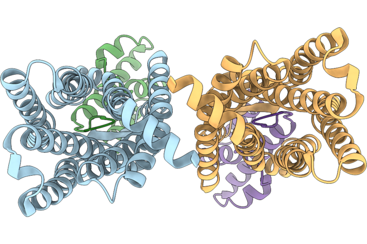
Structure Of C5A Anaphylatoxin Chemotactic Receptor 2, C5Ar2 Bound To R8Y
Organism: Homo sapiens
Method: ELECTRON MICROSCOPY Release Date: 2026-07-01 Classification: SIGNALING PROTEIN |
Organism: Homo sapiens
Method: ELECTRON MICROSCOPY
Release Date: 2026-07-01
 |
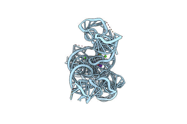
Structure Of Mc5Ar2 In Complex With Mc5A-Desarg
Organism: Mus musculus
Method: ELECTRON MICROSCOPY Release Date: 2026-07-01 Classification: SIGNALING PROTEIN |
Organism: Mus musculus
Method: ELECTRON MICROSCOPY
Release Date: 2026-07-01
 |
Cryoem Structure Of The Candida Albicans Group I Intron-Gmp Complex
Organism: Candida albicans
Method: ELECTRON MICROSCOPY Release Date: 2025-04-30 Classification: RNA Ligands: CA, K, 5GP |
Organism: Candida albicans
Method: ELECTRON MICROSCOPY
Release Date: 2025-04-30
Ligands: CA, K, 5GP
 |
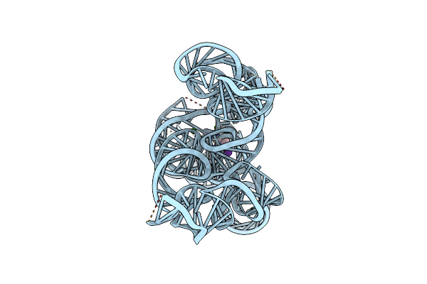
Cryoem Structure Of The Candida Albicans Group I Intron-Compound 11 Complex Under Magnesium Condition
Organism: Candida albicans
Method: ELECTRON MICROSCOPY Release Date: 2025-04-30 Classification: RNA Ligands: A1BNU, MG, K |
Organism: Candida albicans
Method: ELECTRON MICROSCOPY
Release Date: 2025-04-30
Ligands: A1BNU, MG, K
 |
Cryoem Structure Of The Candida Albicans Group I Intron-Compound 11 Complex Under Calcium Condition
Organism: Candida albicans
Method: ELECTRON MICROSCOPY Release Date: 2025-04-30 Classification: RNA Ligands: A1BNU, K, CA |
Organism: Candida albicans
Method: ELECTRON MICROSCOPY
Release Date: 2025-04-30
Ligands: A1BNU, K, CA
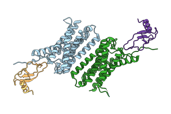 |
Structure Of Duffy Antigen Receptor For Chemokines (Darc)/Ackr1 In Complex With The Chemokine, Ccl7 (Composite Map)
Organism: Homo sapiens
Method: ELECTRON MICROSCOPY Release Date: 2024-07-31 Classification: IMMUNE SYSTEM |
Organism: Homo sapiens
Method: ELECTRON MICROSCOPY
Release Date: 2024-07-31
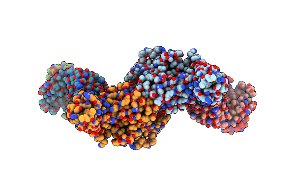 |
Structure Of Beta-Arrestin2 In Complex With A Phosphopeptide Corresponding To The Human Atypical Chemokine Receptor 2, Ackr2 (D6R)
Organism: Bos taurus, Mus musculus, Homo sapiens
Method: ELECTRON MICROSCOPY Release Date: 2023-12-27 Classification: SIGNALING PROTEIN/IMMUNE SYSTEM |
Organism: Bos taurus, Mus musculus, Homo sapiens
Method: ELECTRON MICROSCOPY
Release Date: 2023-12-27
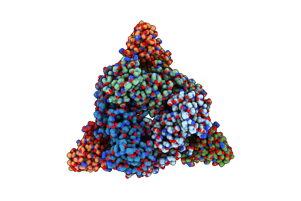 |
Structure Of Beta-Arrestin2 In Complex With M2Rpp
Organism: Bos taurus, Mus musculus, Homo sapiens
Method: ELECTRON MICROSCOPY Release Date: 2023-12-27 Classification: SIGNALING PROTEIN/IMMUNE SYSTEM |
Organism: Bos taurus, Mus musculus, Homo sapiens
Method: ELECTRON MICROSCOPY
Release Date: 2023-12-27
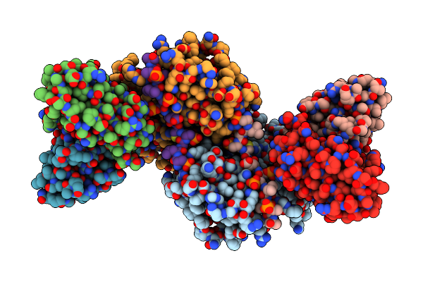 |
Structure Of Beta-Arrestin2 In Complex With D6Rpp (Local Refine)
Organism: Bos taurus, Mus musculus, Homo sapiens
Method: ELECTRON MICROSCOPY Release Date: 2023-12-27 Classification: SIGNALING PROTEIN/IMMUNE SYSTEM |
Organism: Bos taurus, Mus musculus, Homo sapiens
Method: ELECTRON MICROSCOPY
Release Date: 2023-12-27
 |
Structure Of Beta-Arrestin1 In Complex With D6Rpp
Organism: Rattus norvegicus, Mus musculus, Homo sapiens
Method: ELECTRON MICROSCOPY Release Date: 2023-12-27 Classification: SYGNALING PROTEIN/IMMUNE SYSTEM |
Organism: Rattus norvegicus, Mus musculus, Homo sapiens
Method: ELECTRON MICROSCOPY
Release Date: 2023-12-27
 |
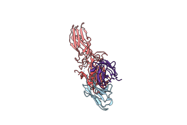
Structure Of Muscarinic Receptor (M2R) In Complex With Beta-Arrestin1 (Local Refine, Cross-Linked)
Organism: Bos taurus, Mus musculus, Homo sapiens
Method: ELECTRON MICROSCOPY Release Date: 2023-12-27 Classification: SIGNALING PROTEIN/IMMUNE SYSTEM |
Organism: Bos taurus, Mus musculus, Homo sapiens
Method: ELECTRON MICROSCOPY
Release Date: 2023-12-27
 |
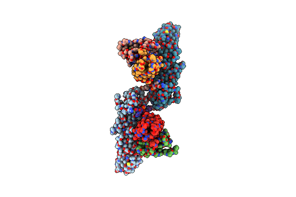
Structure Of Basal Beta-Arrestin2
Organism: Rattus norvegicus, Mus musculus
Method: ELECTRON MICROSCOPY Release Date: 2023-12-27 Classification: SIGNALING PROTEIN/IMMUNE SYSTEM |
Organism: Rattus norvegicus, Mus musculus
Method: ELECTRON MICROSCOPY
Release Date: 2023-12-27
 |
Structure Of Beta-Arrestin1 In Complex With C3Arpp
Organism: Rattus norvegicus, Mus musculus, Homo sapiens
Method: ELECTRON MICROSCOPY Release Date: 2023-12-27 Classification: SIGNALING PROTEIN/IMMUNE SYSTEM |
Organism: Rattus norvegicus, Mus musculus, Homo sapiens
Method: ELECTRON MICROSCOPY
Release Date: 2023-12-27