

Search Count: 29
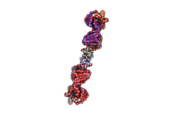 |
Chip U-Box Dimer Bound To Fab 2F1
Organism: Homo sapiens
Method: ELECTRON MICROSCOPY Release Date: 2025-03-12 Classification: LIGASE/IMMUNE SYSTEM |
Organism: Homo sapiens
Method: ELECTRON MICROSCOPY
Release Date: 2025-03-12
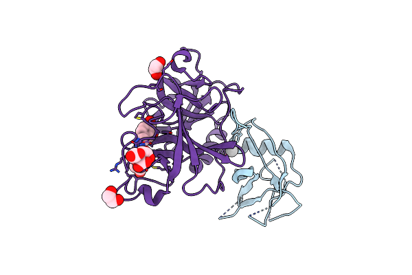 |
Tmprss2 Crystal Structure Following Acylation By Ucsf_157
Organism: Homo sapiens
Method: X-RAY DIFFRACTION Resolution:2.07 Å Release Date: 2024-12-25 Classification: HYDROLASE Ligands: CIT, EDO, VU4, UNX |
Organism: Homo sapiens
Method: X-RAY DIFFRACTION
Release Date: 2024-12-25
Ligands: CIT, EDO, VU4, UNX
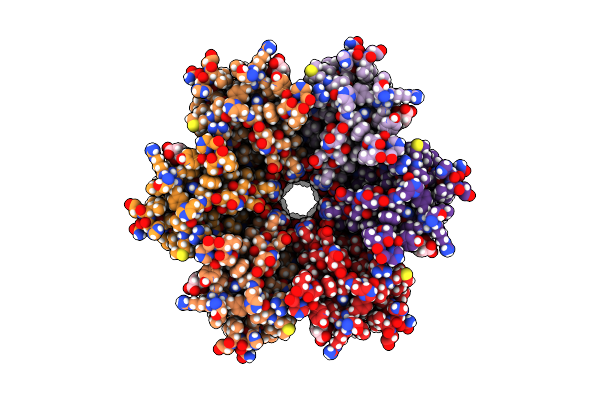 |
Human Cx36/Gjd2 Gap Junction Channel With Pore-Lining N-Terminal Helices In Porcine Brain Lipids.
Organism: Homo sapiens
Method: ELECTRON MICROSCOPY Release Date: 2024-11-06 Classification: MEMBRANE PROTEIN Ligands: MC3, Y01 |
Organism: Homo sapiens
Method: ELECTRON MICROSCOPY
Release Date: 2024-11-06
Ligands: MC3, Y01
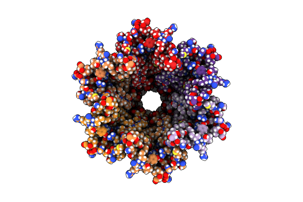 |
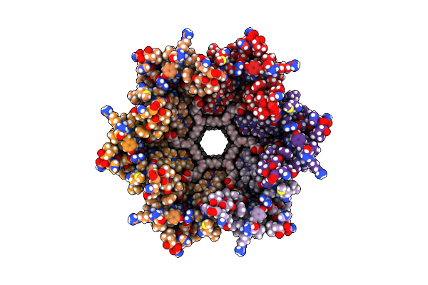
Human Cx36/Gjd2 Gap Junction Channel In Porcine Brain Lipids.
Organism: Homo sapiens
Method: ELECTRON MICROSCOPY Release Date: 2024-11-06 Classification: MEMBRANE PROTEIN Ligands: MC3 |
Organism: Homo sapiens
Method: ELECTRON MICROSCOPY
Release Date: 2024-11-06
Ligands: MC3
 |
Human Cx36/Gjd2 Gap Junction Channel In Complex With Arachidonic Acid.
Organism: Homo sapiens
Method: ELECTRON MICROSCOPY Release Date: 2024-11-06 Classification: MEMBRANE PROTEIN Ligands: MC3, ACD |
Organism: Homo sapiens
Method: ELECTRON MICROSCOPY
Release Date: 2024-11-06
Ligands: MC3, ACD
 |
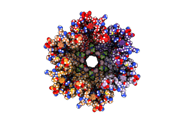
Human Cx36/Gjd2 Gap Junction Channel In Complex With 1-Hexanol.
Organism: Homo sapiens
Method: ELECTRON MICROSCOPY Release Date: 2024-11-06 Classification: MEMBRANE PROTEIN Ligands: MC3, HE2 |
Organism: Homo sapiens
Method: ELECTRON MICROSCOPY
Release Date: 2024-11-06
Ligands: MC3, HE2
 |
Human Cx36/Gjd2 Gap Junction Channel In Complex With Mefloquine.
Organism: Homo sapiens
Method: ELECTRON MICROSCOPY Release Date: 2024-11-06 Classification: MEMBRANE PROTEIN Ligands: YMZ, MC3 |
Organism: Homo sapiens
Method: ELECTRON MICROSCOPY
Release Date: 2024-11-06
Ligands: YMZ, MC3
 |
Human Cx36/Gjd2 (Ala14-Deleted Mutant) Gap Junction Channel In Porcine Brain Lipids
Organism: Homo sapiens
Method: ELECTRON MICROSCOPY Release Date: 2024-11-06 Classification: MEMBRANE PROTEIN Ligands: MC3 |
Organism: Homo sapiens
Method: ELECTRON MICROSCOPY
Release Date: 2024-11-06
Ligands: MC3
 |
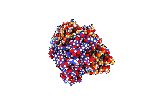
Human Cx36/Gjd2 (Ala14 Deletion Mutant) Gap Junction Channel Prepared With Mefloquine, Showing No Bound Mefloquine
Organism: Homo sapiens
Method: ELECTRON MICROSCOPY Release Date: 2024-11-06 Classification: MEMBRANE PROTEIN Ligands: MC3 |
Organism: Homo sapiens
Method: ELECTRON MICROSCOPY
Release Date: 2024-11-06
Ligands: MC3
 |
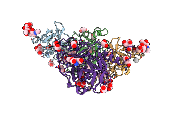
Structure Of The P1B7 Antibody Bound To The Sotorasib-Modified Kras G12C Peptide Presented By The A*03:01 Mhc I Complex
Organism: Homo sapiens
Method: ELECTRON MICROSCOPY Release Date: 2024-10-16 Classification: IMMUNE SYSTEM Ligands: MOV |
Organism: Homo sapiens
Method: ELECTRON MICROSCOPY
Release Date: 2024-10-16
Ligands: MOV
 |
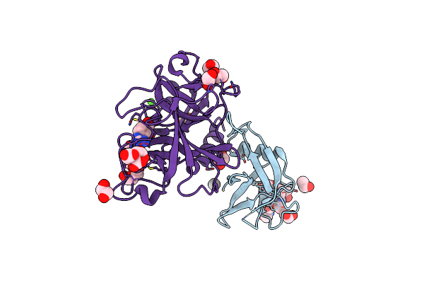
Tmprss2 Complexed With The Noncovalent Inhibitor 6-Amidino-2-Napthol
Organism: Homo sapiens
Method: X-RAY DIFFRACTION Resolution:2.19 Å Release Date: 2024-02-07 Classification: HYDROLASE/HYDROLASE INHIBITOR Ligands: TAM, UNX, EDO, 7R8, PEG, CIT, PG4 |
Organism: Homo sapiens
Method: X-RAY DIFFRACTION
Release Date: 2024-02-07
Ligands: TAM, UNX, EDO, 7R8, PEG, CIT, PG4
 |
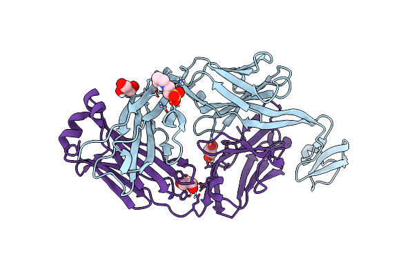
High Resolution Tmprss2 Structure Following Acylation By Nafamostat
Organism: Homo sapiens
Method: X-RAY DIFFRACTION Resolution:1.58 Å Release Date: 2024-01-31 Classification: HYDROLASE/HYDROLASE INHIBITOR Ligands: EDO, UNX, GBS, CIT, CA, PG4 |
Organism: Homo sapiens
Method: X-RAY DIFFRACTION
Release Date: 2024-01-31
Ligands: EDO, UNX, GBS, CIT, CA, PG4
 |
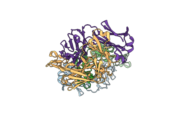
Bovine Fab 2G3
Organism: Bos taurus, Homo sapiens
Method: X-RAY DIFFRACTION Resolution:2.00 Å Release Date: 2023-09-20 Classification: IMMUNE SYSTEM Ligands: GOL, MES |
Organism: Bos taurus, Homo sapiens
Method: X-RAY DIFFRACTION
Release Date: 2023-09-20
Ligands: GOL, MES
 |
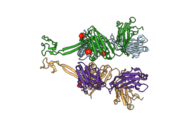
Bovine Fab 2F12
Organism: Bos taurus, Homo sapiens
Method: X-RAY DIFFRACTION Resolution:1.81 Å Release Date: 2023-09-20 Classification: IMMUNE SYSTEM |
Organism: Bos taurus, Homo sapiens
Method: X-RAY DIFFRACTION
Release Date: 2023-09-20
 |
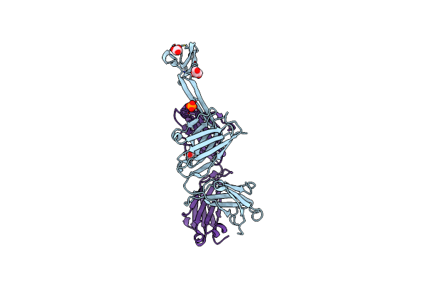
Bovine Fab 4C1
Organism: Bos taurus, Homo sapiens
Method: X-RAY DIFFRACTION Resolution:2.82 Å Release Date: 2023-09-20 Classification: IMMUNE SYSTEM Ligands: PO4 |
Organism: Bos taurus, Homo sapiens
Method: X-RAY DIFFRACTION
Release Date: 2023-09-20
Ligands: PO4
 |
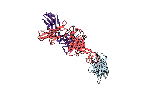
Bovine Fab 5C1
Organism: Bos taurus, Homo sapiens
Method: X-RAY DIFFRACTION Resolution:2.31 Å Release Date: 2023-09-20 Classification: IMMUNE SYSTEM Ligands: GOL, PO4 |
Organism: Bos taurus, Homo sapiens
Method: X-RAY DIFFRACTION
Release Date: 2023-09-20
Ligands: GOL, PO4
 |
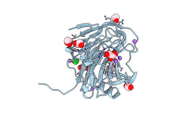
Bovine Fab Skd In Complex With Sars Cov-2 Receptor Binding Domain
Organism: Bos taurus, Homo sapiens, Severe acute respiratory syndrome coronavirus 2
Method: X-RAY DIFFRACTION Resolution:3.40 Å Release Date: 2023-09-20 Classification: IMMUNE SYSTEM Ligands: NAG |
Organism: Bos taurus, Homo sapiens, Severe acute respiratory syndrome coronavirus 2
Method: X-RAY DIFFRACTION
Release Date: 2023-09-20
Ligands: NAG
 |
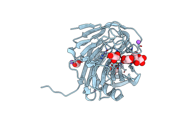
Crystal Structure Of The Gh12 Domain From Acidothermus Cellulolyticus Guxa
Organism: Acidothermus cellulolyticus
Method: X-RAY DIFFRACTION Resolution:1.50 Å Release Date: 2022-05-04 Classification: HYDROLASE |
Organism: Acidothermus cellulolyticus
Method: X-RAY DIFFRACTION
Release Date: 2022-05-04
 |
Crystal Structure Of The Gh12 Domain From Acidothermus Cellulolyticus Guxa Bound To Cellobiose
Organism: Acidothermus cellulolyticus
Method: X-RAY DIFFRACTION Resolution:1.85 Å Release Date: 2022-05-04 Classification: HYDROLASE Ligands: ZN, CL, NA, ACT, GOL |
Organism: Acidothermus cellulolyticus
Method: X-RAY DIFFRACTION
Release Date: 2022-05-04
Ligands: ZN, CL, NA, ACT, GOL
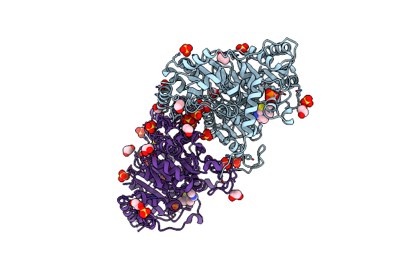 |
Zymomonas Mobilis Pyruvate Decarboxylase Mutant Pdc-2.3
Organism: Zymomonas mobilis
Method: X-RAY DIFFRACTION Resolution:1.67 Å Release Date: 2017-10-18 Classification: CELL ADHESION Ligands: TPP, MG, SO4, EDO |
Organism: Zymomonas mobilis
Method: X-RAY DIFFRACTION
Release Date: 2017-10-18
Ligands: TPP, MG, SO4, EDO