

Search Count: 20
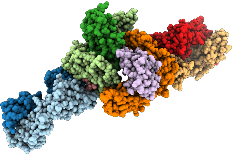 |
An Integrated In Silico-In Vitro Workflow For Discovering High-Affinity, Selective Antibodies To The Kras(G12D)-Mhc I Complex
Organism: Homo sapiens
Method: X-RAY DIFFRACTION Resolution:4.54 Å Release Date: 2026-06-24 Classification: IMMUNE SYSTEM |
Organism: Homo sapiens
Method: X-RAY DIFFRACTION
Release Date: 2026-06-24
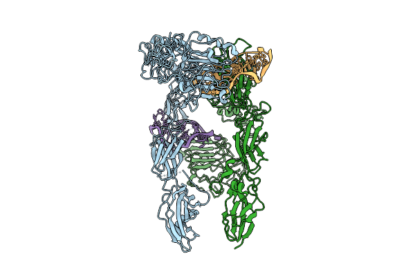 |
Human Insulin Receptor Bound With A62-Dimer, Pseudo-Arrowhead Conformation
Organism: Homo sapiens
Method: ELECTRON MICROSCOPY Release Date: 2025-07-16 Classification: MEMBRANE PROTEIN |
Organism: Homo sapiens
Method: ELECTRON MICROSCOPY
Release Date: 2025-07-16
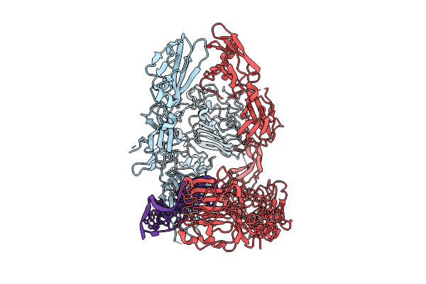 |
Human Insulin Receptor Bound With A62-Dimer, Pseudo-Gamma Conformation
Organism: Homo sapiens
Method: ELECTRON MICROSCOPY Release Date: 2025-07-16 Classification: MEMBRANE PROTEIN |
Organism: Homo sapiens
Method: ELECTRON MICROSCOPY
Release Date: 2025-07-16
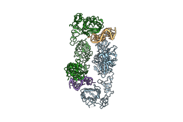 |
Human Insulin Receptor Bound With A62-Dimer, Arrowhead Conformation
Organism: Homo sapiens
Method: ELECTRON MICROSCOPY Release Date: 2025-07-16 Classification: MEMBRANE PROTEIN |
Organism: Homo sapiens
Method: ELECTRON MICROSCOPY
Release Date: 2025-07-16
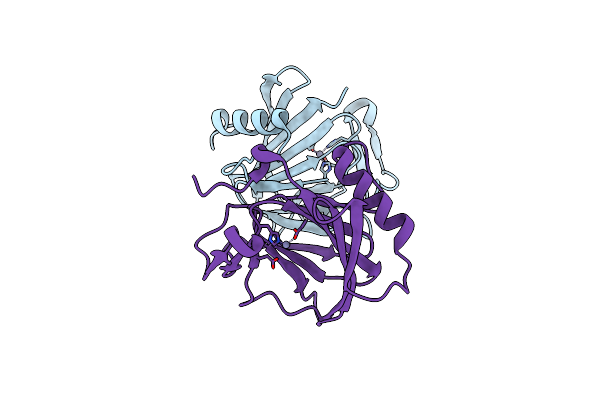 |
Crystal Structure Of Yjgk From Salmonella Typhimurium
Organism: Salmonella enterica subsp. enterica serovar typhimurium
Method: X-RAY DIFFRACTION Resolution:1.76 Å Release Date: 2025-07-02 Classification: STRUCTURAL PROTEIN Ligands: ZN |
Organism: Salmonella enterica subsp. enterica serovar typhimurium
Method: X-RAY DIFFRACTION
Release Date: 2025-07-02
Ligands: ZN
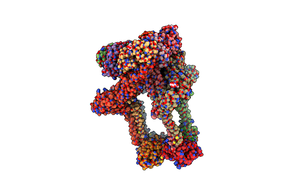 |
Structure Of Western Equine Encephalitis Virus Cba87 Vlp In Complex With Human Pcdh10 Ec1
Organism: Western equine encephalitis virus, Homo sapiens
Method: ELECTRON MICROSCOPY Resolution:3.30 Å Release Date: 2025-03-26 Classification: VIRUS LIKE PARTICLE Ligands: NAG |
Organism: Western equine encephalitis virus, Homo sapiens
Method: ELECTRON MICROSCOPY
Release Date: 2025-03-26
Ligands: NAG
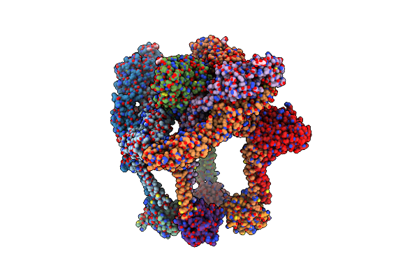 |
Structure Of Western Equine Encephalitis Virus Cba87 Vlp
Organism: Western equine encephalitis virus
Method: ELECTRON MICROSCOPY Resolution:3.40 Å Release Date: 2025-03-26 Classification: VIRUS LIKE PARTICLE Ligands: NAG |
Organism: Western equine encephalitis virus
Method: ELECTRON MICROSCOPY
Release Date: 2025-03-26
Ligands: NAG
 |
Structure Of Western Equine Encephalitis Virus Imperial 181 Vlp In Complex With House Sparrow Pcdh10 Ec1
Organism: Western equine encephalitis virus, Passer domesticus
Method: ELECTRON MICROSCOPY Resolution:2.80 Å Release Date: 2025-03-26 Classification: VIRUS LIKE PARTICLE Ligands: NAG |
Organism: Western equine encephalitis virus, Passer domesticus
Method: ELECTRON MICROSCOPY
Release Date: 2025-03-26
Ligands: NAG
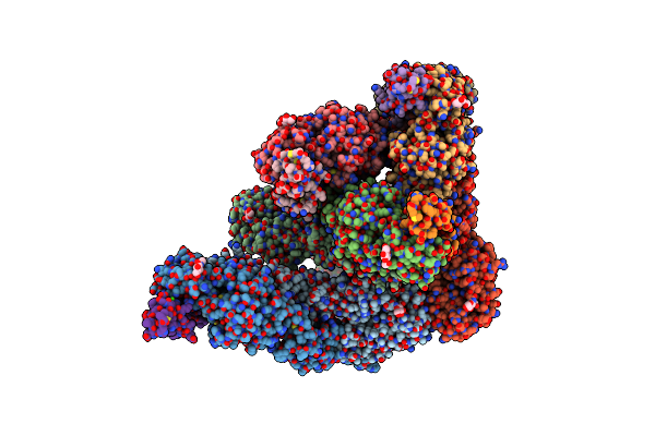 |
Structure Of Western Equine Encephalitis Virus Mcmillan Vlp In Complex With Human Vldlr
Organism: Western equine encephalitis virus, Homo sapiens
Method: ELECTRON MICROSCOPY Resolution:2.90 Å Release Date: 2025-03-26 Classification: VIRUS LIKE PARTICLE Ligands: NAG, CA |
Organism: Western equine encephalitis virus, Homo sapiens
Method: ELECTRON MICROSCOPY
Release Date: 2025-03-26
Ligands: NAG, CA
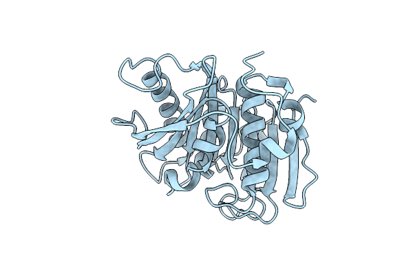 |
Crystal Structure Of Petase S121D, D186H Mutant From Ideonella Sakaiensis
Organism: Ideonella sakaiensis
Method: X-RAY DIFFRACTION Resolution:1.40 Å Release Date: 2019-09-04 Classification: HYDROLASE |
Organism: Ideonella sakaiensis
Method: X-RAY DIFFRACTION
Release Date: 2019-09-04
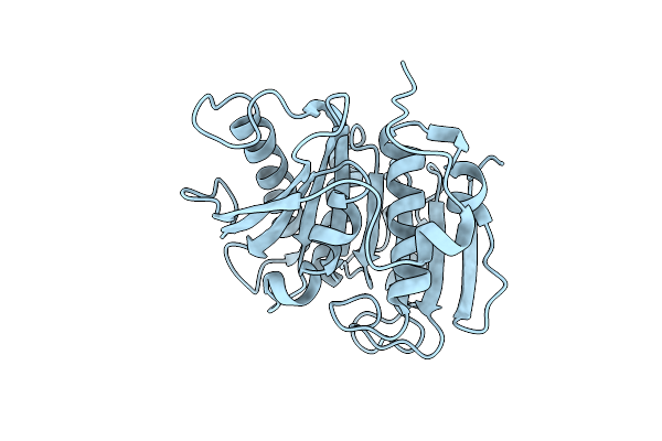 |
Crystal Structure Of Petase S121E, D186H Mutant From Ideonella Sakaiensis
Organism: Ideonella sakaiensis
Method: X-RAY DIFFRACTION Resolution:1.86 Å Release Date: 2019-09-04 Classification: HYDROLASE |
Organism: Ideonella sakaiensis
Method: X-RAY DIFFRACTION
Release Date: 2019-09-04
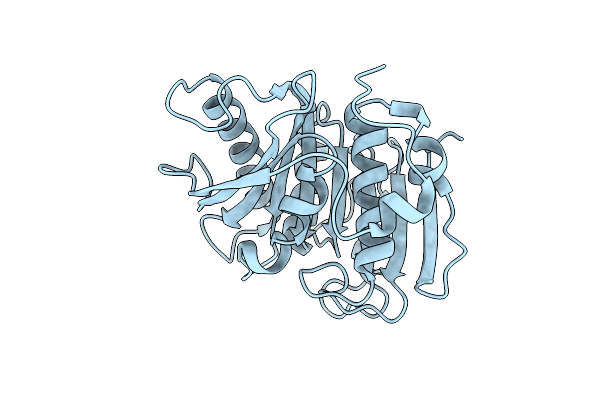 |
Crystal Structure Of Petase P181A Mutant From Ideonella Sakaiensis
Organism: Ideonella sakaiensis
Method: X-RAY DIFFRACTION Resolution:1.72 Å Release Date: 2019-09-04 Classification: HYDROLASE |
Organism: Ideonella sakaiensis
Method: X-RAY DIFFRACTION
Release Date: 2019-09-04
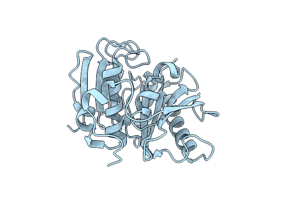 |
Crystal Structure Of Petase S121E, D186H, R280A Mutant From Ideonella Sakaiensis
Organism: Ideonella sakaiensis
Method: X-RAY DIFFRACTION Resolution:1.95 Å Release Date: 2019-09-04 Classification: HYDROLASE |
Organism: Ideonella sakaiensis
Method: X-RAY DIFFRACTION
Release Date: 2019-09-04
 |
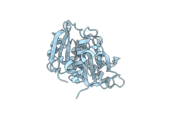
Crystal Structure Of Petase From Ideonella Sakaiensis
Organism: Ideonella sakaiensis
Method: X-RAY DIFFRACTION Resolution:1.54 Å Release Date: 2018-02-14 Classification: HYDROLASE |
Organism: Ideonella sakaiensis
Method: X-RAY DIFFRACTION
Release Date: 2018-02-14
 |
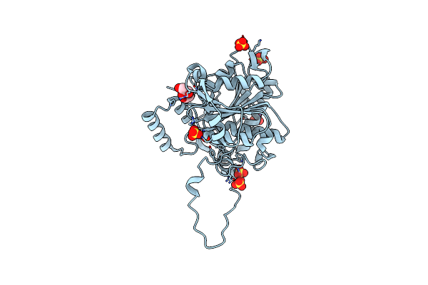
Crystal Structure Of Petase R280A Mutant From Ideonella Sakaiensis
Organism: Ideonella sakaiensis (strain 201-f6)
Method: X-RAY DIFFRACTION Resolution:1.36 Å Release Date: 2018-02-14 Classification: HYDROLASE |
Organism: Ideonella sakaiensis (strain 201-f6)
Method: X-RAY DIFFRACTION
Release Date: 2018-02-14
 |
Crystal Structure Of Phac1 From Ralstonia Eutropha
Organism: Cupriavidus necator
Method: X-RAY DIFFRACTION Resolution:1.80 Å Release Date: 2016-12-07 Classification: TRANSFERASE Ligands: GOL, SO4 |
Organism: Cupriavidus necator
Method: X-RAY DIFFRACTION
Release Date: 2016-12-07
Ligands: GOL, SO4
 |
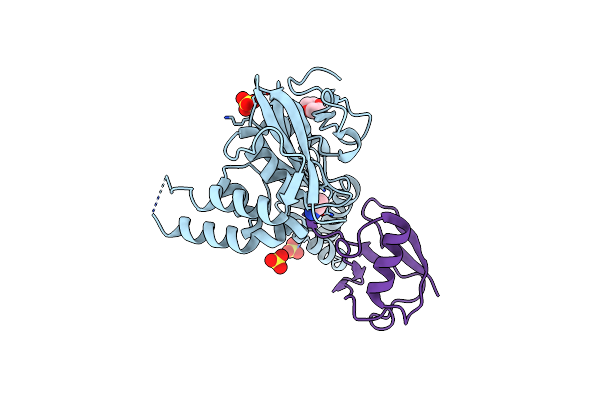
Crystal Structure Of Deubiquitinase Mindy-1
Organism: Homo sapiens
Method: X-RAY DIFFRACTION Resolution:3.00 Å Release Date: 2016-06-22 Classification: HYDROLASE Ligands: PEG, PO4, HG |
Organism: Homo sapiens
Method: X-RAY DIFFRACTION
Release Date: 2016-06-22
Ligands: PEG, PO4, HG
 |
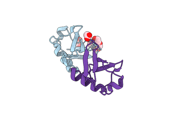
Crystal Structure Of Deubiquitinase Mindy-1 In Complex With Ubiquitin
Organism: Homo sapiens, Bos taurus
Method: X-RAY DIFFRACTION Resolution:2.65 Å Release Date: 2016-06-22 Classification: HYDROLASE Ligands: AYE, SO4, CL, DIO |
Organism: Homo sapiens, Bos taurus
Method: X-RAY DIFFRACTION
Release Date: 2016-06-22
Ligands: AYE, SO4, CL, DIO
 |
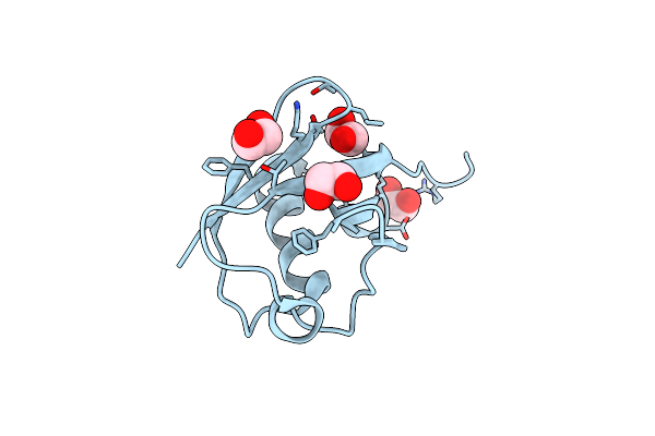
Crystal Structure Of K33 Linked Di-Ubiquitin
Organism: Bos taurus
Method: X-RAY DIFFRACTION Resolution:1.65 Å Release Date: 2015-03-18 Classification: SIGNALING PROTEIN Ligands: EDO, IOD, ACT, PG4 |
Organism: Bos taurus
Method: X-RAY DIFFRACTION
Release Date: 2015-03-18
Ligands: EDO, IOD, ACT, PG4
 |
Crystal Structure Of K33 Linked Tri-Ubiquitin
Organism: Bos taurus
Method: X-RAY DIFFRACTION Resolution:1.40 Å Release Date: 2015-03-18 Classification: SIGNALING PROTEIN Ligands: EDO |
Organism: Bos taurus
Method: X-RAY DIFFRACTION
Release Date: 2015-03-18
Ligands: EDO