
Search Count: 2,000
All
Selected
 |
Organism: Psychrobacter sp. d2
Method: ELECTRON MICROSCOPY Resolution:2.80 Å Release Date: 2026-05-20 Classification: TRANSPORT PROTEIN |
 |
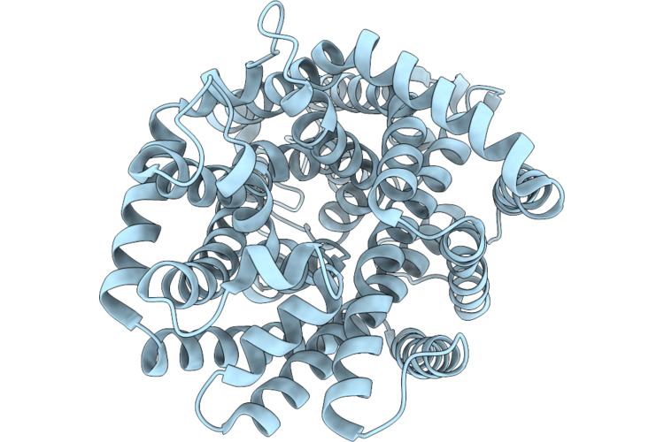
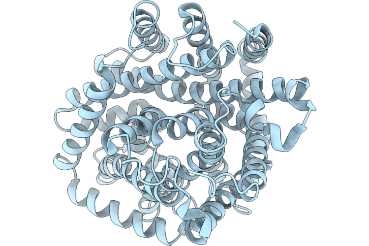
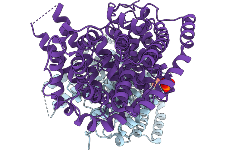
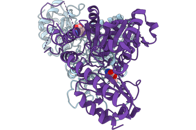
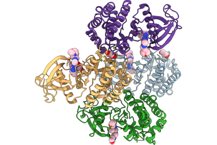
Cryo-Em Structure Of Dddt G101D In Substrate-Free Outward Open Conformation
Organism: Psychrobacter sp. d2
Method: ELECTRON MICROSCOPY Resolution:2.66 Å Release Date: 2026-05-20 Classification: TRANSPORT PROTEIN |
 |
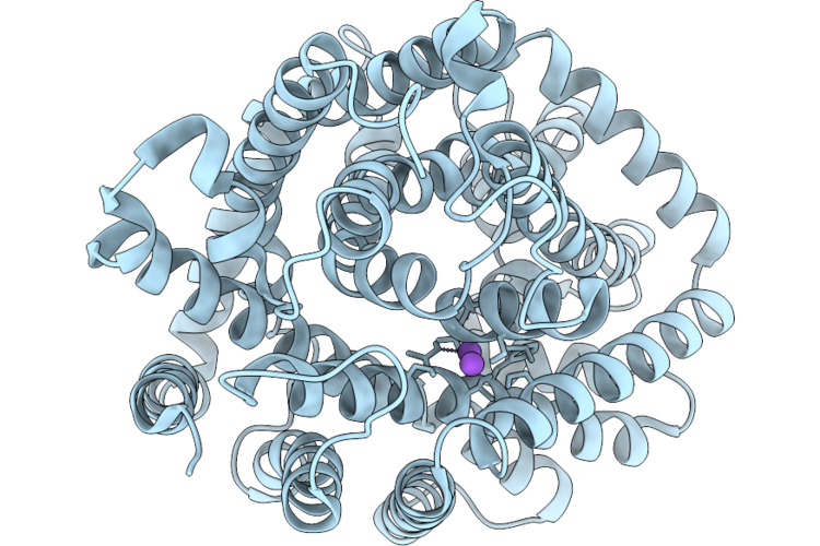
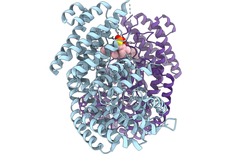
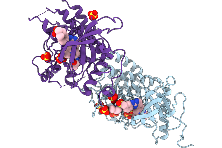
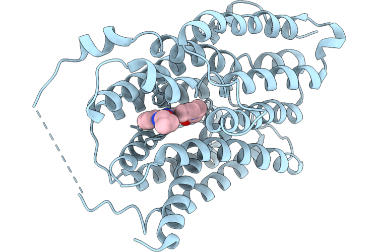
Organism: Psychrobacter sp. d2
Method: ELECTRON MICROSCOPY Resolution:2.52 Å Release Date: 2026-05-20 Classification: TRANSPORT PROTEIN Ligands: DQY, NA |
 |
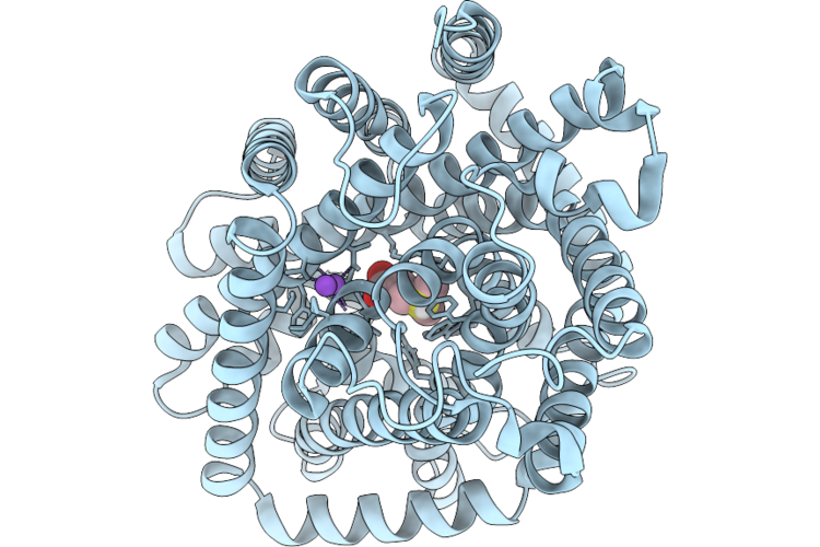
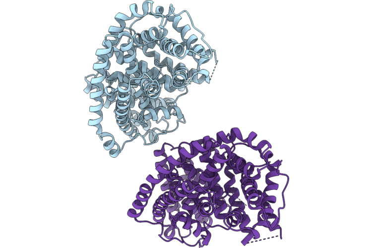
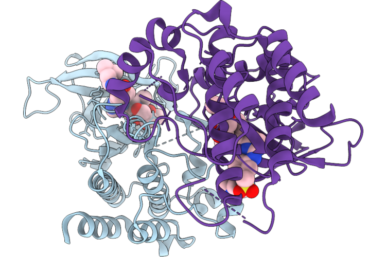
Cryo-Em Structure Of Dddt In Closed Substrate-Free Conformation In The Presence Of Potassium Ions And Dimethylsulfoniopropionate
Organism: Psychrobacter sp. d2
Method: ELECTRON MICROSCOPY Resolution:3.18 Å Release Date: 2026-05-20 Classification: TRANSPORT PROTEIN |
 |
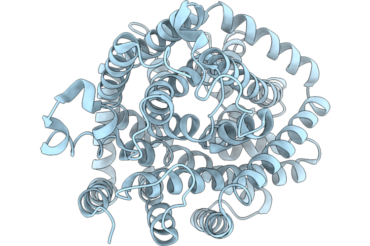
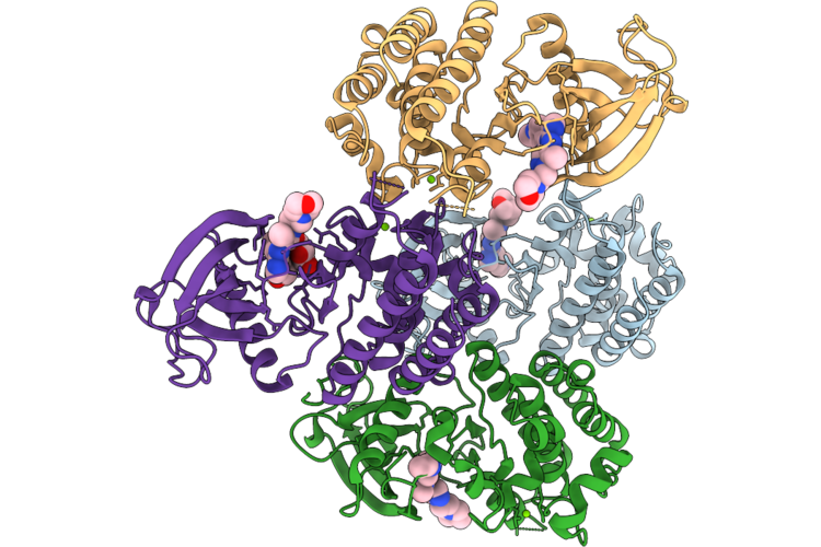
Organism: Psychrobacter sp. d2
Method: ELECTRON MICROSCOPY Resolution:3.29 Å Release Date: 2026-05-20 Classification: TRANSPORT PROTEIN |
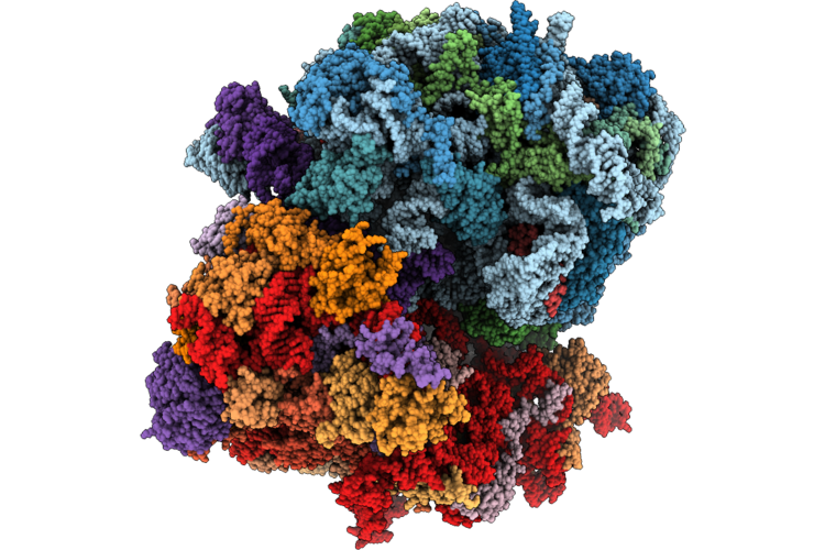 |
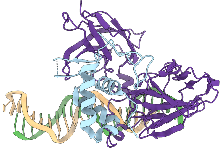
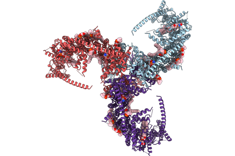
Hypopseudouridylated Yeast 80S Bound With Taura Syndrome Virus (Tsv) Internal Ribosome Entry Site (Ires) And Hygromycin B
Organism: Taura syndrome virus, Saccharomyces cerevisiae
Method: ELECTRON MICROSCOPY Release Date: 2026-05-20 Classification: RIBOSOME Ligands: MG, HYG, ZN |
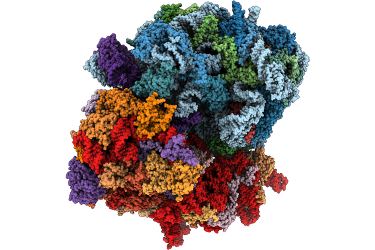 |
Hypopseudouridylated Yeast 80S Bound With Taura Syndrome Virus (Tsv) Internal Ribosome Entry Site (Ires) And Hygromycin B, Class Ii
Organism: Taura syndrome virus, Saccharomyces cerevisiae
Method: ELECTRON MICROSCOPY Release Date: 2026-05-20 Classification: RIBOSOME Ligands: MG, HYG, ZN |
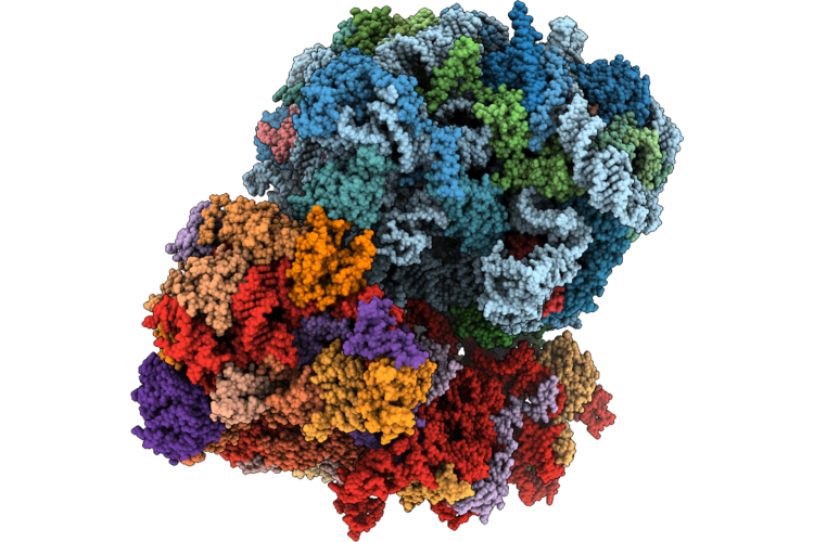 |
Organism: Saccharomyces cerevisiae by4741
Method: ELECTRON MICROSCOPY Release Date: 2026-05-20 Classification: RIBOSOME Ligands: MG, K, ZN |
 |
Organism: Homo sapiens
Method: X-RAY DIFFRACTION Resolution:2.80 Å Release Date: 2026-05-20 Classification: DNA BINDING PROTEIN/DNA |
 |
Organism: Phyla dulcis
Method: X-RAY DIFFRACTION Resolution:2.03 Å Release Date: 2026-05-13 Classification: LYASE Ligands: PO4 |
 |
Organism: Phyla dulcis
Method: X-RAY DIFFRACTION Resolution:2.89 Å Release Date: 2026-05-13 Classification: LYASE Ligands: MG, FPS |
 |
Crystal Structure Of Bicyclogermacrene Synthase Mutant I290V/I385C/V434C/L454C/V476W/L558I
Organism: Phyla dulcis
Method: X-RAY DIFFRACTION Resolution:2.30 Å Release Date: 2026-05-13 Classification: LYASE |
 |
Crystal Structure Of Yarrowia Lipolytica Methionine Gamma-Lyase In Complex With L-Methionine
Organism: Yarrowia lipolytica
Method: X-RAY DIFFRACTION Resolution:3.04 Å Release Date: 2026-05-13 Classification: LYASE Ligands: MET |
 |
Organism: Homo sapiens
Method: X-RAY DIFFRACTION Resolution:1.81 Å Release Date: 2026-05-06 Classification: TRANSFERASE Ligands: A1ESP, SO4 |
 |
Organism: Homo sapiens
Method: X-RAY DIFFRACTION Resolution:2.26 Å Release Date: 2026-05-06 Classification: TRANSFERASE Ligands: A1ESP, GOL |
 |
Organism: Homo sapiens
Method: X-RAY DIFFRACTION Resolution:2.65 Å Release Date: 2026-05-06 Classification: TRANSFERASE Ligands: A1ESW, MG, GOL |
 |
Organism: Homo sapiens
Method: X-RAY DIFFRACTION Resolution:1.97 Å Release Date: 2026-05-06 Classification: TRANSFERASE Ligands: A1ESX, MG, GOL |
 |
Organism: Drosophila
Method: ELECTRON MICROSCOPY Release Date: 2026-05-06 Classification: MEMBRANE PROTEIN Ligands: PLX, PEE, P5S |
 |
Organism: Henipavirus nipahense
Method: ELECTRON MICROSCOPY Resolution:2.92 Å Release Date: 2026-04-29 Classification: VIRAL PROTEIN Ligands: A1E3H, ZN |
 |
Organism: Plasmodium falciparum 3d7
Method: ELECTRON MICROSCOPY Release Date: 2026-04-29 Classification: TRANSPORT PROTEIN Ligands: A1EE2 |