

Search Count: 71
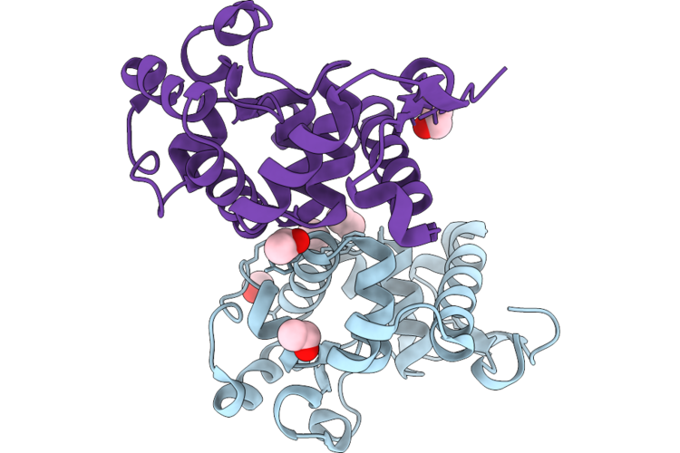 |
Crystal Structure Of The Gh134 Mannanase From Aspergillus Nidulans
Organism: Aspergillus nidulans fgsc a4
Method: X-RAY DIFFRACTION Resolution:1.75 Å Release Date: 2026-09-23 Classification: CARBOHYDRATE Ligands: EOH, POL |
Organism: Aspergillus nidulans fgsc a4
Method: X-RAY DIFFRACTION
Release Date: 2026-09-23
Ligands: EOH, POL
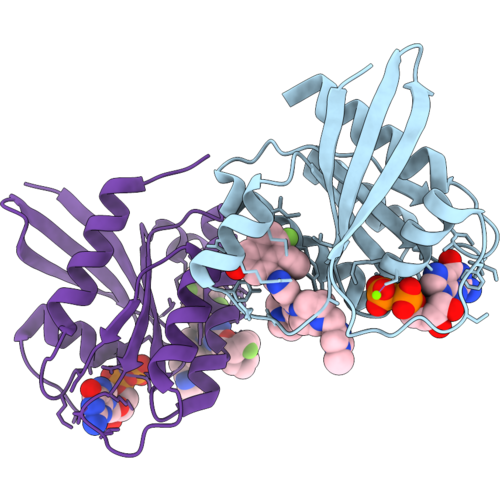 |
Crystal Structure Of Kras(Q61K) Bound To Gdp And Ligand
Organism: Homo sapiens
Method: X-RAY DIFFRACTION Resolution:2.05 Å Release Date: 2026-08-26 Classification: ONCOPROTEIN Ligands: A1DEM, MG, GDP |
Organism: Homo sapiens
Method: X-RAY DIFFRACTION
Release Date: 2026-08-26
Ligands: A1DEM, MG, GDP
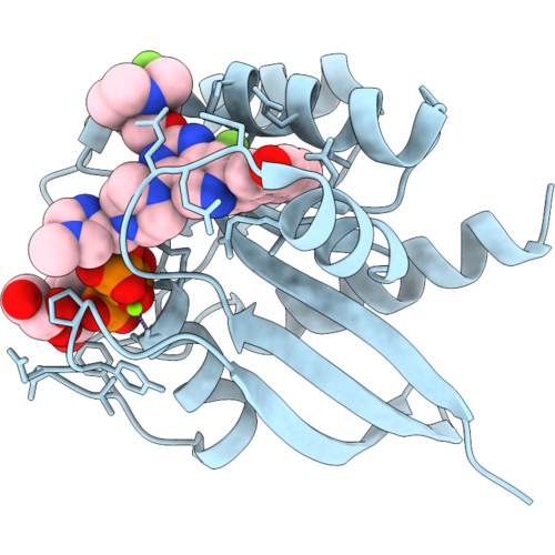 |
Crystal Structure Of Kras(Q61K) Bound To Gppnhp And Ligand
Organism: Homo sapiens
Method: X-RAY DIFFRACTION Resolution:1.01 Å Release Date: 2026-08-26 Classification: ONCOPROTEIN Ligands: A1DEM, GNP, GOL, MG |
Organism: Homo sapiens
Method: X-RAY DIFFRACTION
Release Date: 2026-08-26
Ligands: A1DEM, GNP, GOL, MG
 |
The Structure Of Dutpase From Methanosarcina Mazei
Organism: Methanosarcina mazei
Method: X-RAY DIFFRACTION Resolution:1.45 Å Release Date: 2026-04-29 Classification: HYDROLASE Ligands: PO4 |
Organism: Methanosarcina mazei
Method: X-RAY DIFFRACTION
Release Date: 2026-04-29
Ligands: PO4
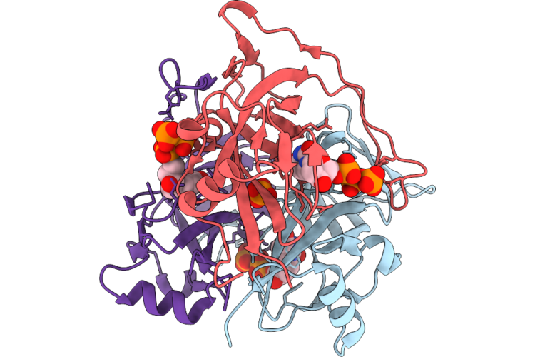 |
The Structure Of Dutpase In Complex With Dutp From Methanosarcina Mazei
Organism: Methanosarcina mazei
Method: X-RAY DIFFRACTION Resolution:1.53 Å Release Date: 2026-04-29 Classification: HYDROLASE Ligands: DUT, PO4 |
Organism: Methanosarcina mazei
Method: X-RAY DIFFRACTION
Release Date: 2026-04-29
Ligands: DUT, PO4
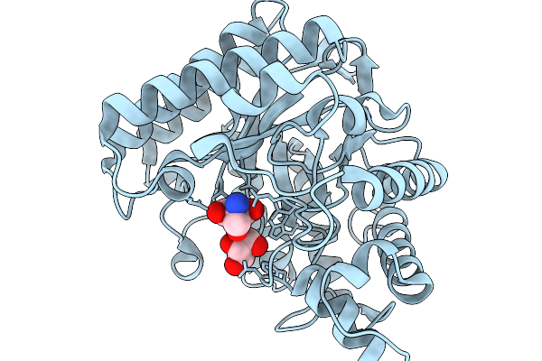 |
Gmman19-1 From Glycine Max
Organism: Glycine max
Method: X-RAY DIFFRACTION Resolution:1.39 Å Release Date: 2026-02-04 Classification: HYDROLASE Ligands: TRS, GOL |
Organism: Glycine max
Method: X-RAY DIFFRACTION
Release Date: 2026-02-04
Ligands: TRS, GOL
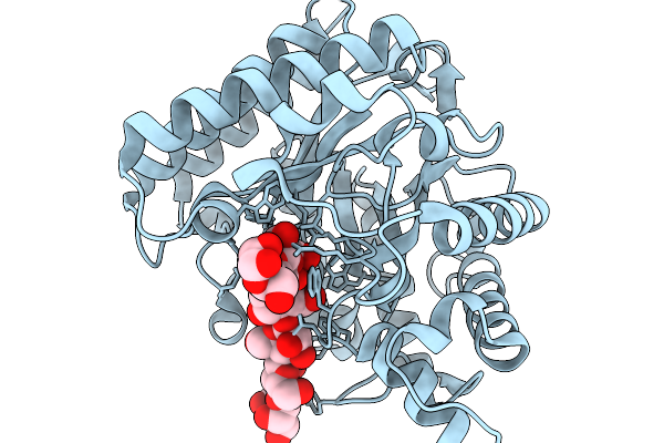 |
Gmman19-1 From Glycine Max In Complex With Mannopentaose
Organism: Glycine max
Method: X-RAY DIFFRACTION Resolution:2.62 Å Release Date: 2026-02-04 Classification: HYDROLASE |
Organism: Glycine max
Method: X-RAY DIFFRACTION
Release Date: 2026-02-04
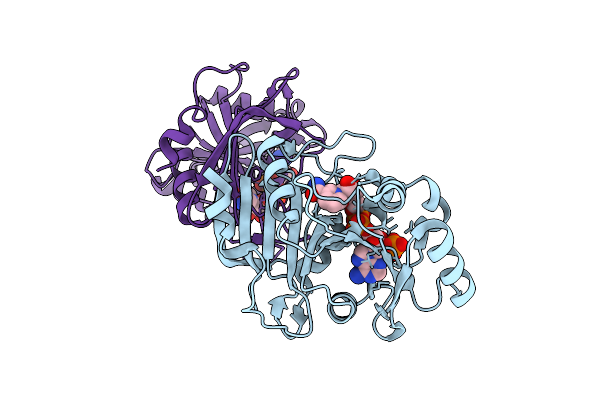 |
Crystal Structure Of Pprib7 In Complex Nadph (P1 Form)
Organism: Komagataella pastoris
Method: X-RAY DIFFRACTION Resolution:1.60 Å Release Date: 2025-12-17 Classification: BIOSYNTHETIC PROTEIN Ligands: NAP |
Organism: Komagataella pastoris
Method: X-RAY DIFFRACTION
Release Date: 2025-12-17
Ligands: NAP
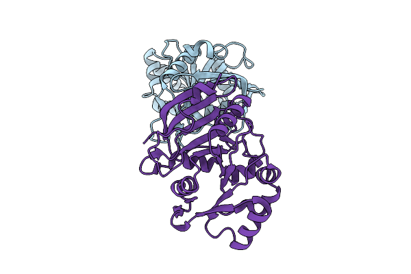 |
Crystal Structure Of Pprib7
Organism: Komagataella pastoris
Method: X-RAY DIFFRACTION Resolution:2.10 Å Release Date: 2025-12-17 Classification: BIOSYNTHETIC PROTEIN |
Organism: Komagataella pastoris
Method: X-RAY DIFFRACTION
Release Date: 2025-12-17
 |
Crystal Structure Of Pprib7 In Complex With Nadph (P212121 Form)
Organism: Komagataella pastoris
Method: X-RAY DIFFRACTION Resolution:2.08 Å Release Date: 2025-12-17 Classification: BIOSYNTHETIC PROTEIN Ligands: NAP |
Organism: Komagataella pastoris
Method: X-RAY DIFFRACTION
Release Date: 2025-12-17
Ligands: NAP
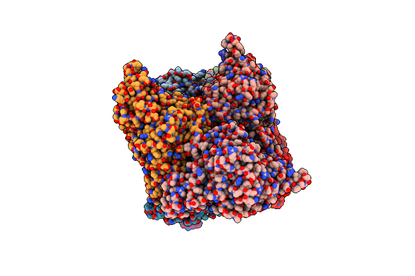 |
Crystal Structure Of Trehalose Synthase Mutant N253C From Deinococcus Radiodurans
Organism: Deinococcus radiodurans
Method: X-RAY DIFFRACTION Resolution:2.65 Å Release Date: 2025-03-19 Classification: ISOMERASE Ligands: CA, MG, TRS |
Organism: Deinococcus radiodurans
Method: X-RAY DIFFRACTION
Release Date: 2025-03-19
Ligands: CA, MG, TRS
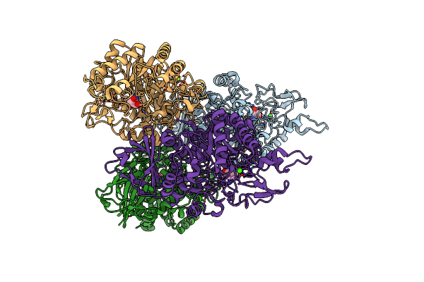 |
Crystal Structure Of Trehalose Synthase Mutant N253E From Deinococcus Radiodurans
Organism: Deinococcus radiodurans (strain atcc 13939 / dsm 20539 / jcm 16871 / ccug 27074 / lmg 4051 / nbrc 15346 / ncimb 9279 / vkm b-1422 / r1)
Method: X-RAY DIFFRACTION Resolution:3.04 Å Release Date: 2025-01-01 Classification: ISOMERASE Ligands: CA, MG, TRS |
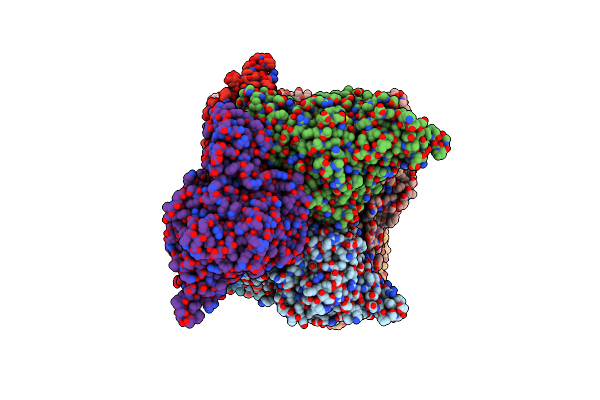 |
Crystal Structure Of Trehalose Synthase Mutant N253Q From Deinococcus Radiodurans
Organism: Deinococcus radiodurans (strain atcc 13939 / dsm 20539 / jcm 16871 / ccug 27074 / lmg 4051 / nbrc 15346 / ncimb 9279 / vkm b-1422 / r1)
Method: X-RAY DIFFRACTION Resolution:2.99 Å Release Date: 2025-01-01 Classification: ISOMERASE Ligands: CA, MG, TRS |
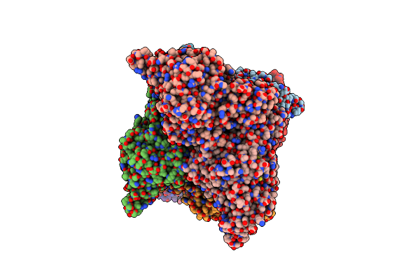 |
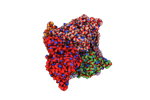
Crystal Structure Of Trehalose Synthase Mutant N253T From Deinococcus Radiodurans
Organism: Deinococcus radiodurans (strain atcc 13939 / dsm 20539 / jcm 16871 / ccug 27074 / lmg 4051 / nbrc 15346 / ncimb 9279 / vkm b-1422 / r1)
Method: X-RAY DIFFRACTION Resolution:2.53 Å Release Date: 2025-01-01 Classification: ISOMERASE Ligands: CA, MG, TRS |
 |
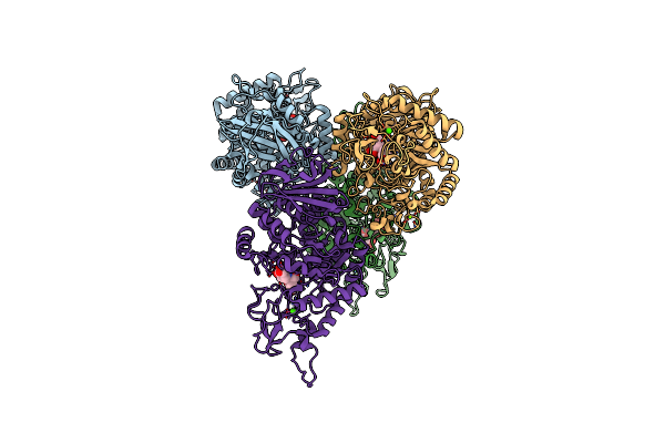
Crystal Structure Of Trehalose Synthase Mutant R148A From Deinococcus Radiodurans
Organism: Deinococcus radiodurans
Method: X-RAY DIFFRACTION Resolution:2.32 Å Release Date: 2025-01-01 Classification: ISOMERASE Ligands: CA, MG, TRS |
Organism: Deinococcus radiodurans
Method: X-RAY DIFFRACTION
Release Date: 2025-01-01
Ligands: CA, MG, TRS
 |
Crystal Structure Of Trehalose Synthase From Deinococcus Radiodurans Complexed With Validoxylamine A (Vaa)
Organism: Deinococcus radiodurans r1 = atcc 13939 = dsm 20539
Method: X-RAY DIFFRACTION Resolution:2.83 Å Release Date: 2025-01-01 Classification: ISOMERASE Ligands: CA, MG, VDM |
Organism: Deinococcus radiodurans r1 = atcc 13939 = dsm 20539
Method: X-RAY DIFFRACTION
Release Date: 2025-01-01
Ligands: CA, MG, VDM
 |
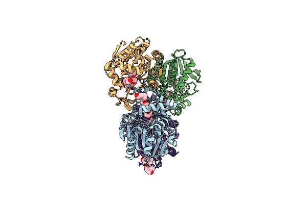
Crystal Structure Of Trehalose Synthase Mutamt E324D From Deinococcus Radiodurans Complexed With Validoxylamine A (Vaa)
Organism: Deinococcus radiodurans (strain atcc 13939 / dsm 20539 / jcm 16871 / ccug 27074 / lmg 4051 / nbrc 15346 / ncimb 9279 / vkm b-1422 / r1)
Method: X-RAY DIFFRACTION Resolution:2.90 Å Release Date: 2025-01-01 Classification: ISOMERASE Ligands: CA, MG, VDM |
 |
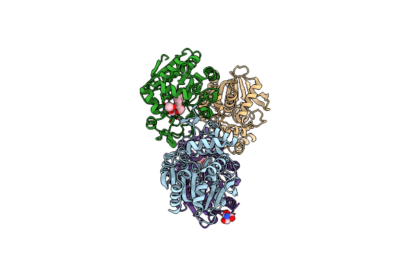
Poly(3-Hydroxybutyrate) Depolymerase Phaz From Bacillus Thuringiensis
Organism: Bacillus thuringiensis
Method: X-RAY DIFFRACTION Resolution:1.42 Å Release Date: 2024-12-11 Classification: HYDROLASE Ligands: P33, BTB |
Organism: Bacillus thuringiensis
Method: X-RAY DIFFRACTION
Release Date: 2024-12-11
Ligands: P33, BTB
 |
S102A Mutant Of Poly(3-Hydroxybutyrate) Depolymerase Phaz From Bacillus Thuringiensis
Organism: Bacillus thuringiensis
Method: X-RAY DIFFRACTION Resolution:1.90 Å Release Date: 2024-12-11 Classification: HYDROLASE Ligands: P33, TRS |
Organism: Bacillus thuringiensis
Method: X-RAY DIFFRACTION
Release Date: 2024-12-11
Ligands: P33, TRS
 |
Crystal Structure Of Trehalose Synthase Mutant N253H From Deinococcus Radiodurans
Organism: Deinococcus radiodurans
Method: X-RAY DIFFRACTION Resolution:2.97 Å Release Date: 2024-09-04 Classification: ISOMERASE Ligands: CA, MG, TRS |
Organism: Deinococcus radiodurans
Method: X-RAY DIFFRACTION
Release Date: 2024-09-04
Ligands: CA, MG, TRS