
Search Count: 37
All
Selected
 |
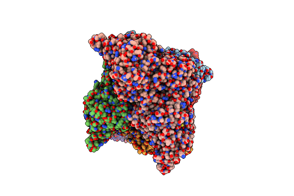
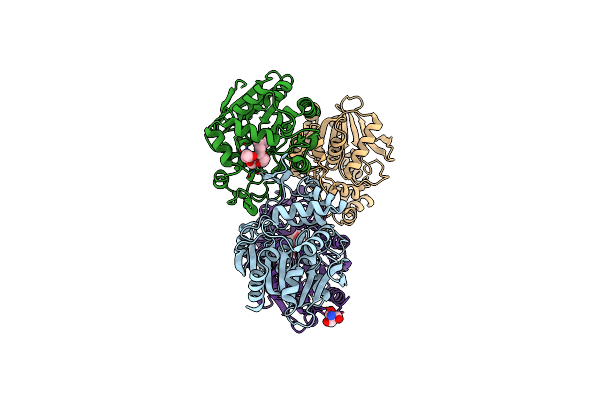
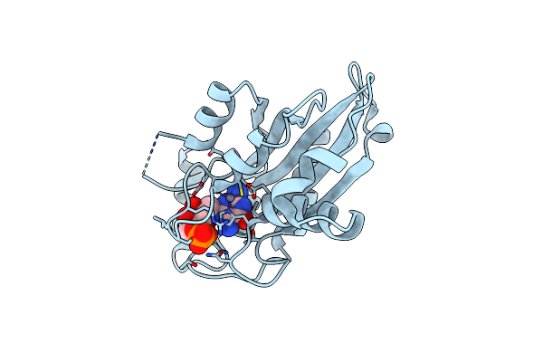
Organism: Methanosarcina mazei
Method: X-RAY DIFFRACTION Resolution:1.45 Å Release Date: 2026-04-29 Classification: HYDROLASE Ligands: PO4 |
 |
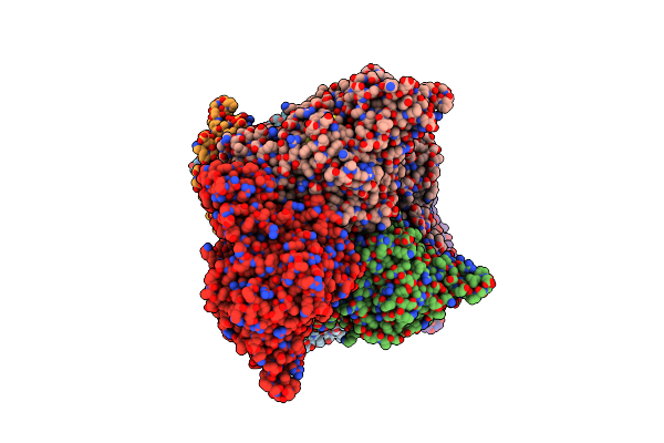
Organism: Methanosarcina mazei
Method: X-RAY DIFFRACTION Resolution:1.53 Å Release Date: 2026-04-29 Classification: HYDROLASE Ligands: DUT, PO4 |
 |
Crystal Structure Of Trehalose Synthase Mutant N253C From Deinococcus Radiodurans
Organism: Deinococcus radiodurans
Method: X-RAY DIFFRACTION Resolution:2.65 Å Release Date: 2025-03-19 Classification: ISOMERASE Ligands: CA, MG, TRS |
 |
Crystal Structure Of Trehalose Synthase Mutant N253E From Deinococcus Radiodurans
Organism: Deinococcus radiodurans (strain atcc 13939 / dsm 20539 / jcm 16871 / ccug 27074 / lmg 4051 / nbrc 15346 / ncimb 9279 / vkm b-1422 / r1)
Method: X-RAY DIFFRACTION Resolution:3.04 Å Release Date: 2025-01-01 Classification: ISOMERASE Ligands: CA, MG, TRS |
 |
Crystal Structure Of Trehalose Synthase Mutant N253Q From Deinococcus Radiodurans
Organism: Deinococcus radiodurans (strain atcc 13939 / dsm 20539 / jcm 16871 / ccug 27074 / lmg 4051 / nbrc 15346 / ncimb 9279 / vkm b-1422 / r1)
Method: X-RAY DIFFRACTION Resolution:2.99 Å Release Date: 2025-01-01 Classification: ISOMERASE Ligands: CA, MG, TRS |
 |
Crystal Structure Of Trehalose Synthase Mutant N253T From Deinococcus Radiodurans
Organism: Deinococcus radiodurans (strain atcc 13939 / dsm 20539 / jcm 16871 / ccug 27074 / lmg 4051 / nbrc 15346 / ncimb 9279 / vkm b-1422 / r1)
Method: X-RAY DIFFRACTION Resolution:2.53 Å Release Date: 2025-01-01 Classification: ISOMERASE Ligands: CA, MG, TRS |
 |
Crystal Structure Of Trehalose Synthase Mutant R148A From Deinococcus Radiodurans
Organism: Deinococcus radiodurans
Method: X-RAY DIFFRACTION Resolution:2.32 Å Release Date: 2025-01-01 Classification: ISOMERASE Ligands: CA, MG, TRS |
 |
Crystal Structure Of Trehalose Synthase From Deinococcus Radiodurans Complexed With Validoxylamine A (Vaa)
Organism: Deinococcus radiodurans r1 = atcc 13939 = dsm 20539
Method: X-RAY DIFFRACTION Resolution:2.83 Å Release Date: 2025-01-01 Classification: ISOMERASE Ligands: CA, MG, VDM |
 |
Crystal Structure Of Trehalose Synthase Mutamt E324D From Deinococcus Radiodurans Complexed With Validoxylamine A (Vaa)
Organism: Deinococcus radiodurans (strain atcc 13939 / dsm 20539 / jcm 16871 / ccug 27074 / lmg 4051 / nbrc 15346 / ncimb 9279 / vkm b-1422 / r1)
Method: X-RAY DIFFRACTION Resolution:2.90 Å Release Date: 2025-01-01 Classification: ISOMERASE Ligands: CA, MG, VDM |
 |
Organism: Bacillus thuringiensis
Method: X-RAY DIFFRACTION Resolution:1.42 Å Release Date: 2024-12-11 Classification: HYDROLASE Ligands: P33, BTB |
 |
S102A Mutant Of Poly(3-Hydroxybutyrate) Depolymerase Phaz From Bacillus Thuringiensis
Organism: Bacillus thuringiensis
Method: X-RAY DIFFRACTION Resolution:1.90 Å Release Date: 2024-12-11 Classification: HYDROLASE Ligands: P33, TRS |
 |
Crystal Structure Of Trehalose Synthase Mutant N253H From Deinococcus Radiodurans
Organism: Deinococcus radiodurans
Method: X-RAY DIFFRACTION Resolution:2.97 Å Release Date: 2024-09-04 Classification: ISOMERASE Ligands: CA, MG, TRS |
 |
Organism: Aspergillus oryzae rib40
Method: X-RAY DIFFRACTION Resolution:1.44 Å Release Date: 2021-07-14 Classification: BIOSYNTHETIC PROTEIN Ligands: ZN, SO4 |
 |
Crystal Structure Of Aspergillus Oryzae Rib2 Deaminase (C-Terminal Deletion Mutant) At Ph 4.6
Organism: Aspergillus oryzae rib40
Method: X-RAY DIFFRACTION Resolution:1.70 Å Release Date: 2021-07-14 Classification: BIOSYNTHETIC PROTEIN Ligands: ZN |
 |
Crystal Structure Of Aspergillus Oryzae Rib2 Deaminase (C-Terminal Deletion Mutant) At Ph 6.5
Organism: Aspergillus oryzae rib40
Method: X-RAY DIFFRACTION Resolution:1.69 Å Release Date: 2021-07-14 Classification: BIOSYNTHETIC PROTEIN Ligands: ZN, SO4 |
 |
Crystal Structure Of Aspergillus Oryzae Rib2 Deaminase In Complex With Daripp (C-Terminal Deletion Mutant At Ph 6.5)
Organism: Aspergillus oryzae rib40
Method: X-RAY DIFFRACTION Resolution:1.58 Å Release Date: 2021-07-14 Classification: BIOSYNTHETIC PROTEIN Ligands: ZN, HJL |
 |
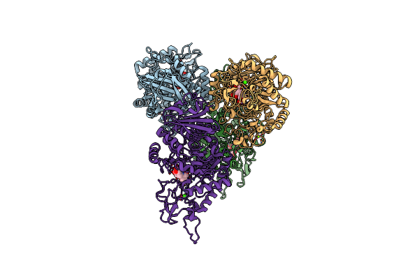
Organism: Severe acute respiratory syndrome coronavirus 2
Method: X-RAY DIFFRACTION Resolution:3.83 Å Release Date: 2020-11-11 Classification: HYDROLASE Ligands: APR |
 |
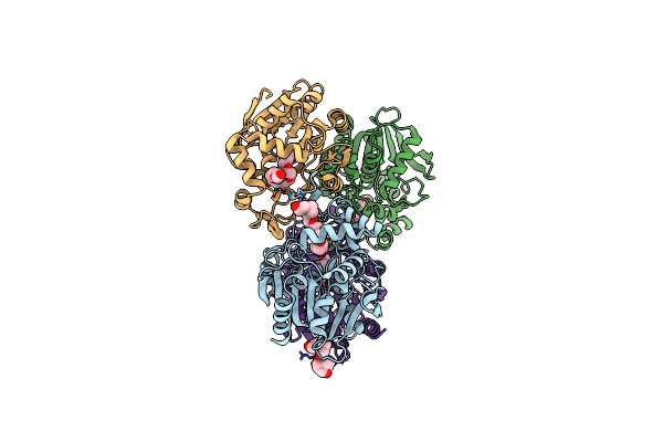
Organism: Severe acute respiratory syndrome coronavirus 2
Method: X-RAY DIFFRACTION Resolution:2.64 Å Release Date: 2020-11-11 Classification: VIRAL PROTEIN Ligands: APR |
 |
Organism: Vitis vinifera
Method: X-RAY DIFFRACTION Resolution:1.30 Å Release Date: 2020-09-16 Classification: ANTIMICROBIAL PROTEIN |
 |
Organism: Elaeis guineensis
Method: X-RAY DIFFRACTION Resolution:2.10 Å Release Date: 2020-09-09 Classification: PROTEIN BINDING |