

Search Count: 333
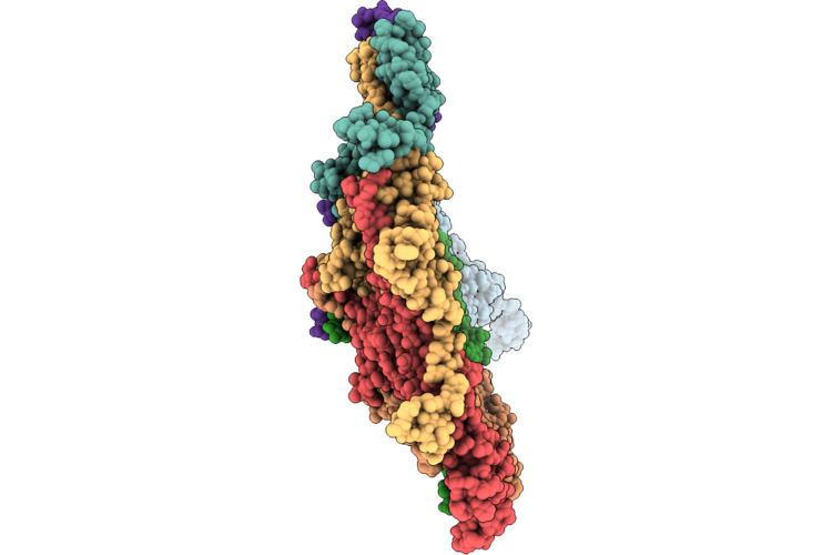 |
Capsid Of The Oenococcus Oeni Phage Oe33Pa
Organism: Oenococcus phage phioe33pa
Method: ELECTRON MICROSCOPY Release Date: 2026-06-17 Classification: VIRAL PROTEIN |
Organism: Oenococcus phage phioe33pa
Method: ELECTRON MICROSCOPY
Release Date: 2026-06-17
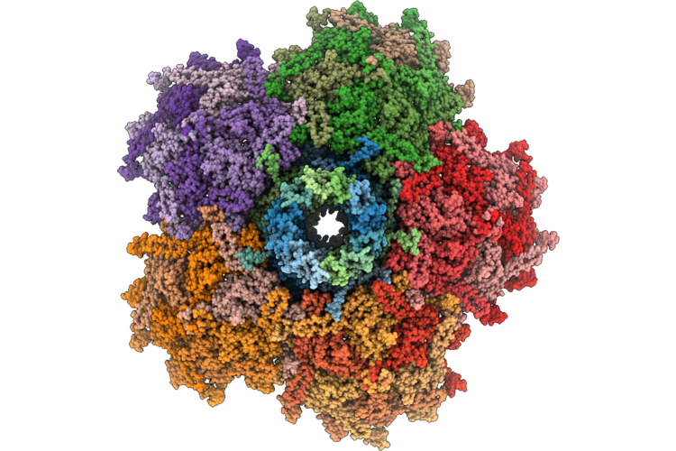 |
Capsid-Connector Assembly Of The Phage Oe33Pa
Organism: Oenococcus phage phioe33pa
Method: ELECTRON MICROSCOPY Release Date: 2026-06-17 Classification: VIRAL PROTEIN |
Organism: Oenococcus phage phioe33pa
Method: ELECTRON MICROSCOPY
Release Date: 2026-06-17
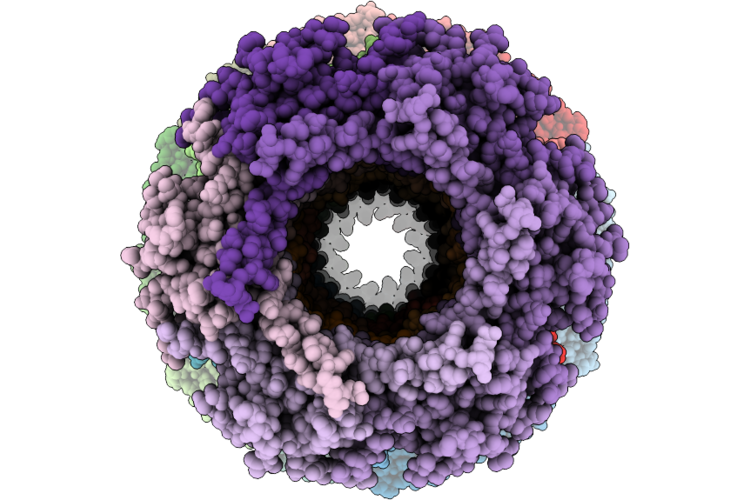 |
Connector And Major Tail Protein Of The Phage Oe33Pa
Organism: Oenococcus phage phioe33pa
Method: ELECTRON MICROSCOPY Release Date: 2026-06-17 Classification: VIRAL PROTEIN Ligands: CA |
Organism: Oenococcus phage phioe33pa
Method: ELECTRON MICROSCOPY
Release Date: 2026-06-17
Ligands: CA
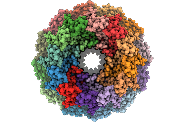 |
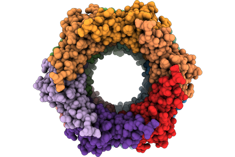
Portal-Adaptor Asssembly Of The Phage Oe33Pa
Organism: Oenococcus phage phioe33pa
Method: ELECTRON MICROSCOPY Release Date: 2026-06-17 Classification: VIRAL PROTEIN |
Organism: Oenococcus phage phioe33pa
Method: ELECTRON MICROSCOPY
Release Date: 2026-06-17
 |
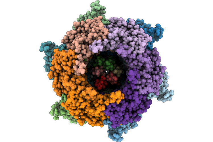
Stopper And Tail Terminator Assembly Of The Phage Oe33Pa
Organism: Oenococcus phage phioe33pa
Method: ELECTRON MICROSCOPY Release Date: 2026-06-17 Classification: VIRAL PROTEIN |
Organism: Oenococcus phage phioe33pa
Method: ELECTRON MICROSCOPY
Release Date: 2026-06-17
 |
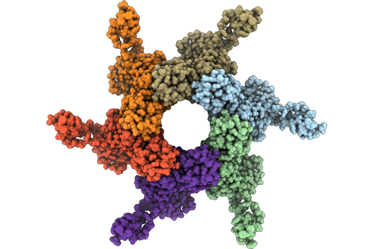
Adhesion Device (C3 Symmetrized Reconstruction) Of The Phage Oe33Pa
Organism: Oenococcus phage phioe33pa
Method: ELECTRON MICROSCOPY Release Date: 2026-06-17 Classification: VIRAL PROTEIN Ligands: CA |
Organism: Oenococcus phage phioe33pa
Method: ELECTRON MICROSCOPY
Release Date: 2026-06-17
Ligands: CA
 |
C6 Symmetrized Reconstruction Of The Adhesion Device Of The Phage Oe33Pa
Organism: Oenococcus phage phioe33pa
Method: ELECTRON MICROSCOPY Release Date: 2026-06-17 Classification: VIRAL PROTEIN |
Organism: Oenococcus phage phioe33pa
Method: ELECTRON MICROSCOPY
Release Date: 2026-06-17
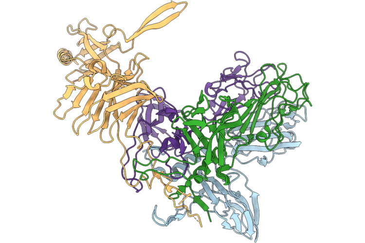 |
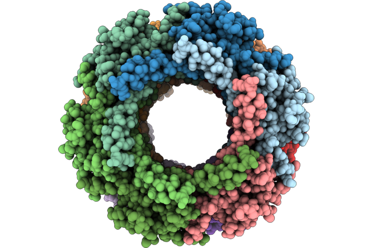
Rbp Trimer Bound To A Monomer Of The Distal Tail Protein Of The Phage Oe33Pa
Organism: Oenococcus phage phioe33pa
Method: ELECTRON MICROSCOPY Release Date: 2026-06-17 Classification: VIRAL PROTEIN |
Organism: Oenococcus phage phioe33pa
Method: ELECTRON MICROSCOPY
Release Date: 2026-06-17
 |
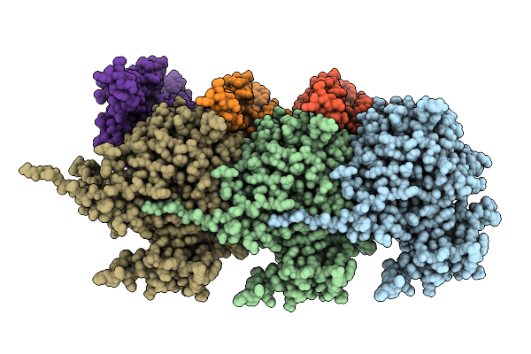
Part Of The Tail Tube Of The Phage Oe33Pa
Organism: Oenococcus phage phioe33pa
Method: ELECTRON MICROSCOPY Release Date: 2026-06-17 Classification: VIRAL PROTEIN Ligands: CA |
Organism: Oenococcus phage phioe33pa
Method: ELECTRON MICROSCOPY
Release Date: 2026-06-17
Ligands: CA
 |
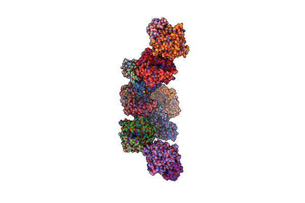
Cryo-Em Structure Of The T9Ss Porkn Ring Complex Of P. Gingivalis
Organism: Porphyromonas gingivalis atcc 33277
Method: ELECTRON MICROSCOPY Resolution:3.20 Å Release Date: 2025-08-06 Classification: MEMBRANE PROTEIN |
Organism: Porphyromonas gingivalis atcc 33277
Method: ELECTRON MICROSCOPY
Release Date: 2025-08-06
 |
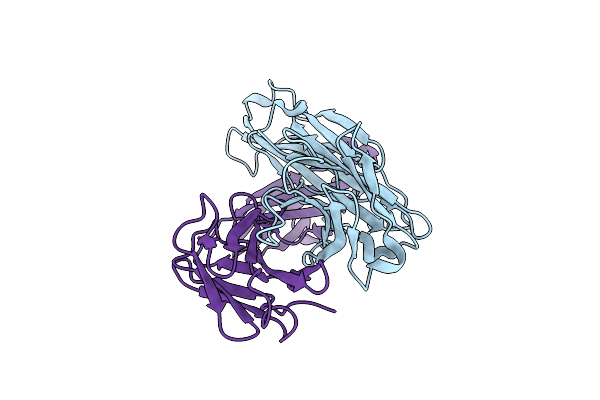
Structure Of The Adherent-Invasive Escherichia Coli Tle3/Tli3 T6Ss Effector/Immunity Complex
Organism: Escherichia coli
Method: X-RAY DIFFRACTION Resolution:3.60 Å Release Date: 2023-02-08 Classification: HYDROLASE Ligands: CA |
Organism: Escherichia coli
Method: X-RAY DIFFRACTION
Release Date: 2023-02-08
Ligands: CA
 |
Highly Potent Il6 Antagonistic Antibody Selected From A Camelid Immune Phage Display Repertoire
Organism: Lama glama
Method: X-RAY DIFFRACTION Resolution:2.77 Å Release Date: 2022-09-07 Classification: IMMUNE SYSTEM |
Organism: Lama glama
Method: X-RAY DIFFRACTION
Release Date: 2022-09-07
 |
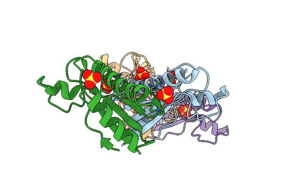
Crystal Structure Of Pseudomonas Aeruginosa Lytic Polysaccharide Monooxygenase Cbpd
Organism: Pseudomonas aeruginosa (strain atcc 15692 / dsm 22644 / cip 104116 / jcm 14847 / lmg 12228 / 1c / prs 101 / pao1)
Method: X-RAY DIFFRACTION Resolution:3.00 Å Release Date: 2022-07-20 Classification: OXIDOREDUCTASE Ligands: NH4 |
 |
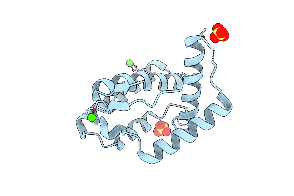
Structure Of The Tagl Peptidoglycan Binding Domain From Eaec T6Ss
Organism: Escherichia coli
Method: X-RAY DIFFRACTION Resolution:2.43 Å Release Date: 2021-08-04 Classification: PROTEIN BINDING Ligands: SO4 |
Organism: Escherichia coli
Method: X-RAY DIFFRACTION
Release Date: 2021-08-04
Ligands: SO4
 |
Structure Of Obp1 From Varroa Destructor, Form P3<2>21
Organism: Varroa destructor
Method: X-RAY DIFFRACTION Resolution:1.81 Å Release Date: 2021-07-07 Classification: TRANSPORT PROTEIN Ligands: SO4, CA, NA |
Organism: Varroa destructor
Method: X-RAY DIFFRACTION
Release Date: 2021-07-07
Ligands: SO4, CA, NA
 |
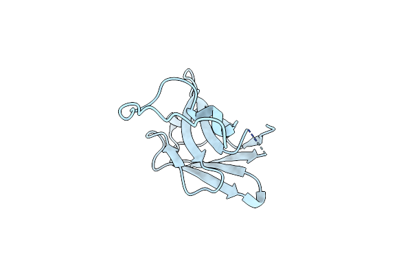
Structure Of Obp1 From Varroa Destructor, Form P2<1>
Organism: Varroa destructor
Method: X-RAY DIFFRACTION Resolution:1.20 Å Release Date: 2021-07-07 Classification: TRANSPORT PROTEIN Ligands: SO4, NHE, NA |
Organism: Varroa destructor
Method: X-RAY DIFFRACTION
Release Date: 2021-07-07
Ligands: SO4, NHE, NA
 |
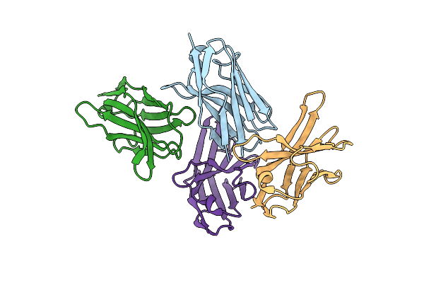
Llama Nanobody Nb17 Raised Against Gldl From Flavobacterium Jonhsoniae
Organism: Lama glama
Method: X-RAY DIFFRACTION Resolution:1.70 Å Release Date: 2021-05-26 Classification: IMMUNE SYSTEM |
Organism: Lama glama
Method: X-RAY DIFFRACTION
Release Date: 2021-05-26
 |
Llama Nanobody Nb18 Raised Against Gldl From Flavobacterium Jonhsoniae
Organism: Lama glama
Method: X-RAY DIFFRACTION Resolution:2.59 Å Release Date: 2021-05-26 Classification: IMMUNE SYSTEM |
Organism: Lama glama
Method: X-RAY DIFFRACTION
Release Date: 2021-05-26
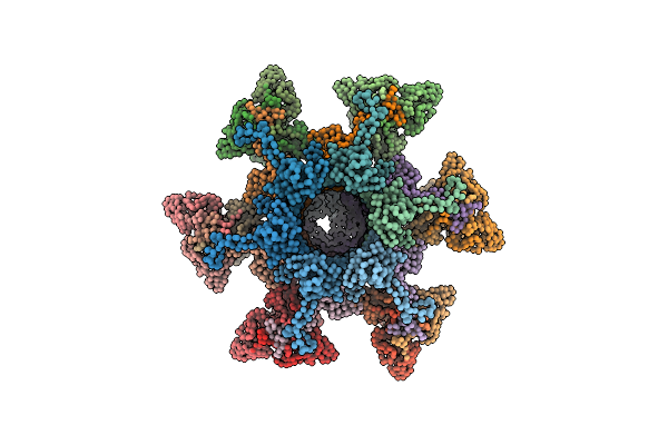 |
Topological Model Of The P2 Virion Baseplate In Activated Conformation (Closed Tal Trimer)
Organism: Lactococcus phage p2
Method: ELECTRON MICROSCOPY Release Date: 2020-08-26 Classification: VIRAL PROTEIN |
Organism: Lactococcus phage p2
Method: ELECTRON MICROSCOPY
Release Date: 2020-08-26
 |
Topological Model Of P2 Virion Baseplate In Activated Conformation
Organism: Lactococcus phage p2
Method: ELECTRON MICROSCOPY Release Date: 2020-08-26 Classification: VIRAL PROTEIN |
Organism: Lactococcus phage p2
Method: ELECTRON MICROSCOPY
Release Date: 2020-08-26