

Search Count: 571
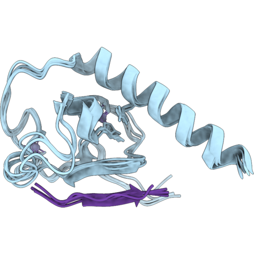 |
Solution Nmr Structure Of Set-9/26 Phd Finger In The Peptide (H3K4Me3)-Bound State
Organism: Caenorhabditis elegans, Homo sapiens
Method: SOLUTION NMR Release Date: 2026-10-14 Classification: GENE REGULATION Ligands: ZN |
Organism: Caenorhabditis elegans, Homo sapiens
Method: SOLUTION NMR
Release Date: 2026-10-14
Ligands: ZN
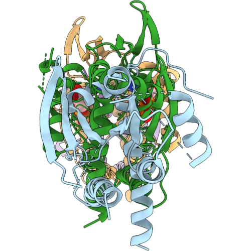 |
Septin Tetrameric Complex From Caenorhabditis Elegans By Cryo-Em
Organism: Caenorhabditis elegans
Method: ELECTRON MICROSCOPY Release Date: 2026-09-09 Classification: CELL CYCLE Ligands: GDP |
Organism: Caenorhabditis elegans
Method: ELECTRON MICROSCOPY
Release Date: 2026-09-09
Ligands: GDP
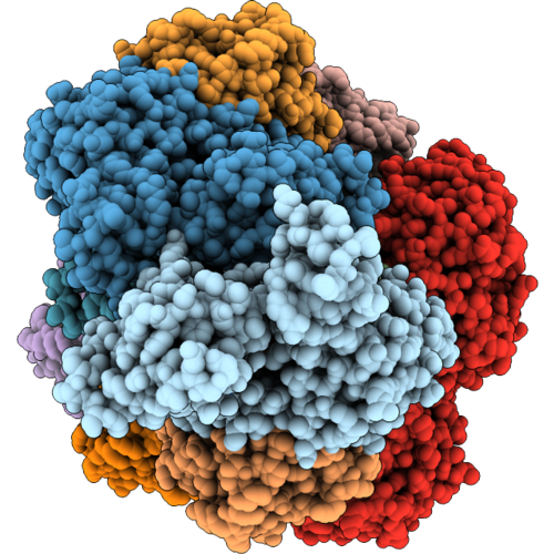 |
Cofilactin Filament With One Aip1 Bound To Middle Of The Filament
Organism: Caenorhabditis elegans, Oryctolagus cuniculus
Method: ELECTRON MICROSCOPY Resolution:2.29 Å Release Date: 2026-09-09 Classification: STRUCTURAL PROTEIN Ligands: ADP, MG |
Organism: Caenorhabditis elegans, Oryctolagus cuniculus
Method: ELECTRON MICROSCOPY
Release Date: 2026-09-09
Ligands: ADP, MG
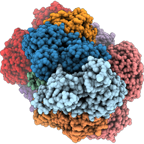 |
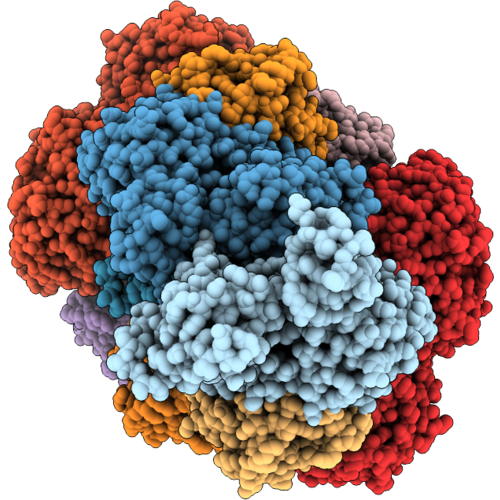
Two Aip1 Bound To The Barbed End Of Cofilin Actin
Organism: Caenorhabditis elegans, Oryctolagus cuniculus
Method: ELECTRON MICROSCOPY Resolution:2.69 Å Release Date: 2026-09-09 Classification: STRUCTURAL PROTEIN Ligands: ADP, MG |
Organism: Caenorhabditis elegans, Oryctolagus cuniculus
Method: ELECTRON MICROSCOPY
Release Date: 2026-09-09
Ligands: ADP, MG
 |
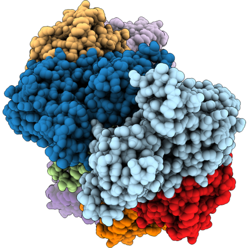
Cofilactin Filament With Two Aip1 Bound To Middle Of The Filament
Organism: Caenorhabditis elegans, Oryctolagus cuniculus
Method: ELECTRON MICROSCOPY Resolution:2.30 Å Release Date: 2026-09-09 Classification: STRUCTURAL PROTEIN Ligands: ADP, MG |
Organism: Caenorhabditis elegans, Oryctolagus cuniculus
Method: ELECTRON MICROSCOPY
Release Date: 2026-09-09
Ligands: ADP, MG
 |
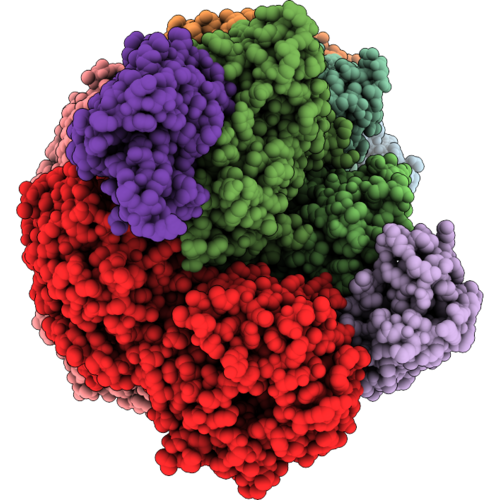
Cofilactin Filament
Organism: Caenorhabditis elegans, Oryctolagus cuniculus
Method: ELECTRON MICROSCOPY Resolution:1.99 Å Release Date: 2026-09-09 Classification: STRUCTURAL PROTEIN Ligands: ADP, MG |
Organism: Caenorhabditis elegans, Oryctolagus cuniculus
Method: ELECTRON MICROSCOPY
Release Date: 2026-09-09
Ligands: ADP, MG
 |
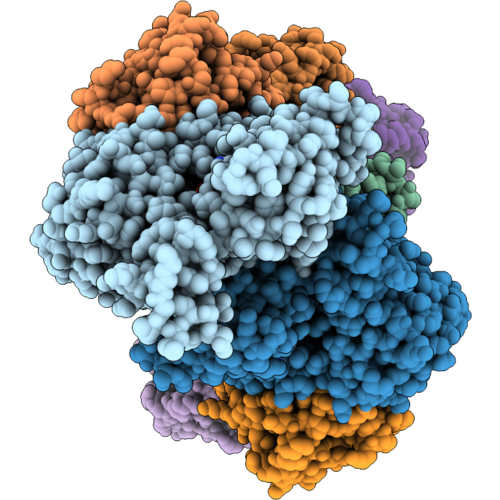
One Aip1 Bound To The Barbed End Of Cofilin Actin
Organism: Caenorhabditis elegans, Oryctolagus cuniculus
Method: ELECTRON MICROSCOPY Resolution:3.97 Å Release Date: 2026-09-09 Classification: STRUCTURAL PROTEIN Ligands: ADP, MG |
Organism: Caenorhabditis elegans, Oryctolagus cuniculus
Method: ELECTRON MICROSCOPY
Release Date: 2026-09-09
Ligands: ADP, MG
 |
Pointed End Of Cofilin Bound F-Actin
Organism: Caenorhabditis elegans, Oryctolagus cuniculus
Method: ELECTRON MICROSCOPY Resolution:2.83 Å Release Date: 2026-09-09 Classification: STRUCTURAL PROTEIN Ligands: ADP, MG |
Organism: Caenorhabditis elegans, Oryctolagus cuniculus
Method: ELECTRON MICROSCOPY
Release Date: 2026-09-09
Ligands: ADP, MG
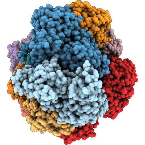 |
Two Aip1 Bound To Same Side Of Cofilin Filament (Stacked Aip1)
Organism: Caenorhabditis elegans, Oryctolagus cuniculus
Method: ELECTRON MICROSCOPY Resolution:2.86 Å Release Date: 2026-09-09 Classification: STRUCTURAL PROTEIN Ligands: ADP, MG |
Organism: Caenorhabditis elegans, Oryctolagus cuniculus
Method: ELECTRON MICROSCOPY
Release Date: 2026-09-09
Ligands: ADP, MG
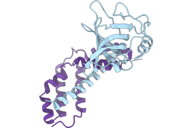 |
Crystal Structure Of The Caenorhabditis Elegans Telomeric Pot-1-Tebp-1 Complex Interface
Organism: Caenorhabditis elegans
Method: X-RAY DIFFRACTION Resolution:1.48 Å Release Date: 2026-08-12 Classification: DNA BINDING PROTEIN |
Organism: Caenorhabditis elegans
Method: X-RAY DIFFRACTION
Release Date: 2026-08-12
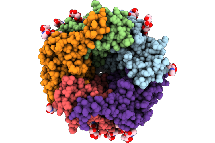 |
Cryo-Em Structure Of The Deg-3/Des-2 Apo State
Organism: Caenorhabditis elegans
Method: ELECTRON MICROSCOPY Resolution:2.57 Å Release Date: 2026-08-12 Classification: MEMBRANE PROTEIN Ligands: NAG |
Organism: Caenorhabditis elegans
Method: ELECTRON MICROSCOPY
Release Date: 2026-08-12
Ligands: NAG
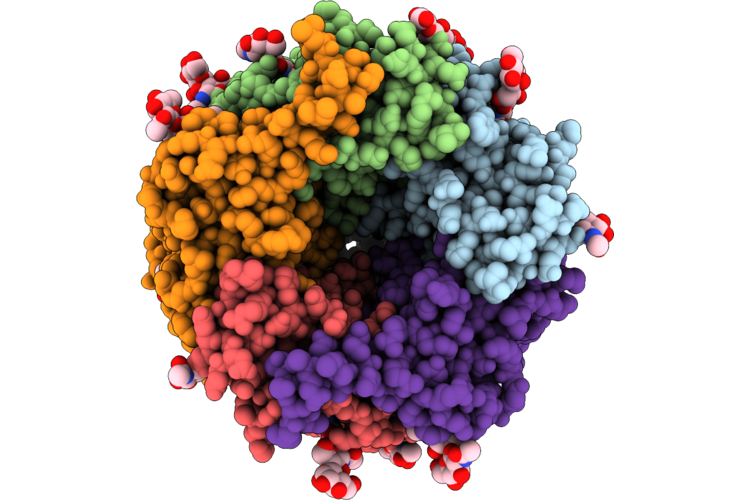 |
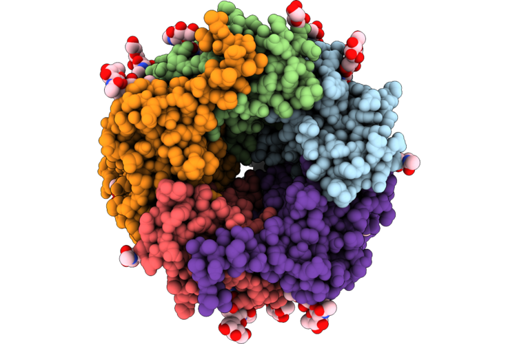
Cryo-Em Structure Of The Deg-3/Des-2 Betaine-Bound Intermediate State
Organism: Caenorhabditis elegans
Method: ELECTRON MICROSCOPY Resolution:2.66 Å Release Date: 2026-08-12 Classification: MEMBRANE PROTEIN Ligands: BET, NAG |
Organism: Caenorhabditis elegans
Method: ELECTRON MICROSCOPY
Release Date: 2026-08-12
Ligands: BET, NAG
 |
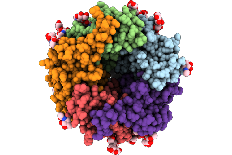
Cryo-Em Structure Of The Deg-3/Des-2 Betaine-Bound Open State
Organism: Caenorhabditis elegans
Method: ELECTRON MICROSCOPY Resolution:2.50 Å Release Date: 2026-08-12 Classification: MEMBRANE PROTEIN Ligands: BET, NAG |
Organism: Caenorhabditis elegans
Method: ELECTRON MICROSCOPY
Release Date: 2026-08-12
Ligands: BET, NAG
 |
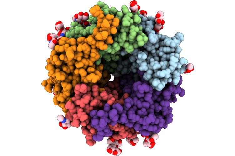
Cryo-Em Structure Of The Deg-3/Des-2 Choline-Bound Intermediate State
Organism: Caenorhabditis elegans
Method: ELECTRON MICROSCOPY Resolution:2.67 Å Release Date: 2026-08-12 Classification: MEMBRANE PROTEIN Ligands: CHT, NAG |
Organism: Caenorhabditis elegans
Method: ELECTRON MICROSCOPY
Release Date: 2026-08-12
Ligands: CHT, NAG
 |
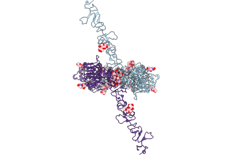
Cryo-Em Structure Of The Deg-3/Des-2 Choline-Bound Open State
Organism: Caenorhabditis elegans
Method: ELECTRON MICROSCOPY Resolution:2.77 Å Release Date: 2026-08-12 Classification: MEMBRANE PROTEIN Ligands: CHT, NAG |
Organism: Caenorhabditis elegans
Method: ELECTRON MICROSCOPY
Release Date: 2026-08-12
Ligands: CHT, NAG
 |
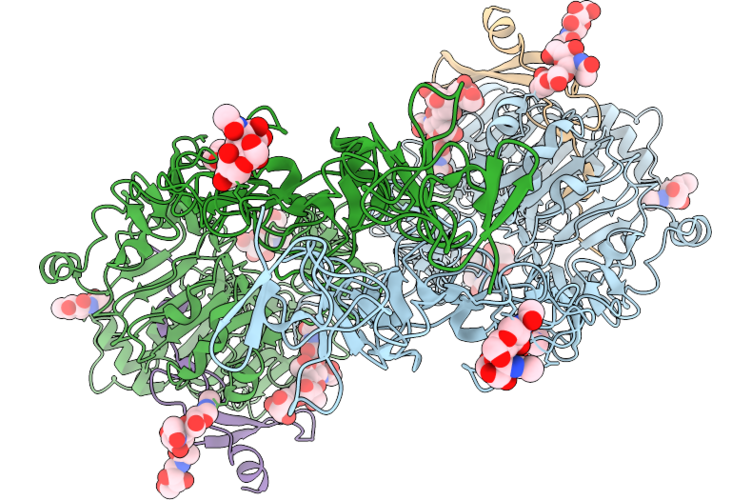
Cryo-Em Structure Of A Preformed Dimer Of The C. Elegans Egfr (Let-23) Extracellular Region
Organism: Caenorhabditis elegans
Method: ELECTRON MICROSCOPY Release Date: 2026-07-29 Classification: SIGNALING PROTEIN Ligands: NAG |
Organism: Caenorhabditis elegans
Method: ELECTRON MICROSCOPY
Release Date: 2026-07-29
Ligands: NAG
 |
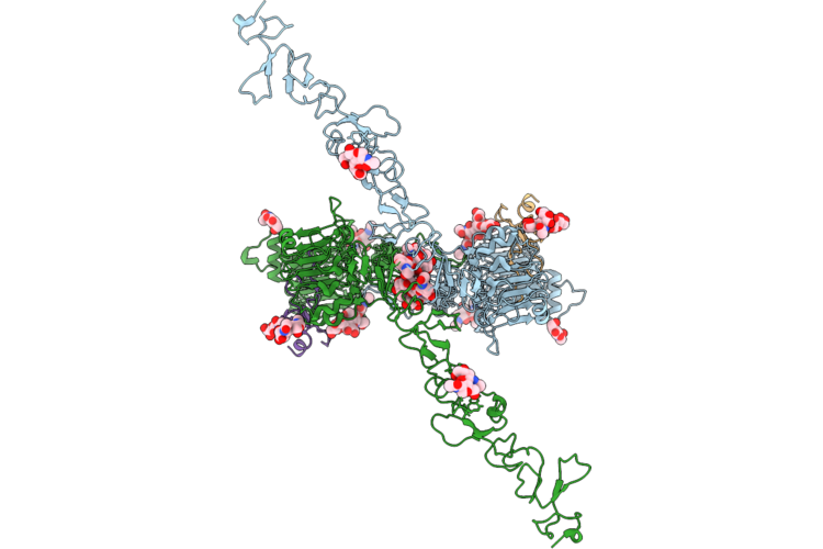
Cryo-Em Structure Of An Active Dimer Of The C. Elegans Egfr (Let-23) Extracellular Region Bound To Lin-3
Organism: Caenorhabditis elegans, Drosophila melanogaster
Method: ELECTRON MICROSCOPY Release Date: 2026-07-29 Classification: SIGNALING PROTEIN Ligands: NAG |
Organism: Caenorhabditis elegans, Drosophila melanogaster
Method: ELECTRON MICROSCOPY
Release Date: 2026-07-29
Ligands: NAG
 |
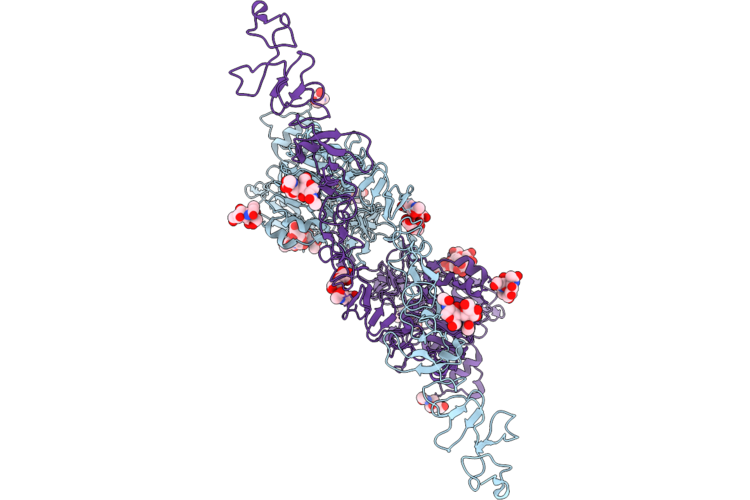
Cryo-Em Structure Of An Inactive Dimer Of The C. Elegans Egfr (Let-23) Extracellular Region Bound To Lin-3.
Organism: Caenorhabditis elegans, Drosophila melanogaster
Method: ELECTRON MICROSCOPY Release Date: 2026-07-29 Classification: SIGNALING PROTEIN Ligands: NAG |
Organism: Caenorhabditis elegans, Drosophila melanogaster
Method: ELECTRON MICROSCOPY
Release Date: 2026-07-29
Ligands: NAG
 |
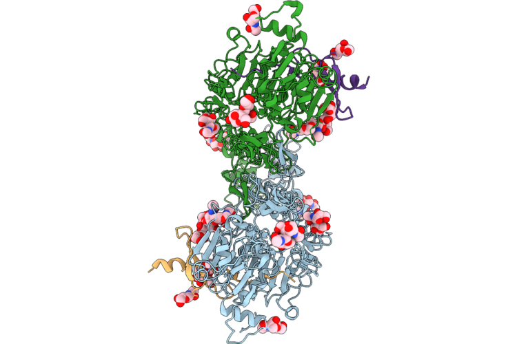
Cryo-Em Structure Of A Weak Dimer Of The C. Elegans Egfr (Let-23) Extracellular Region With A Domain Iv Loop Deletion
Organism: Caenorhabditis elegans
Method: ELECTRON MICROSCOPY Release Date: 2026-07-29 Classification: SIGNALING PROTEIN Ligands: NAG |
Organism: Caenorhabditis elegans
Method: ELECTRON MICROSCOPY
Release Date: 2026-07-29
Ligands: NAG
 |
Cryo-Em Structure Of An Active Dimer Of The C. Elegans Egfr (Let-23) Extracellular Region With A Domain Iv Loop Deletion Bound To Lin-3.
Organism: Caenorhabditis elegans
Method: ELECTRON MICROSCOPY Release Date: 2026-07-29 Classification: SIGNALING PROTEIN Ligands: NAG |
Organism: Caenorhabditis elegans
Method: ELECTRON MICROSCOPY
Release Date: 2026-07-29
Ligands: NAG