

Search Count: 2,329
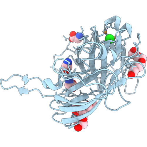 |
Crystal Structure Of A Tower-Slac, A Laccase From Streptomyces Coelicolor With A Tower Loop Mutation
Organism: Streptomyces coelicolor
Method: X-RAY DIFFRACTION Resolution:2.85 Å Release Date: 2026-10-14 Classification: OXIDOREDUCTASE Ligands: CU, OH, GLY, PG4, CL |
Organism: Streptomyces coelicolor
Method: X-RAY DIFFRACTION
Release Date: 2026-10-14
Ligands: CU, OH, GLY, PG4, CL
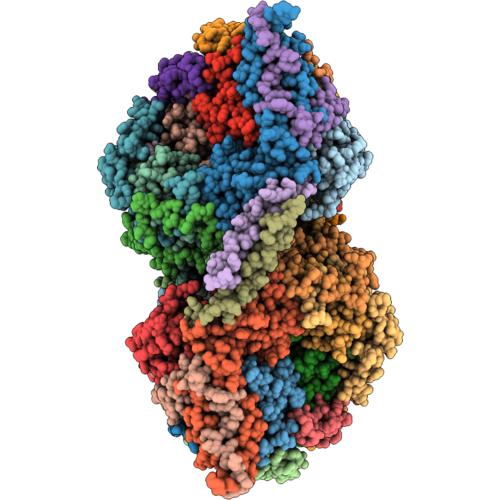 |
Cryo-Em Structure Of Leishmania Tarentolae Respiratory Complex Iv (Cytochrome C Oxidase Dimer)
Organism: Leishmania tarentolae
Method: ELECTRON MICROSCOPY Resolution:3.21 Å Release Date: 2026-10-07 Classification: ELECTRON TRANSPORT Ligands: CU, HEA |
Organism: Leishmania tarentolae
Method: ELECTRON MICROSCOPY
Release Date: 2026-10-07
Ligands: CU, HEA
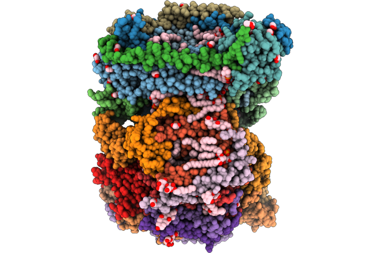 |
Crystal Structure Of Cyanate Bound Bovine Cytochrome C Oxidase
Organism: Bos taurus
Method: X-RAY DIFFRACTION Resolution:2.10 Å Release Date: 2026-09-23 Classification: MEMBRANE PROTEIN Ligands: CU, MG, NA, PGV, HEA, EDO, 0NM, CUA, PSC, CHD, PEK, CDL, DMU, TGL, ZN, SAC |
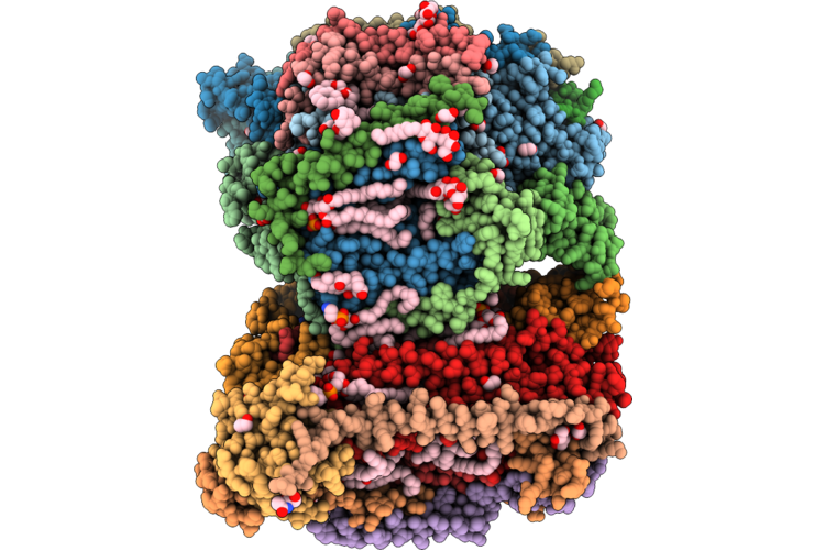 |
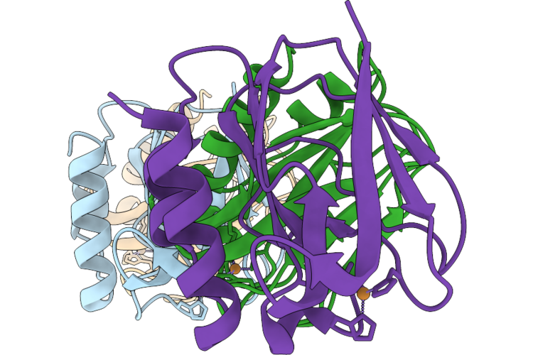
Crystal Structure Of Thiocyanate Bound Bovine Cytochrome C Oxidase
Organism: Bos taurus
Method: X-RAY DIFFRACTION Resolution:2.39 Å Release Date: 2026-09-23 Classification: MEMBRANE PROTEIN Ligands: CU, MG, NA, PGV, HEA, EDO, SCN, CUA, TGL, DMU, CHD, PEK, CDL, ZN, PSC, SAC |
 |
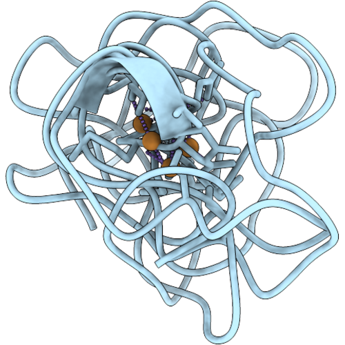
X-Ray Crystal Structure Of Pseudoazurin Met16Ala Variant
Organism: Achromobacter cycloclastes
Method: X-RAY DIFFRACTION Resolution:1.80 Å Release Date: 2026-09-23 Classification: ELECTRON TRANSPORT Ligands: CU |
Organism: Achromobacter cycloclastes
Method: X-RAY DIFFRACTION
Release Date: 2026-09-23
Ligands: CU
 |
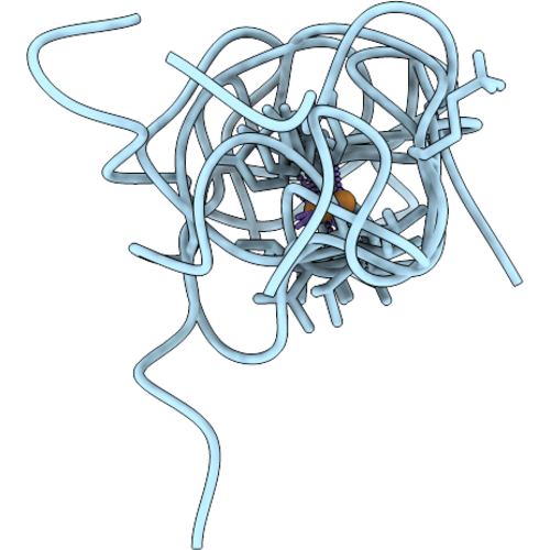
Cu(Ii)-Bound Alpha-Synuclein Monomer Complex - Local Structure Of N-Terminal Binding Site
Organism: Homo sapiens
Method: SOLUTION NMR Release Date: 2026-09-16 Classification: METAL BINDING PROTEIN Ligands: CU |
Organism: Homo sapiens
Method: SOLUTION NMR
Release Date: 2026-09-16
Ligands: CU
 |
Cu(Ii) - Alpha-Synuclein Monomer Complex - Local Structure Of Binding Site His50 With Gly47 As Ligand
Organism: Homo sapiens
Method: SOLUTION NMR Release Date: 2026-09-16 Classification: METAL BINDING PROTEIN Ligands: CU |
Organism: Homo sapiens
Method: SOLUTION NMR
Release Date: 2026-09-16
Ligands: CU
 |
Cu(Ii) - Alpha-Synuclein Monomer Complex - Local Structure Of Binding Site His50 With Glu46 As Ligand
Organism: Homo sapiens
Method: SOLUTION NMR Release Date: 2026-09-16 Classification: METAL BINDING PROTEIN Ligands: CU |
Organism: Homo sapiens
Method: SOLUTION NMR
Release Date: 2026-09-16
Ligands: CU
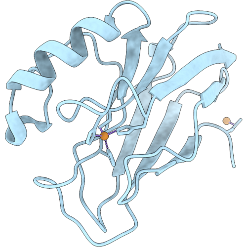 |
Pseudomonas Aeruginosa Azurin Mutant - M121H
Organism: Pseudomonas aeruginosa
Method: X-RAY DIFFRACTION Resolution:2.47 Å Release Date: 2026-09-09 Classification: ELECTRON TRANSPORT Ligands: CU |
Organism: Pseudomonas aeruginosa
Method: X-RAY DIFFRACTION
Release Date: 2026-09-09
Ligands: CU
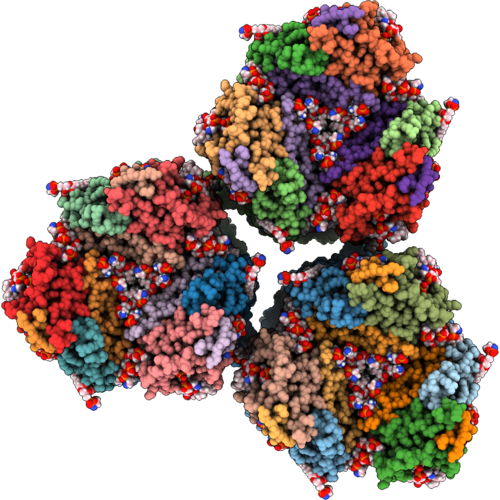 |
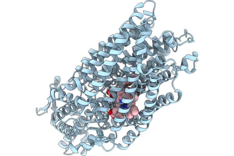
Particulate Methane Monooxygenase In Membrane Arrays
Organism: Methylococcus capsulatus str. bath
Method: ELECTRON MICROSCOPY Release Date: 2026-08-26 Classification: OXIDOREDUCTASE Ligands: CU, A1A0P |
Organism: Methylococcus capsulatus str. bath
Method: ELECTRON MICROSCOPY
Release Date: 2026-08-26
Ligands: CU, A1A0P
 |
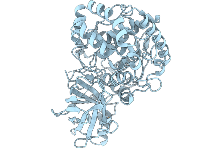
Cryo-Em Structure Of Native Monomeric Quinol-Dependent Nitric Oxide Reductase From Achromobacter Xylosoxidans.
Organism: Achromobacter xylosoxidans
Method: ELECTRON MICROSCOPY Release Date: 2026-08-26 Classification: MEMBRANE PROTEIN Ligands: CA, HEM, CU, FE |
Organism: Achromobacter xylosoxidans
Method: ELECTRON MICROSCOPY
Release Date: 2026-08-26
Ligands: CA, HEM, CU, FE
 |
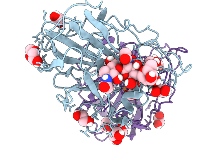
A Tyrosinase With Hydroxylation Activity
Organism: Sinorhizobium saheli
Method: X-RAY DIFFRACTION Resolution:1.84 Å Release Date: 2026-08-26 Classification: METAL BINDING PROTEIN Ligands: CU |
Organism: Sinorhizobium saheli
Method: X-RAY DIFFRACTION
Release Date: 2026-08-26
Ligands: CU
 |
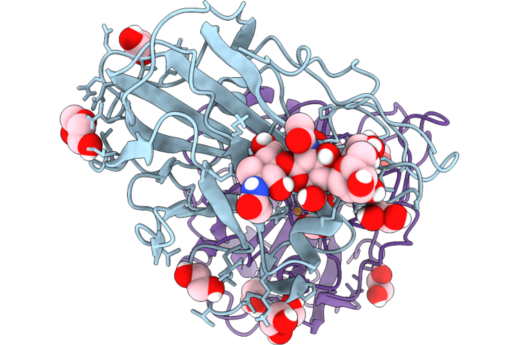
Neurospora Crassa Polysaccharide Monooxygenase 9D Dose Series - Pseudohelix 1 (1.22 Mgy)
Organism: Neurospora crassa
Method: X-RAY DIFFRACTION Resolution:1.10 Å Release Date: 2026-08-12 Classification: OXIDOREDUCTASE Ligands: EDO, PEG, CU, OXY, CO2 |
Organism: Neurospora crassa
Method: X-RAY DIFFRACTION
Release Date: 2026-08-12
Ligands: EDO, PEG, CU, OXY, CO2
 |
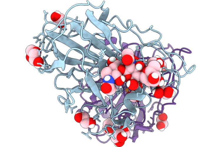
Neurospora Crassa Polysaccharide Monooxygenase 9D Dose Series - Pseudohelix 2 (1.74 Mgy)
Organism: Neurospora crassa
Method: X-RAY DIFFRACTION Resolution:1.10 Å Release Date: 2026-08-12 Classification: OXIDOREDUCTASE Ligands: EDO, PEG, CU, OXY, CO2 |
Organism: Neurospora crassa
Method: X-RAY DIFFRACTION
Release Date: 2026-08-12
Ligands: EDO, PEG, CU, OXY, CO2
 |
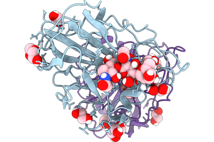
Neurospora Crassa Polysaccharide Monooxygenase 9D Dose Series - Pseudohelix 3 (2.07 Mgy)
Organism: Neurospora crassa
Method: X-RAY DIFFRACTION Resolution:1.10 Å Release Date: 2026-08-12 Classification: OXIDOREDUCTASE Ligands: EDO, PEG, CU, OXY, CO2 |
Organism: Neurospora crassa
Method: X-RAY DIFFRACTION
Release Date: 2026-08-12
Ligands: EDO, PEG, CU, OXY, CO2
 |
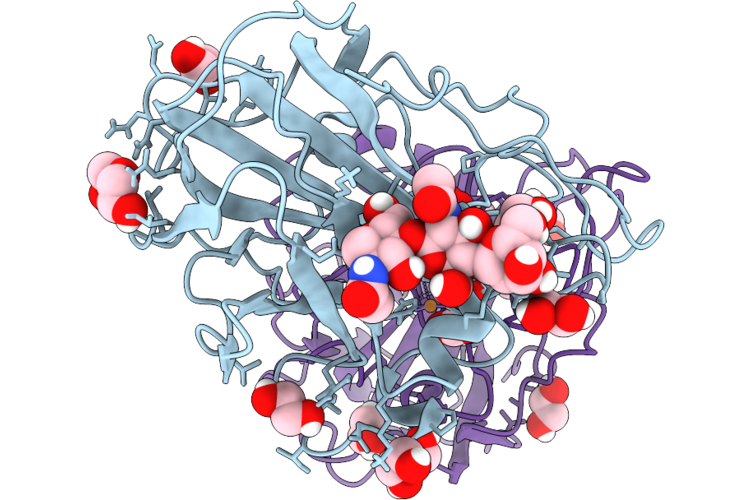
Neurospora Crassa Polysaccharide Monooxygenase 9D Dose Series - Pseudohelix 4 (2.37 Mgy)
Organism: Neurospora crassa
Method: X-RAY DIFFRACTION Resolution:1.10 Å Release Date: 2026-08-12 Classification: OXIDOREDUCTASE Ligands: EDO, PEG, CU, OXY, CO2 |
Organism: Neurospora crassa
Method: X-RAY DIFFRACTION
Release Date: 2026-08-12
Ligands: EDO, PEG, CU, OXY, CO2
 |
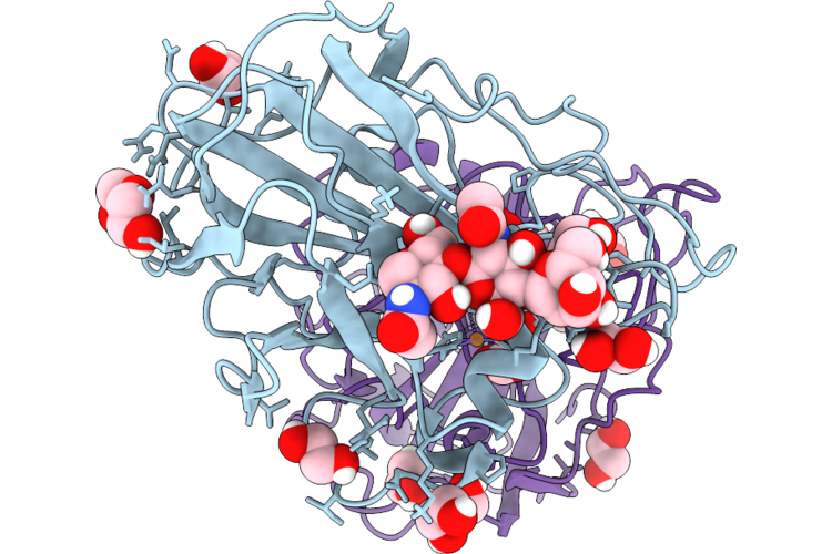
Neurospora Crassa Polysaccharide Monooxygenase 9D Dose Series - Pseudohelix 5 (2.74 Mgy)
Organism: Neurospora crassa
Method: X-RAY DIFFRACTION Resolution:1.10 Å Release Date: 2026-08-12 Classification: OXIDOREDUCTASE Ligands: EDO, PEG, CU, OXY, CO2 |
Organism: Neurospora crassa
Method: X-RAY DIFFRACTION
Release Date: 2026-08-12
Ligands: EDO, PEG, CU, OXY, CO2
 |
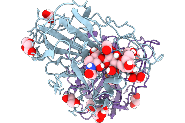
Neurospora Crassa Polysaccharide Monooxygenase 9D Dose Series - Pseudohelix 6 (2.98 Mgy)
Organism: Neurospora crassa
Method: X-RAY DIFFRACTION Resolution:1.10 Å Release Date: 2026-08-12 Classification: OXIDOREDUCTASE Ligands: EDO, PEG, CU, OXY, CO2 |
Organism: Neurospora crassa
Method: X-RAY DIFFRACTION
Release Date: 2026-08-12
Ligands: EDO, PEG, CU, OXY, CO2
 |
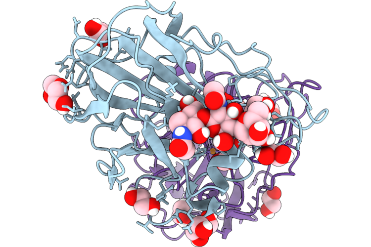
Neurospora Crassa Polysaccharide Monooxygenase 9D Dose Series - Pseudohelix 7 (3.39 Mgy)
Organism: Neurospora crassa
Method: X-RAY DIFFRACTION Resolution:1.10 Å Release Date: 2026-08-12 Classification: OXIDOREDUCTASE Ligands: EDO, PEG, CU, OXY, CO2 |
Organism: Neurospora crassa
Method: X-RAY DIFFRACTION
Release Date: 2026-08-12
Ligands: EDO, PEG, CU, OXY, CO2
 |
Neurospora Crassa Polysaccharide Monooxygenase 9D Dose Series - Pseudohelix 8 (3.56 Mgy)
Organism: Neurospora crassa
Method: X-RAY DIFFRACTION Resolution:1.10 Å Release Date: 2026-08-12 Classification: OXIDOREDUCTASE Ligands: EDO, PEG, CU, OXY, CO2 |
Organism: Neurospora crassa
Method: X-RAY DIFFRACTION
Release Date: 2026-08-12
Ligands: EDO, PEG, CU, OXY, CO2