

Search Count: 16,477
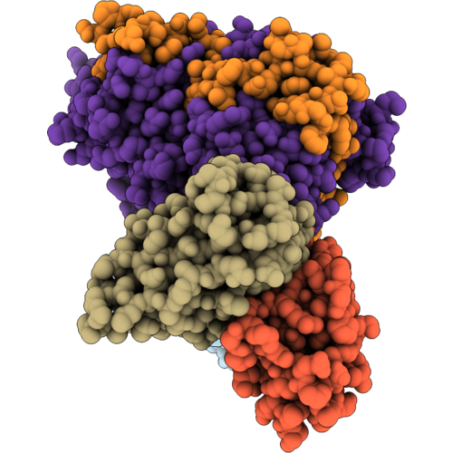 |
Cryo-Em Structure Of Mouse Myeloperoxidase In Complex With Fab Fragments Of Antibodies Mab-A24 And Mab-A46
Organism: Mus musculus
Method: ELECTRON MICROSCOPY Release Date: 2026-09-16 Classification: IMMUNE SYSTEM Ligands: HEM, NAG, CA |
Organism: Mus musculus
Method: ELECTRON MICROSCOPY
Release Date: 2026-09-16
Ligands: HEM, NAG, CA
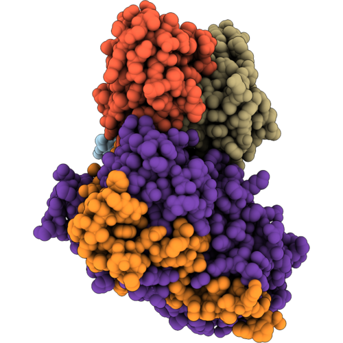 |
Cryo-Em Structure Of Mouse Myeloperoxidase In Complex With Fab Fragments Of Antibodies Mab-A46 And Mab-B88
Organism: Mus musculus
Method: ELECTRON MICROSCOPY Release Date: 2026-09-16 Classification: IMMUNE SYSTEM Ligands: HEM, NAG, CA |
Organism: Mus musculus
Method: ELECTRON MICROSCOPY
Release Date: 2026-09-16
Ligands: HEM, NAG, CA
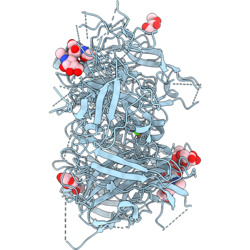 |
Coagulation Factor V Dtqq (B-Domain Region 811-1491 Truncated, R709Q, R1545Q) In Solution-Phase
Organism: Homo sapiens
Method: ELECTRON MICROSCOPY Release Date: 2026-09-16 Classification: BLOOD CLOTTING Ligands: NAG, CA, CU1 |
Organism: Homo sapiens
Method: ELECTRON MICROSCOPY
Release Date: 2026-09-16
Ligands: NAG, CA, CU1
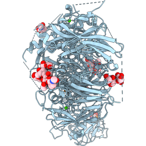 |
Coagulation Factor V Dtqq, Membrane-Bound, On Ptd-Choline : Ptd-Serine 75:25 Vesicles
Organism: Homo sapiens
Method: ELECTRON MICROSCOPY Resolution:2.90 Å Release Date: 2026-09-16 Classification: MEMBRANE PROTEIN Ligands: NAG, CA, CU1, SEP |
Organism: Homo sapiens
Method: ELECTRON MICROSCOPY
Release Date: 2026-09-16
Ligands: NAG, CA, CU1, SEP
 |
Coagulation Factor Viii, Full-Length, In Solution-Phase
Organism: Homo sapiens
Method: ELECTRON MICROSCOPY Resolution:3.26 Å Release Date: 2026-09-16 Classification: BLOOD CLOTTING Ligands: NAG, CA, CU1, CPS |
Organism: Homo sapiens
Method: ELECTRON MICROSCOPY
Release Date: 2026-09-16
Ligands: NAG, CA, CU1, CPS
 |
Coagulation Factor Viii, Full-Length, Membrane-Bound, On Ptd-Choline : Ptd-Serine 75:25 Vesicles
Organism: Homo sapiens
Method: ELECTRON MICROSCOPY Resolution:3.15 Å Release Date: 2026-09-16 Classification: MEMBRANE PROTEIN Ligands: NAG, CA, CU1, SEP, CPS |
Organism: Homo sapiens
Method: ELECTRON MICROSCOPY
Release Date: 2026-09-16
Ligands: NAG, CA, CU1, SEP, CPS
 |
Calcium-Coupled Proline Ring Puckering Defines A Structural Switch In A Thermophilic Lipase (Geobacillus Kaustophilus)
Organism: Geobacillus kaustophilus nbrc 102445
Method: X-RAY DIFFRACTION Resolution:2.47 Å Release Date: 2026-09-16 Classification: HYDROLASE Ligands: CA, ZN |
Organism: Geobacillus kaustophilus nbrc 102445
Method: X-RAY DIFFRACTION
Release Date: 2026-09-16
Ligands: CA, ZN
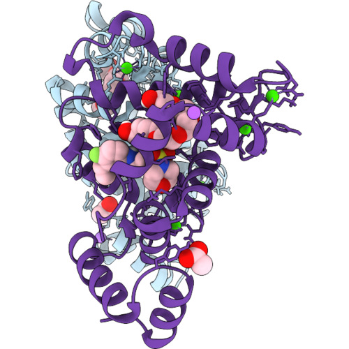 |
Ncs-1 Bound To Xchem Ligand 1
Organism: Homo sapiens
Method: X-RAY DIFFRACTION Resolution:2.15 Å Release Date: 2026-09-16 Classification: METAL BINDING PROTEIN Ligands: CA, ACT, A1I63, P6G, NA, 1PE |
Organism: Homo sapiens
Method: X-RAY DIFFRACTION
Release Date: 2026-09-16
Ligands: CA, ACT, A1I63, P6G, NA, 1PE
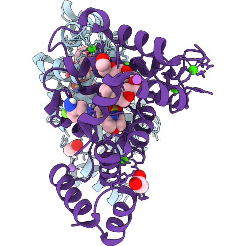 |
Ncs-1 Bound To Xchem Ligand 2
Organism: Homo sapiens
Method: X-RAY DIFFRACTION Resolution:2.27 Å Release Date: 2026-09-16 Classification: METAL BINDING PROTEIN Ligands: CA, ACT, A1I68, P6G, NA |
Organism: Homo sapiens
Method: X-RAY DIFFRACTION
Release Date: 2026-09-16
Ligands: CA, ACT, A1I68, P6G, NA
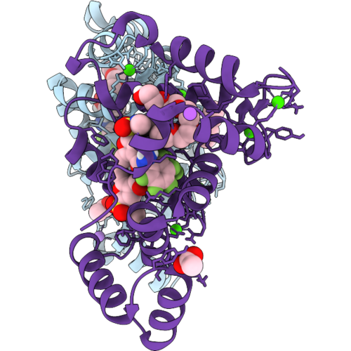 |
Ncs-1 Bound To Xchem Ligand 3
Organism: Homo sapiens
Method: X-RAY DIFFRACTION Resolution:2.33 Å Release Date: 2026-09-16 Classification: METAL BINDING PROTEIN Ligands: CA, ACT, 1PE, A1I7M, NA |
Organism: Homo sapiens
Method: X-RAY DIFFRACTION
Release Date: 2026-09-16
Ligands: CA, ACT, 1PE, A1I7M, NA
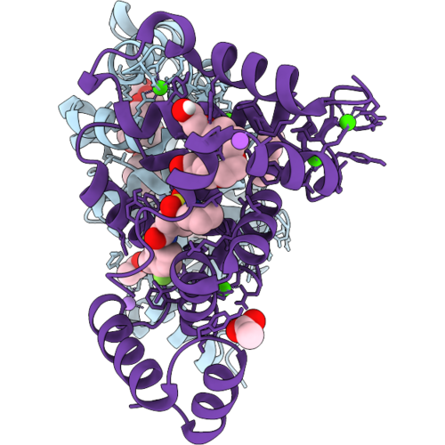 |
Ncs-1 Bound To Xchem Ligand 4
Organism: Homo sapiens
Method: X-RAY DIFFRACTION Resolution:2.82 Å Release Date: 2026-09-16 Classification: METAL BINDING PROTEIN Ligands: CA, ACT, A1I7N, P6G, NA, 1PE |
Organism: Homo sapiens
Method: X-RAY DIFFRACTION
Release Date: 2026-09-16
Ligands: CA, ACT, A1I7N, P6G, NA, 1PE
 |
Ncs-1 Bound To Xchem Ligand 5
Organism: Homo sapiens
Method: X-RAY DIFFRACTION Resolution:2.01 Å Release Date: 2026-09-16 Classification: METAL BINDING PROTEIN Ligands: CA, ACT, 1PE, A1I7A, NA |
Organism: Homo sapiens
Method: X-RAY DIFFRACTION
Release Date: 2026-09-16
Ligands: CA, ACT, 1PE, A1I7A, NA
 |
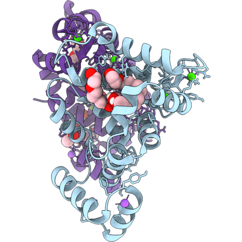
Ncs-1 Bound To Xchem Ligand 6
Organism: Homo sapiens
Method: X-RAY DIFFRACTION Resolution:1.97 Å Release Date: 2026-09-16 Classification: METAL BINDING PROTEIN Ligands: CA, ACT, 1PE, A1I7D, NA |
Organism: Homo sapiens
Method: X-RAY DIFFRACTION
Release Date: 2026-09-16
Ligands: CA, ACT, 1PE, A1I7D, NA
 |
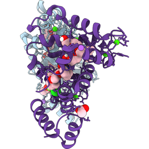
Ncs-1 Bound To Xchem Ligand 7
Organism: Homo sapiens
Method: X-RAY DIFFRACTION Resolution:2.00 Å Release Date: 2026-09-16 Classification: METAL BINDING PROTEIN Ligands: CA, P6G, NA, ACT, 1PE, A1I7C |
Organism: Homo sapiens
Method: X-RAY DIFFRACTION
Release Date: 2026-09-16
Ligands: CA, P6G, NA, ACT, 1PE, A1I7C
 |
Ncs-1 Bound To Xchem Ligand 8
Organism: Homo sapiens
Method: X-RAY DIFFRACTION Resolution:2.93 Å Release Date: 2026-09-16 Classification: METAL BINDING PROTEIN Ligands: CA, ACT, 1PE, A1I7O, NA, P6G |
Organism: Homo sapiens
Method: X-RAY DIFFRACTION
Release Date: 2026-09-16
Ligands: CA, ACT, 1PE, A1I7O, NA, P6G
 |
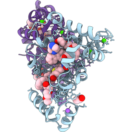
Ncs-1 Bound To Xchem Ligand 9
Organism: Homo sapiens
Method: X-RAY DIFFRACTION Resolution:2.15 Å Release Date: 2026-09-16 Classification: METAL BINDING PROTEIN Ligands: CA, ACT, 1PE, A1I7P, NA |
Organism: Homo sapiens
Method: X-RAY DIFFRACTION
Release Date: 2026-09-16
Ligands: CA, ACT, 1PE, A1I7P, NA
 |
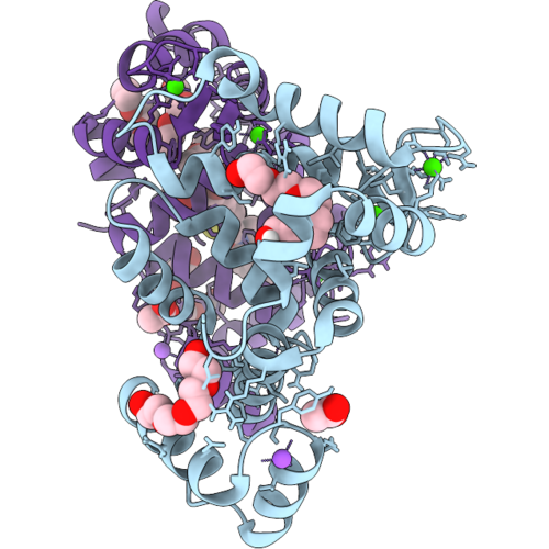
Ncs-1 Bound To Xchem Ligand 10
Organism: Homo sapiens
Method: X-RAY DIFFRACTION Resolution:2.10 Å Release Date: 2026-09-16 Classification: METAL BINDING PROTEIN Ligands: CA, P6G, NA, ACT, 1PE, A1I7V |
Organism: Homo sapiens
Method: X-RAY DIFFRACTION
Release Date: 2026-09-16
Ligands: CA, P6G, NA, ACT, 1PE, A1I7V
 |
Ncs-1 Bound To Xchem Ligand 11
Organism: Homo sapiens
Method: X-RAY DIFFRACTION Resolution:1.73 Å Release Date: 2026-09-16 Classification: METAL BINDING PROTEIN Ligands: CA, P6G, NA, ACT, 1PE, A1I7Q |
Organism: Homo sapiens
Method: X-RAY DIFFRACTION
Release Date: 2026-09-16
Ligands: CA, P6G, NA, ACT, 1PE, A1I7Q
 |
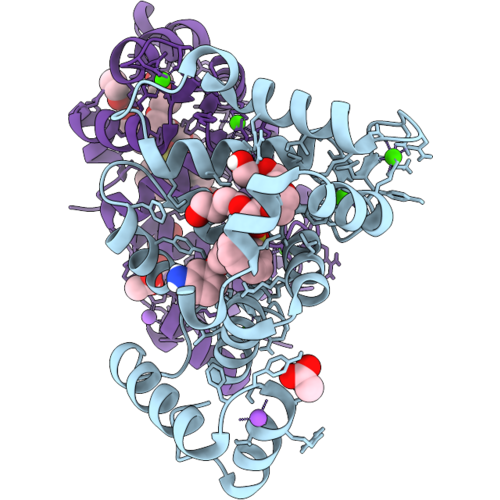
Ncs-1 Bound To Xchem Ligand 12
Organism: Homo sapiens
Method: X-RAY DIFFRACTION Resolution:1.90 Å Release Date: 2026-09-16 Classification: METAL BINDING PROTEIN Ligands: CA, P6G, NA, ACT, 1PE, A1I7R |
Organism: Homo sapiens
Method: X-RAY DIFFRACTION
Release Date: 2026-09-16
Ligands: CA, P6G, NA, ACT, 1PE, A1I7R
 |
Ncs-1 Bound To Xchem Ligand 13
Organism: Homo sapiens
Method: X-RAY DIFFRACTION Resolution:1.78 Å Release Date: 2026-09-16 Classification: METAL BINDING PROTEIN Ligands: CA, P6G, NA, ACT, 1PE, A1I7B |
Organism: Homo sapiens
Method: X-RAY DIFFRACTION
Release Date: 2026-09-16
Ligands: CA, P6G, NA, ACT, 1PE, A1I7B