

Search Count: 215
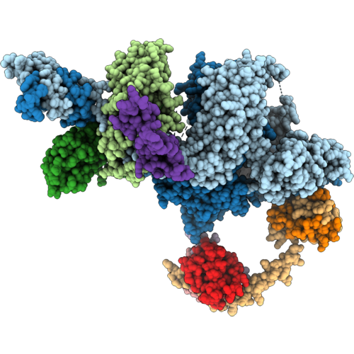 |
In Situ Subtomogram Average Of The Transition Zone Linker In Human Airway Cilia (Composite Map)
Organism: Homo sapiens
Method: ELECTRON MICROSCOPY Release Date: 2026-09-09 Classification: STRUCTURAL PROTEIN |
Organism: Homo sapiens
Method: ELECTRON MICROSCOPY
Release Date: 2026-09-09
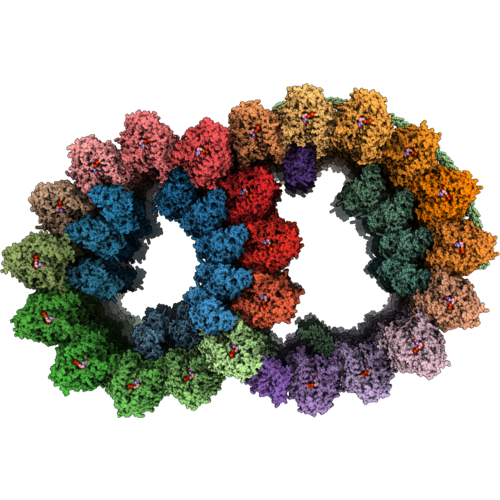 |
In Situ Subtomogram Average Of The Transition Zone Doublet Microtubule In Human Airway Cilia (Composite Map)
Organism: Homo sapiens
Method: ELECTRON MICROSCOPY Release Date: 2026-09-09 Classification: STRUCTURAL PROTEIN Ligands: GTP, GDP |
Organism: Homo sapiens
Method: ELECTRON MICROSCOPY
Release Date: 2026-09-09
Ligands: GTP, GDP
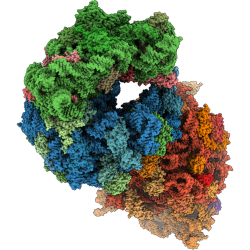 |
Crystal Structure Of The Wild-Type Thermus Thermophilus 70S Ribosome In Complex With Pleuromutilin And Protein Y At 2.70A Resolution
Organism: Escherichia coli k-12, Thermus thermophilus hb8
Method: X-RAY DIFFRACTION Resolution:2.70 Å Release Date: 2026-09-09 Classification: RIBOSOME Ligands: MG, A1CQ9, MPD, ARG, ZN, SF4 |
Organism: Escherichia coli k-12, Thermus thermophilus hb8
Method: X-RAY DIFFRACTION
Release Date: 2026-09-09
Ligands: MG, A1CQ9, MPD, ARG, ZN, SF4
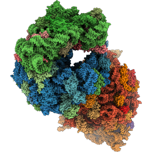 |
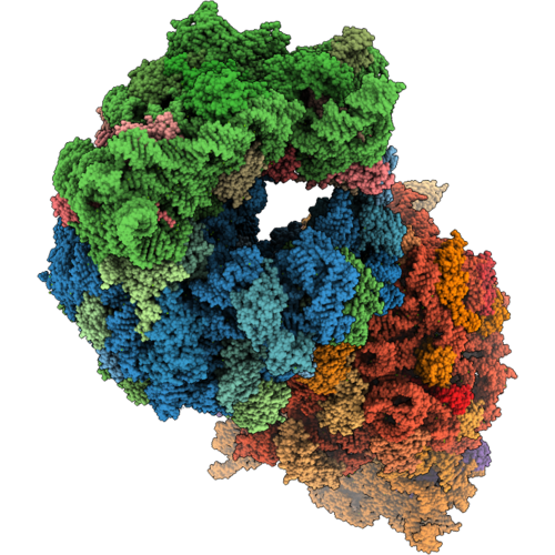
Crystal Structure Of The Wild-Type Thermus Thermophilus 70S Ribosome In Complex With Pleuromutilin-18 Triazole Derivative And Protein Y At 2.90A Resolution
Organism: Escherichia coli k-12, Thermus thermophilus hb8
Method: X-RAY DIFFRACTION Resolution:2.90 Å Release Date: 2026-09-09 Classification: RIBOSOME Ligands: MG, A1CRE, MPD, ARG, ZN, SF4 |
Organism: Escherichia coli k-12, Thermus thermophilus hb8
Method: X-RAY DIFFRACTION
Release Date: 2026-09-09
Ligands: MG, A1CRE, MPD, ARG, ZN, SF4
 |
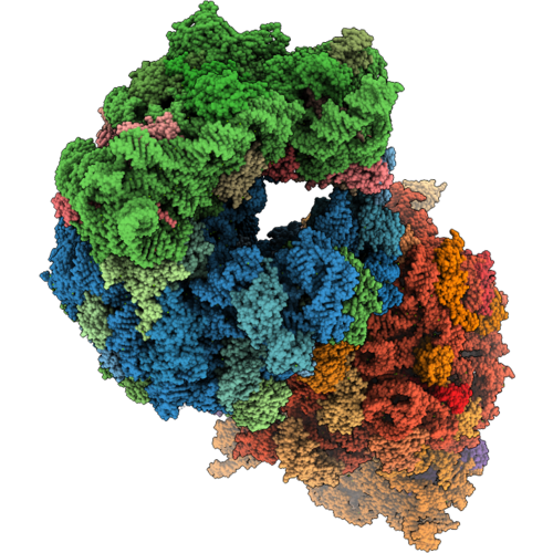
Crystal Structure Of The Wild-Type Thermus Thermophilus 70S Ribosome In Complex With Pleuromutilin-27 Triazole Derivative And Protein Y At 2.85A Resolution
Organism: Escherichia coli k-12, Thermus thermophilus hb8
Method: X-RAY DIFFRACTION Resolution:2.85 Å Release Date: 2026-09-09 Classification: RIBOSOME Ligands: MG, A1CQ7, MPD, ARG, ZN, SF4 |
Organism: Escherichia coli k-12, Thermus thermophilus hb8
Method: X-RAY DIFFRACTION
Release Date: 2026-09-09
Ligands: MG, A1CQ7, MPD, ARG, ZN, SF4
 |
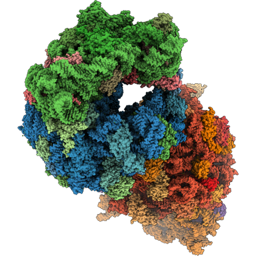
Crystal Structure Of The Wild-Type Thermus Thermophilus 70S Ribosome In Complex With Pleuromutilin-30 Triazole Derivative And Protein Y At 2.90A Resolution
Organism: Escherichia coli k-12, Thermus thermophilus hb8
Method: X-RAY DIFFRACTION Resolution:2.90 Å Release Date: 2026-09-09 Classification: RIBOSOME Ligands: MG, A1CQ8, ARG, MPD, ZN, SF4 |
Organism: Escherichia coli k-12, Thermus thermophilus hb8
Method: X-RAY DIFFRACTION
Release Date: 2026-09-09
Ligands: MG, A1CQ8, ARG, MPD, ZN, SF4
 |
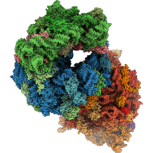
Crystal Structure Of The Wild-Type Thermus Thermophilus 70S Ribosome In Complex With Tiamulin And Protein Y At 2.65A Resolution
Organism: Escherichia coli k-12, Thermus thermophilus hb8
Method: X-RAY DIFFRACTION Resolution:2.65 Å Release Date: 2026-09-09 Classification: RIBOSOME Ligands: MG, MUL, ARG, MPD, ZN, SF4 |
Organism: Escherichia coli k-12, Thermus thermophilus hb8
Method: X-RAY DIFFRACTION
Release Date: 2026-09-09
Ligands: MG, MUL, ARG, MPD, ZN, SF4
 |
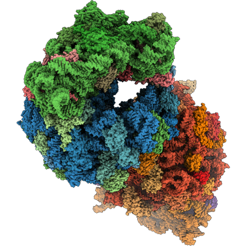
Crystal Structure Of The Wild-Type Thermus Thermophilus 70S Ribosome In Complex With Tiamulin-26 Triazole Derivative And Protein Y At 2.80A Resolution
Organism: Escherichia coli k-12, Thermus thermophilus hb8
Method: X-RAY DIFFRACTION Resolution:2.80 Å Release Date: 2026-09-09 Classification: RIBOSOME Ligands: MG, A1CRD, MPD, ARG, ZN, SF4 |
Organism: Escherichia coli k-12, Thermus thermophilus hb8
Method: X-RAY DIFFRACTION
Release Date: 2026-09-09
Ligands: MG, A1CRD, MPD, ARG, ZN, SF4
 |
Crystal Structure Of The Wild-Type Thermus Thermophilus 70S Ribosome In Complex With Tiamulin-31 Triazole Derivative And Protein Y At 2.85A Resolution
Organism: Escherichia coli k-12, Thermus thermophilus hb8
Method: X-RAY DIFFRACTION Resolution:2.85 Å Release Date: 2026-09-09 Classification: RIBOSOME Ligands: MG, A1CQ5, MPD, ARG, ZN, SF4 |
Organism: Escherichia coli k-12, Thermus thermophilus hb8
Method: X-RAY DIFFRACTION
Release Date: 2026-09-09
Ligands: MG, A1CQ5, MPD, ARG, ZN, SF4
 |
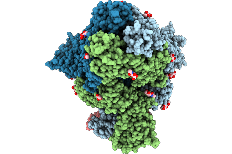
Hucov-Hku1 C S 2P In Complex With H501-018 Fab (State 1, Global Cryoem)
Organism: Human coronavirus hku1, Homo sapiens
Method: ELECTRON MICROSCOPY Release Date: 2026-07-01 Classification: VIRAL PROTEIN/IMMUNE SYSTEM Ligands: NAG |
Organism: Human coronavirus hku1, Homo sapiens
Method: ELECTRON MICROSCOPY
Release Date: 2026-07-01
Ligands: NAG
 |
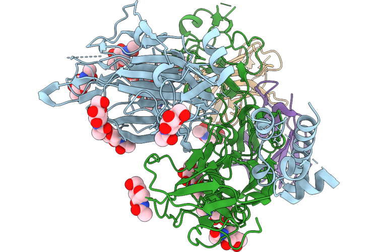
Hucov-Hku1 C S 2P In Complex With H501-018 Fab (State 2, Global Cryoem)
Organism: Human coronavirus hku1 (isolate n5), Homo sapiens
Method: ELECTRON MICROSCOPY Release Date: 2026-07-01 Classification: VIRAL PROTEIN/IMMUNE SYSTEM Ligands: NAG |
Organism: Human coronavirus hku1 (isolate n5), Homo sapiens
Method: ELECTRON MICROSCOPY
Release Date: 2026-07-01
Ligands: NAG
 |
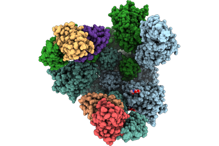
Hcov-Hku1 C S 2P In Complex With H501-018 Fab (Local Cryoem)
Organism: Human coronavirus hku1, Homo sapiens
Method: ELECTRON MICROSCOPY Release Date: 2026-07-01 Classification: VIRAL PROTEIN/IMMUNE SYSTEM Ligands: NAG |
Organism: Human coronavirus hku1, Homo sapiens
Method: ELECTRON MICROSCOPY
Release Date: 2026-07-01
Ligands: NAG
 |
Hcov-Hku1 C S 2P In Complex With H501-022 Fab (Global Cryoem)
Organism: Human coronavirus hku1 (isolate n5), Homo sapiens
Method: ELECTRON MICROSCOPY Release Date: 2026-07-01 Classification: VIRAL PROTEIN/IMMUNE SYSTEM Ligands: NAG |
Organism: Human coronavirus hku1 (isolate n5), Homo sapiens
Method: ELECTRON MICROSCOPY
Release Date: 2026-07-01
Ligands: NAG
 |
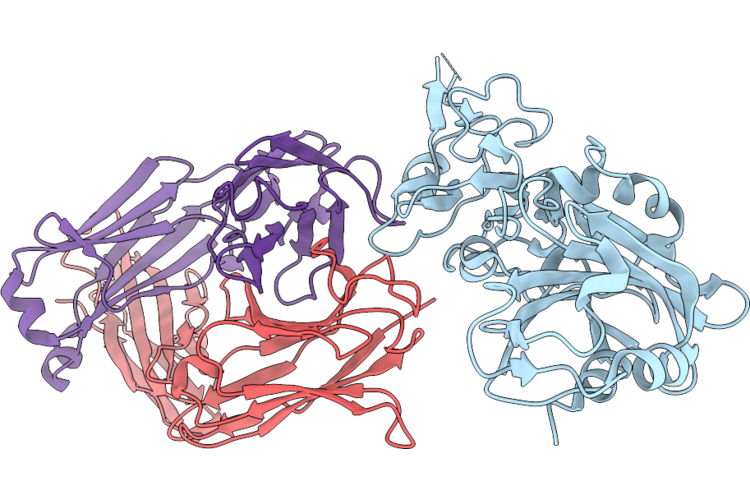
Hcov-Hku1 C S 2P In Complex With H501-022 Fab (Local Cryoem)
Organism: Human coronavirus hku1, Homo sapiens
Method: ELECTRON MICROSCOPY Release Date: 2026-07-01 Classification: VIRAL PROTEIN/IMMUNE SYSTEM |
Organism: Human coronavirus hku1, Homo sapiens
Method: ELECTRON MICROSCOPY
Release Date: 2026-07-01
 |
Crystal Structure Of Hcov-Hku1 Rbd Bound By H501-008 Fab
Organism: Human coronavirus hku1, Homo sapiens
Method: X-RAY DIFFRACTION Resolution:2.60 Å Release Date: 2026-07-01 Classification: VIRAL PROTEIN |
Organism: Human coronavirus hku1, Homo sapiens
Method: X-RAY DIFFRACTION
Release Date: 2026-07-01
 |
96-Nm Repeat Of The Leishmania Tarentolae Doublet Microtubule
Organism: Leishmania tarentolae, Leishmania mexicana
Method: ELECTRON MICROSCOPY Release Date: 2025-12-17 Classification: STRUCTURAL PROTEIN Ligands: GDP, MG, GTP, ZN, CA, ATP |
Organism: Leishmania tarentolae, Leishmania mexicana
Method: ELECTRON MICROSCOPY
Release Date: 2025-12-17
Ligands: GDP, MG, GTP, ZN, CA, ATP
 |
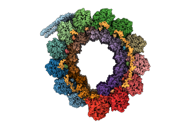
Cryo-Em Structure Of Conoid Fiber From Toxoplasma Gondii (24-Nm Repeat)
Organism: Toxoplasma gondii
Method: ELECTRON MICROSCOPY Release Date: 2025-12-03 Classification: STRUCTURAL PROTEIN Ligands: GTP, MG, GDP |
Organism: Toxoplasma gondii
Method: ELECTRON MICROSCOPY
Release Date: 2025-12-03
Ligands: GTP, MG, GDP
 |
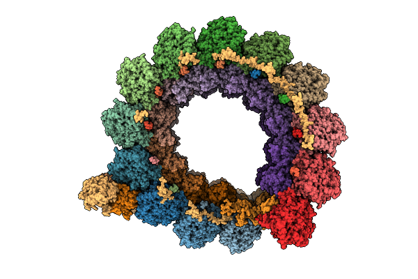
Cryo-Em Structure Of Intraconoidal Microtubule 2 (Icmt2) From Toxoplasma Gondii (8-Nm Repeat)
Organism: Toxoplasma gondii
Method: ELECTRON MICROSCOPY Release Date: 2025-12-03 Classification: STRUCTURAL PROTEIN Ligands: GTP, MG, GDP |
Organism: Toxoplasma gondii
Method: ELECTRON MICROSCOPY
Release Date: 2025-12-03
Ligands: GTP, MG, GDP
 |
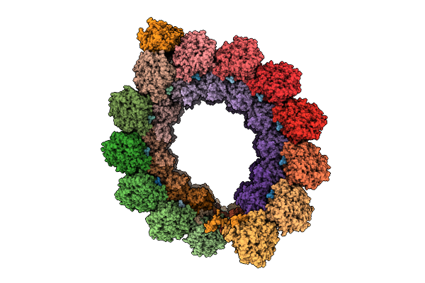
Cryo-Em Structure Of Intraconoidal Microtubule 1 (Icmt1) From Toxoplasma Gondii (8-Nm Repeat)
Organism: Toxoplasma gondii
Method: ELECTRON MICROSCOPY Release Date: 2025-12-03 Classification: STRUCTURAL PROTEIN Ligands: GTP, MG, GDP |
Organism: Toxoplasma gondii
Method: ELECTRON MICROSCOPY
Release Date: 2025-12-03
Ligands: GTP, MG, GDP
 |
Cryo-Em Structure Of The Apical Region Of Subpellicular Microtubule (Spmt) From Toxoplasma Gondii (8-Nm Repeat)
Organism: Toxoplasma gondii
Method: ELECTRON MICROSCOPY Release Date: 2025-12-03 Classification: STRUCTURAL PROTEIN Ligands: GDP, GTP, MG |
Organism: Toxoplasma gondii
Method: ELECTRON MICROSCOPY
Release Date: 2025-12-03
Ligands: GDP, GTP, MG