9YGP image
Deposition Date 2025-09-29
Release Date 2026-07-01
Last Version Date 2026-07-01
Entry Detail
PDB ID:
9YGP
Title:
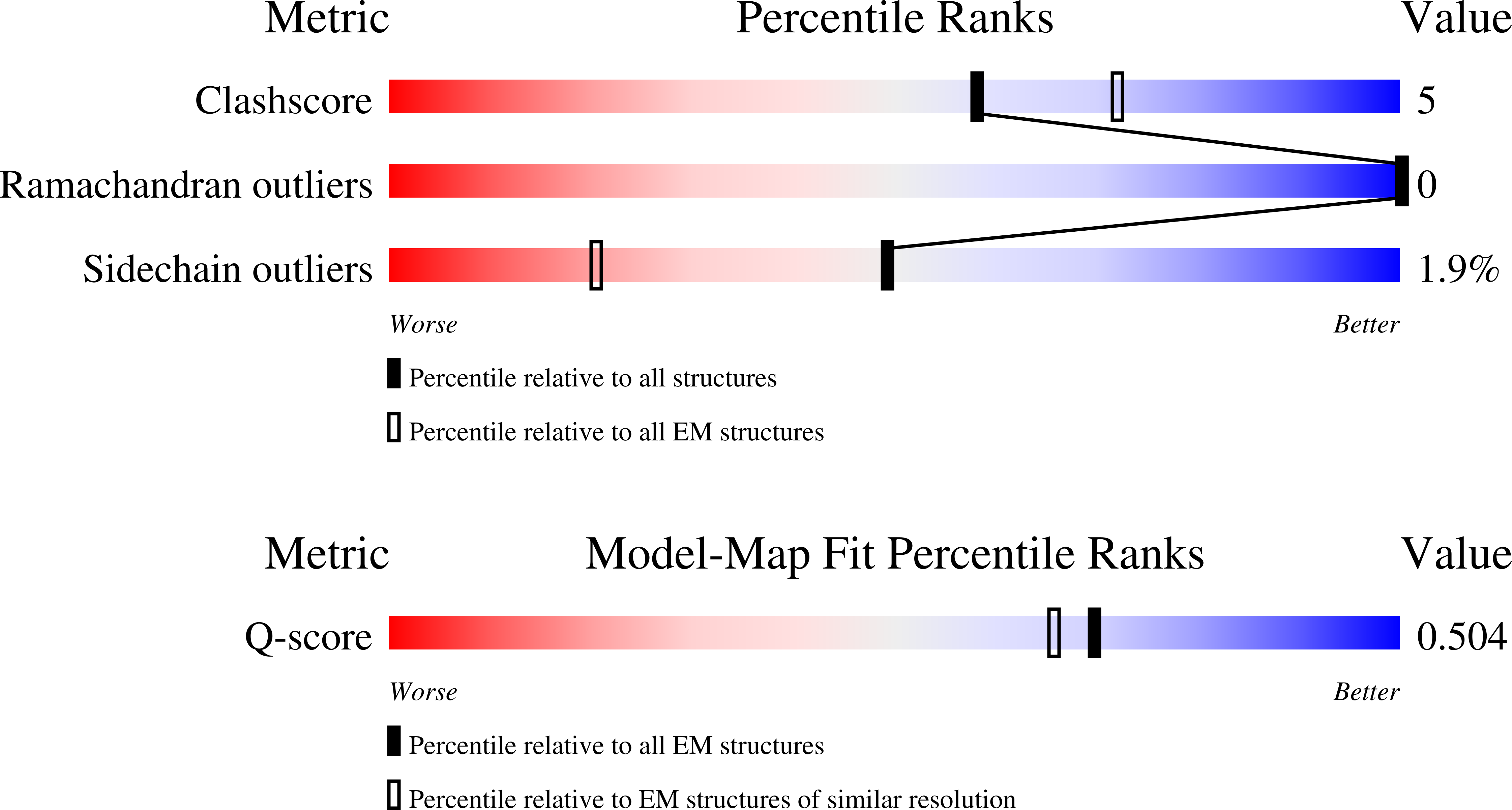
HCoV-HKU1 C S 2P in complex with H501-018 Fab (local cryoEM)
Biological Source:
Source Organism(s):
Expression System(s):
Method Details:
Experimental Method:
Resolution:
3.20 Å
Aggregation State:
PARTICLE
Reconstruction Method:
SINGLE PARTICLE
Macromolecular Entities
Structures with similar UniProt ID
Protein Blast
Polymer Type:polypeptide(L)
Molecule:Spike glycoprotein
Gene (Uniprot):S
Mutagens:752-756 RRKRR to GGSGS, and N1067P, L1068P
Chain IDs:A (auth: B), B (auth: C)
Chain Length:1334
Number of Molecules:2
Biological Source:Human coronavirus HKU1
Protein Blast
Polymer Type:polypeptide(L)
Molecule:H501-018 Fab heavy chain
Chain IDs:C (auth: H)
Chain Length:224
Number of Molecules:1
Biological Source:Homo sapiens
Protein Blast
Polymer Type:polypeptide(L)
Molecule:H501-018 Fab light chain
Chain IDs:D (auth: L)
Chain Length:216
Number of Molecules:1
Biological Source:Homo sapiens
Ligand Molecules
Primary Citation
Human Coronavirus HKU1 Neutralizing Monoclonal Antibodies Target Diverse Epitopes Within and Around the TMPRSS2 Receptor Binding Site.
Biorxiv ? ? ? (2025)
PMID: 41279056 DOI: 10.1101/2025.10.29.685445

Abstact

Endemic human coronaviruses (HCoVs), like HCoV-HKU1, account for ~30% of common cold/year and can cause serious upper and lower respiratory infections, yet no licensed vaccines target HCoVs. In fact, little is known about HCoV-HKU1's antigenic landscape. Thus, we characterized key interactions between HCoV-HKU1 spike (S) with monoclonal antibodies (mAbs) isolated from pre-pandemic HCoV-HKU1 convalescent PBMCs. We isolated 14 mAbs, which bound distinct S regions: receptor binding domain (RBD), N-terminal domain (NTD), and S2 subunit. Structural and functional studies revealed three groups of RBD-specific mAbs targeting diverse footprints within and around the TMPRSS2 receptor binding site, exemplified by: (1) The most potently neutralizing mAb, H501-022 (IC(50) = 0.01 mug/mL), which recognizes the TMPRSS2 binding motif, thereby blocking receptor engagement; (2) mAb H501-008 (IC(50) = 0.05 mug/mL) that binds a conserved, cross-reactive epitope outside of the TMPRSS2 binding site that is shared with HCoV-OC43; and (3) H501-018 (IC(50) = 0.28 mug/mL) that recognizes both "up" and "down" RBD conformations at a distinct, non-overlapping site outside of the TMPRSS2 binding motif, distinguishing itself from H501-022 and H501-008, which bind exclusively to the "up" RBD conformation. These mAbs represent the first type-specific HCoV-HKU1 mAbs isolated from a convalescent donor. Our findings provide molecular insight into HCoV-HKU1 antibody recognition and neutralization mechanisms, importantly highlighting antigenic differences comparing HCoVs and pandemic CoVs - a critical step towards advancing universal CoV vaccine design.

Legend

Protein

Chemical

Disease

Primary Citation of related structures
Feedback Form
Name
Email
Institute
Feedback