

Search Count: 34
 |
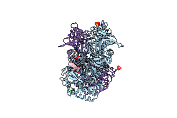
Txgh116 Complex With A Beta-D-Arabinofuranose-Configured Cyclitol Aziridine
Organism: Thermoanaerobacterium xylanolyticum lx-11
Method: X-RAY DIFFRACTION Resolution:1.75 Å Release Date: 2026-09-30 Classification: HYDROLASE Ligands: EDO, A1J9P, SO4, CA |
Organism: Thermoanaerobacterium xylanolyticum lx-11
Method: X-RAY DIFFRACTION
Release Date: 2026-09-30
Ligands: EDO, A1J9P, SO4, CA
 |
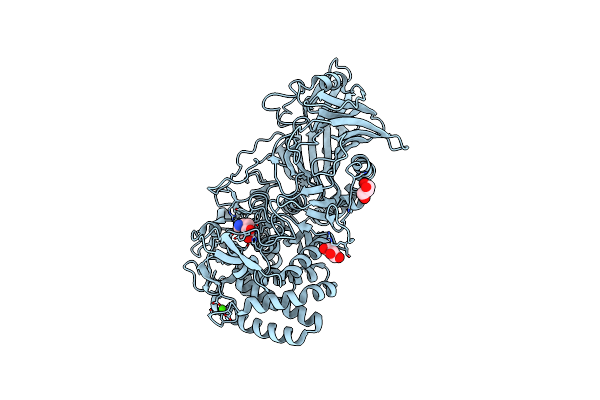
Structure Of Txgh116 With Covalently Bound N-Azido-Octyl Aziridine
Organism: Thermoanaerobacterium xylanolyticum lx-11
Method: X-RAY DIFFRACTION Resolution:1.90 Å Release Date: 2024-10-02 Classification: HYDROLASE Ligands: CA, CL, SO4, XHR |
Organism: Thermoanaerobacterium xylanolyticum lx-11
Method: X-RAY DIFFRACTION
Release Date: 2024-10-02
Ligands: CA, CL, SO4, XHR
 |
Crystal Structure Of Thermoanaerobacterium Xylanolyticum Gh116 Beta-Glucosidase With A Covalently Bound Cyclophellitol Aziridine
Organism: Thermoanaerobacterium xylanolyticum lx-11
Method: X-RAY DIFFRACTION Resolution:1.70 Å Release Date: 2023-11-22 Classification: HYDROLASE Ligands: PO8, CA, GOL, CL |
Organism: Thermoanaerobacterium xylanolyticum lx-11
Method: X-RAY DIFFRACTION
Release Date: 2023-11-22
Ligands: PO8, CA, GOL, CL
 |
Cjgh35 With A Galactosidase Activity-Based Probe
Organism: Cellvibrio japonicus ueda107
Method: X-RAY DIFFRACTION Resolution:1.50 Å Release Date: 2023-10-04 Classification: HYDROLASE |
Organism: Cellvibrio japonicus ueda107
Method: X-RAY DIFFRACTION
Release Date: 2023-10-04
 |
Golgi Alpha-Mannosidase Ii
Organism: Drosophila melanogaster
Method: X-RAY DIFFRACTION Resolution:1.69 Å Release Date: 2020-07-08 Classification: HYDROLASE Ligands: EDO, SIN, ZN |
Organism: Drosophila melanogaster
Method: X-RAY DIFFRACTION
Release Date: 2020-07-08
Ligands: EDO, SIN, ZN
 |
Golgi Alpha-Mannosidase Ii Complex With Manno-Epi-Cyclophellitol Aziridine
Organism: Drosophila melanogaster
Method: X-RAY DIFFRACTION Resolution:1.53 Å Release Date: 2020-07-08 Classification: HYDROLASE Ligands: EDO, SIN, KGH, ZN |
Organism: Drosophila melanogaster
Method: X-RAY DIFFRACTION
Release Date: 2020-07-08
Ligands: EDO, SIN, KGH, ZN
 |
Golgi Alpha-Mannosidase Ii
Organism: Drosophila melanogaster
Method: X-RAY DIFFRACTION Resolution:2.07 Å Release Date: 2020-07-08 Classification: HYDROLASE Ligands: EDO, ZN |
Organism: Drosophila melanogaster
Method: X-RAY DIFFRACTION
Release Date: 2020-07-08
Ligands: EDO, ZN
 |
Golgi Alpha-Mannosidase Ii In Complex With 5-(Adamantan-1Yl-Methoxy)-Pentyl 2,5-Dideoxy-2,5-Imino-D-Talo-Hexonamide
Organism: Drosophila melanogaster
Method: X-RAY DIFFRACTION Resolution:1.95 Å Release Date: 2020-07-08 Classification: HYDROLASE Ligands: KFE, EDO, ZN |
Organism: Drosophila melanogaster
Method: X-RAY DIFFRACTION
Release Date: 2020-07-08
Ligands: KFE, EDO, ZN
 |
Golgi Alpha-Mannosidase Ii In Complex With Pentyl 2,5-Dideoxy-2,5-Imino-D-Talo-Hexonamide
Organism: Drosophila melanogaster
Method: X-RAY DIFFRACTION Resolution:1.59 Å Release Date: 2020-07-08 Classification: HYDROLASE Ligands: EDO, SIN, KFT, ZN |
Organism: Drosophila melanogaster
Method: X-RAY DIFFRACTION
Release Date: 2020-07-08
Ligands: EDO, SIN, KFT, ZN
 |
Golgi Alpha-Mannosidase Ii In Complex With (5R,6R,7S,8S)-5,6,7,8-Tetrahydro-5-(Hydroxymethyl)-3-(3-Phenylpropyl)Imidazo[1,2-A]Pyridine-6,7,8-Triol
Organism: Drosophila melanogaster
Method: X-RAY DIFFRACTION Resolution:1.90 Å Release Date: 2020-07-08 Classification: HYDROLASE Ligands: KG8, ZN |
Organism: Drosophila melanogaster
Method: X-RAY DIFFRACTION
Release Date: 2020-07-08
Ligands: KG8, ZN
 |
Golgi Alpha-Mannosidase Ii In Complex With (2R,3R,4R,5S)-1-(5-{[4-(3,4-Dihydro-2H-1,5-Benzodioxepin-7-Yl)Benzyl]Oxy}Pentyl)-2-(Hydroxymethyl)-3,4,5-Piperidinetriol
Organism: Drosophila melanogaster
Method: X-RAY DIFFRACTION Resolution:2.15 Å Release Date: 2020-07-08 Classification: HYDROLASE Ligands: KG5, ZN |
Organism: Drosophila melanogaster
Method: X-RAY DIFFRACTION
Release Date: 2020-07-08
Ligands: KG5, ZN
 |
Golgi Alpha-Mannosidase Ii In Complex With (2S,3R)-2-(Hydroxymethyl)-3-Piperidinol
Organism: Drosophila melanogaster
Method: X-RAY DIFFRACTION Resolution:1.84 Å Release Date: 2020-07-08 Classification: HYDROLASE Ligands: ZN, EDO, KFW |
Organism: Drosophila melanogaster
Method: X-RAY DIFFRACTION
Release Date: 2020-07-08
Ligands: ZN, EDO, KFW
 |
Golgi Alpha-Mannosidase Ii In Complex With (2S,3R)-2-(Hydroxymethyl)-1,2,3,6-Tetrahydro-3-Pyridinol
Organism: Drosophila melanogaster
Method: X-RAY DIFFRACTION Resolution:1.86 Å Release Date: 2020-07-08 Classification: HYDROLASE Ligands: EDO, KGE, SIN, ZN |
Organism: Drosophila melanogaster
Method: X-RAY DIFFRACTION
Release Date: 2020-07-08
Ligands: EDO, KGE, SIN, ZN
 |
Golgi Alpha-Mannosidase Ii In Complex With (2S,3S,4R,5R)-1-(2-(Benzyloxy)Ethyl)-2-(Hydroxymethyl)Piperidine-3,4,5-Triol
Organism: Drosophila melanogaster
Method: X-RAY DIFFRACTION Resolution:1.80 Å Release Date: 2020-07-08 Classification: HYDROLASE Ligands: ZN, EDO, KGB |
Organism: Drosophila melanogaster
Method: X-RAY DIFFRACTION
Release Date: 2020-07-08
Ligands: ZN, EDO, KGB
 |
Crystal Structure Of Recombinant Human Beta-Glucocerebrosidase In Complex With Cyclophellitol Activity Based Probe With Cy5 Tag (Me569)
Organism: Homo sapiens
Method: X-RAY DIFFRACTION Resolution:1.92 Å Release Date: 2019-03-27 Classification: HYDROLASE Ligands: ACT, SO4, HKW, NAG |
Organism: Homo sapiens
Method: X-RAY DIFFRACTION
Release Date: 2019-03-27
Ligands: ACT, SO4, HKW, NAG
 |
Crystal Structure Of Recombinant Human Beta-Glucocerebrosidase In Complex With Adamantyl-Cyclophellitol Inhibitor (Me656)
Organism: Homo sapiens
Method: X-RAY DIFFRACTION Resolution:1.81 Å Release Date: 2019-03-27 Classification: HYDROLASE Ligands: SO4, ACT, RJR, NAG |
Organism: Homo sapiens
Method: X-RAY DIFFRACTION
Release Date: 2019-03-27
Ligands: SO4, ACT, RJR, NAG
 |
Crystal Structure Of Recombinant Human Beta-Glucocerebrosidase In Complex With Biphenyl-Cyclophellitol Inhibitor (Me655)
Organism: Homo sapiens
Method: X-RAY DIFFRACTION Resolution:1.63 Å Release Date: 2019-03-27 Classification: HYDROLASE Ligands: SO4, ACT, HK2, NAG |
Organism: Homo sapiens
Method: X-RAY DIFFRACTION
Release Date: 2019-03-27
Ligands: SO4, ACT, HK2, NAG
 |
Crystal Structure Of Agd31B, Alpha-Transglucosylase In Glycoside Hydrolase Family 31, In Complex With Cyclophellitol Aziridine Probe Cf022
Organism: Cellvibrio japonicus (strain ueda107)
Method: X-RAY DIFFRACTION Resolution:1.95 Å Release Date: 2016-05-04 Classification: TRANSFERASE Ligands: PG4, SO4, EDO, 66V |
Organism: Cellvibrio japonicus (strain ueda107)
Method: X-RAY DIFFRACTION
Release Date: 2016-05-04
Ligands: PG4, SO4, EDO, 66V
 |
Crystal Structure Of Agd31B, Alpha-Transglucosylase In Glycoside Hydrolase Family 31, In Complex With Cyclophellitol Aziridine Probe Cf021
Organism: Cellvibrio japonicus (strain ueda107)
Method: X-RAY DIFFRACTION Resolution:1.85 Å Release Date: 2016-05-04 Classification: HYDROLASE Ligands: SO4, PG4, 66U, EDO, TRS |
Organism: Cellvibrio japonicus (strain ueda107)
Method: X-RAY DIFFRACTION
Release Date: 2016-05-04
Ligands: SO4, PG4, 66U, EDO, TRS
 |
Crystal Structure Of A Bacterial Fucosidase With Phenyl((1R,2R,3R,4R,5R,6R)-2,3,4-Trihydroxy-5-Methyl-7-Azabicyclo[4.1.0]Heptan-7-Yl)Methanone
Organism: Bacteroides thetaiotaomicron
Method: X-RAY DIFFRACTION Resolution:1.92 Å Release Date: 2014-11-12 Classification: HYDROLASE Ligands: SO4, 3U2, IMD |
Organism: Bacteroides thetaiotaomicron
Method: X-RAY DIFFRACTION
Release Date: 2014-11-12
Ligands: SO4, 3U2, IMD