

Search Count: 32
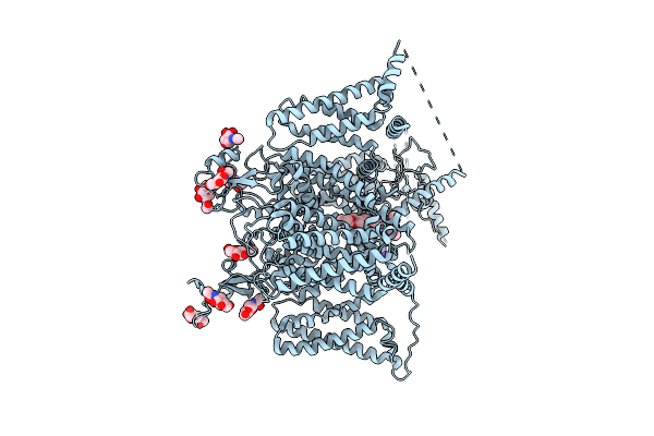 |
Structure Of Human Cardiac Sodium Channel Nav1.5 In Intermediate Open State
Organism: Homo sapiens
Method: ELECTRON MICROSCOPY Resolution:3.48 Å Release Date: 2026-04-08 Classification: MEMBRANE PROTEIN Ligands: 9Z9, NAG, NA |
Organism: Homo sapiens
Method: ELECTRON MICROSCOPY
Release Date: 2026-04-08
Ligands: 9Z9, NAG, NA
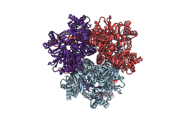 |
Cryo-Em Structure Of Mdtf
Organism: Escherichia coli k-12
Method: ELECTRON MICROSCOPY Release Date: 2025-12-10 Classification: TRANSPORT PROTEIN Ligands: LMT |
Organism: Escherichia coli k-12
Method: ELECTRON MICROSCOPY
Release Date: 2025-12-10
Ligands: LMT
 |
Structure Of Rnd Efflux Pump Mdtb
Organism: Escherichia coli k-12
Method: ELECTRON MICROSCOPY Release Date: 2025-12-10 Classification: TRANSPORT PROTEIN |
Organism: Escherichia coli k-12
Method: ELECTRON MICROSCOPY
Release Date: 2025-12-10
 |
Cryoem Structure Of De Novo Antibody Fragment Scfv 6 With C. Difficile Toxin B (Tcdb)
Organism: Clostridioides difficile, Synthetic construct
Method: ELECTRON MICROSCOPY Release Date: 2025-11-12 Classification: DE NOVO PROTEIN |
Organism: Clostridioides difficile, Synthetic construct
Method: ELECTRON MICROSCOPY
Release Date: 2025-11-12
 |
Cryoem Structure Of De Novo Vhh, Vhh_Flu_01, Bound To Influenza Ha, Strain A/Usa:Iowa/1943 H1N1.
Organism: Synthetic construct, Influenza a virus (strain a/usa:iowa/1943 h1n1)
Method: ELECTRON MICROSCOPY Resolution:3.02 Å Release Date: 2025-11-12 Classification: DE NOVO PROTEIN Ligands: NAG |
Organism: Synthetic construct, Influenza a virus (strain a/usa:iowa/1943 h1n1)
Method: ELECTRON MICROSCOPY
Release Date: 2025-11-12
Ligands: NAG
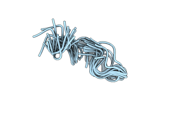 |
Solution Nmr Structure Of The Bound Antimicrobial Peptide Kg18 To Pseudomonas Aeruginosa Lps Bicelle At Ph 4
Organism: Dengue virus
Method: SOLUTION NMR Release Date: 2025-09-24 Classification: ANTIMICROBIAL PROTEIN |
Organism: Dengue virus
Method: SOLUTION NMR
Release Date: 2025-09-24
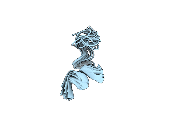 |
Solution Nmr Structure Of The Bound Antimicrobial Peptide Vr18 To Pseudomonas Aeruginosa Lps Bicelle At Ph 4
Organism: Dengue virus
Method: SOLUTION NMR Release Date: 2025-09-17 Classification: ANTIMICROBIAL PROTEIN |
Organism: Dengue virus
Method: SOLUTION NMR
Release Date: 2025-09-17
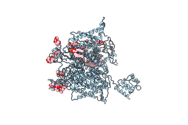 |
Structure Of Full-Length Human Cardiac Sodium Channel - Class-I.
Organism: Homo sapiens
Method: ELECTRON MICROSCOPY Resolution:3.60 Å Release Date: 2025-02-12 Classification: MEMBRANE PROTEIN Ligands: 6OU, Y01, NAG |
Organism: Homo sapiens
Method: ELECTRON MICROSCOPY
Release Date: 2025-02-12
Ligands: 6OU, Y01, NAG
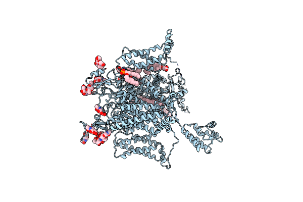 |
Structure Of Full-Length Human Cardiac Sodium Channel - Class-Ii.
Organism: Homo sapiens
Method: ELECTRON MICROSCOPY Resolution:3.90 Å Release Date: 2025-02-12 Classification: MEMBRANE PROTEIN Ligands: 6OU, Y01, NAG |
Organism: Homo sapiens
Method: ELECTRON MICROSCOPY
Release Date: 2025-02-12
Ligands: 6OU, Y01, NAG
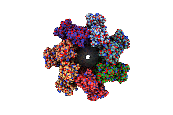 |
Cryo-Em Structure Of Complete Transmembrane Channel E289A Mutant Vibrio Cholerae Cytolysin
Organism: Vibrio cholerae
Method: ELECTRON MICROSCOPY Release Date: 2022-08-31 Classification: TOXIN |
Organism: Vibrio cholerae
Method: ELECTRON MICROSCOPY
Release Date: 2022-08-31
 |
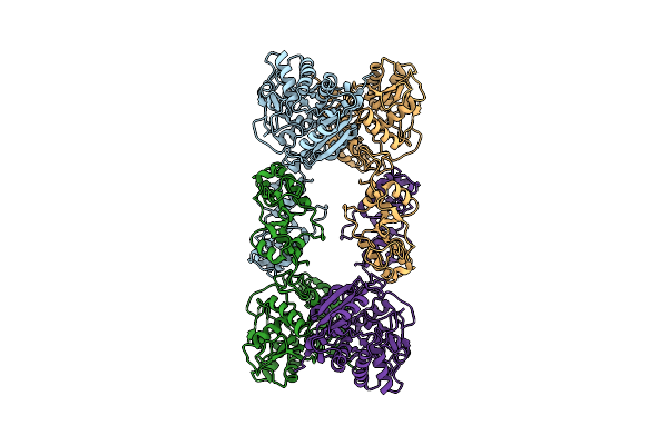
Native Cystathionine Beta-Synthase Of Mycobacterium Tuberculosis.
Organism: Mycobacterium tuberculosis h37rv
Method: ELECTRON MICROSCOPY Release Date: 2022-05-25 Classification: LYASE Ligands: PLP |
Organism: Mycobacterium tuberculosis h37rv
Method: ELECTRON MICROSCOPY
Release Date: 2022-05-25
Ligands: PLP
 |
Cystathionine Beta-Synthase Of Mycobacterium Tuberculosis In The Presence Of S-Adenosylmethionine.
Organism: Mycobacterium tuberculosis h37rv
Method: ELECTRON MICROSCOPY Release Date: 2022-05-25 Classification: LYASE |
Organism: Mycobacterium tuberculosis h37rv
Method: ELECTRON MICROSCOPY
Release Date: 2022-05-25
 |
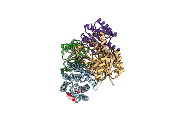
Cystathionine Beta-Synthase Of Mycobacterium Tuberculosis In The Presence Of S-Adenosylmethionine And Serine.
Organism: Mycobacterium tuberculosis h37rv
Method: ELECTRON MICROSCOPY Release Date: 2022-05-25 Classification: LYASE Ligands: PLS |
Organism: Mycobacterium tuberculosis h37rv
Method: ELECTRON MICROSCOPY
Release Date: 2022-05-25
Ligands: PLS
 |
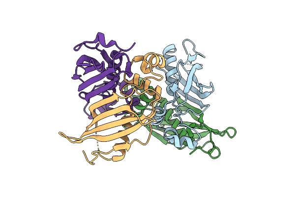
Crystal Structure Of Apo-Fabg From Vibrio Harveyi
Organism: Vibrio harveyi
Method: X-RAY DIFFRACTION Resolution:2.00 Å Release Date: 2021-09-08 Classification: OXIDOREDUCTASE Ligands: PEG |
Organism: Vibrio harveyi
Method: X-RAY DIFFRACTION
Release Date: 2021-09-08
Ligands: PEG
 |
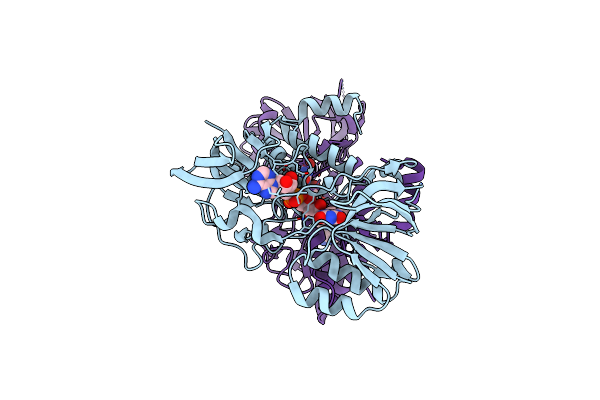
Crystal Structure Of C Terminal Truncated Hadbc (3R-Hydroxyacyl-Acp Dehydratase) Complex From Mycobacterium Tuberculosis
Organism: Mycobacterium tuberculosis (strain atcc 25618 / h37rv)
Method: X-RAY DIFFRACTION Resolution:2.90 Å Release Date: 2019-06-12 Classification: LYASE |
Organism: Mycobacterium tuberculosis (strain atcc 25618 / h37rv)
Method: X-RAY DIFFRACTION
Release Date: 2019-06-12
 |
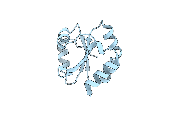
Crystal Structure Of Thioredoxin Reductase 2 From Staphylococcus Aureus
Organism: Staphylococcus aureus
Method: X-RAY DIFFRACTION Resolution:3.40 Å Release Date: 2018-07-11 Classification: OXIDOREDUCTASE Ligands: FAD |
Organism: Staphylococcus aureus
Method: X-RAY DIFFRACTION
Release Date: 2018-07-11
Ligands: FAD
 |
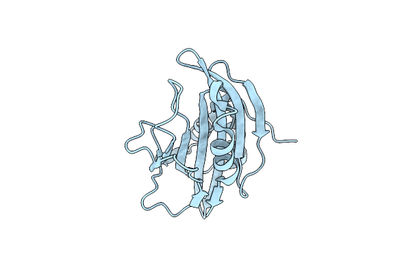
Crystal Structure Of Thioredoxin 2 From Staphylococcus Aureus Nctc8325
Organism: Staphylococcus aureus subsp. aureus nctc 8325
Method: X-RAY DIFFRACTION Resolution:1.93 Å Release Date: 2015-12-09 Classification: OXIDOREDUCTASE |
Organism: Staphylococcus aureus subsp. aureus nctc 8325
Method: X-RAY DIFFRACTION
Release Date: 2015-12-09
 |
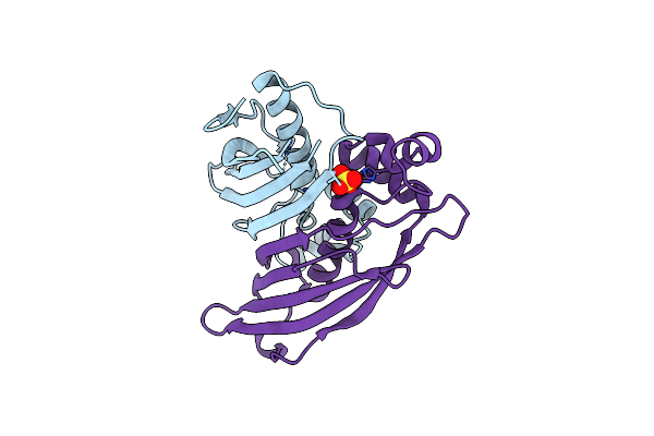
Crystal Structure Of Secreted Proline Rich Antigen Mtc28 (Rv0040C) At 2.15 A With Bound Chloride From Mycobacterium Tuberculosis
Organism: Mycobacterium tuberculosis
Method: X-RAY DIFFRACTION Resolution:2.15 Å Release Date: 2015-03-25 Classification: IMMUNE SYSTEM Ligands: CL |
Organism: Mycobacterium tuberculosis
Method: X-RAY DIFFRACTION
Release Date: 2015-03-25
Ligands: CL
 |
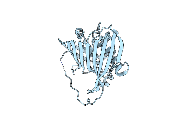
Crystal Structure Of (3R)-Hydroxyacyl-Acp Dehydratase Hadab Hetero-Dimer From Mycobacterium Smegmatis
Organism: Mycobacterium smegmatis str. mc2 155
Method: X-RAY DIFFRACTION Resolution:2.70 Å Release Date: 2015-02-11 Classification: LYASE Ligands: SO4 |
Organism: Mycobacterium smegmatis str. mc2 155
Method: X-RAY DIFFRACTION
Release Date: 2015-02-11
Ligands: SO4
 |
Crystal Structure Of Htdx(Rv0241C) From Mycobacterium Tuberculosis
Organism: Mycobacterium tuberculosis
Method: X-RAY DIFFRACTION Resolution:2.30 Å Release Date: 2015-02-04 Classification: OXIDOREDUCTASE Ligands: CL |
Organism: Mycobacterium tuberculosis
Method: X-RAY DIFFRACTION
Release Date: 2015-02-04
Ligands: CL