

Search Count: 36
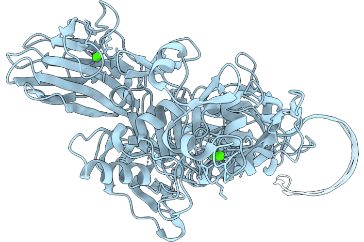 |
Single Full-Length Subunit Of The Vpb4Aa2 Pore Complex
Organism: Bacillus thuringiensis
Method: ELECTRON MICROSCOPY Resolution:2.20 Å Release Date: 2026-06-24 Classification: TOXIN Ligands: CA |
Organism: Bacillus thuringiensis
Method: ELECTRON MICROSCOPY
Release Date: 2026-06-24
Ligands: CA
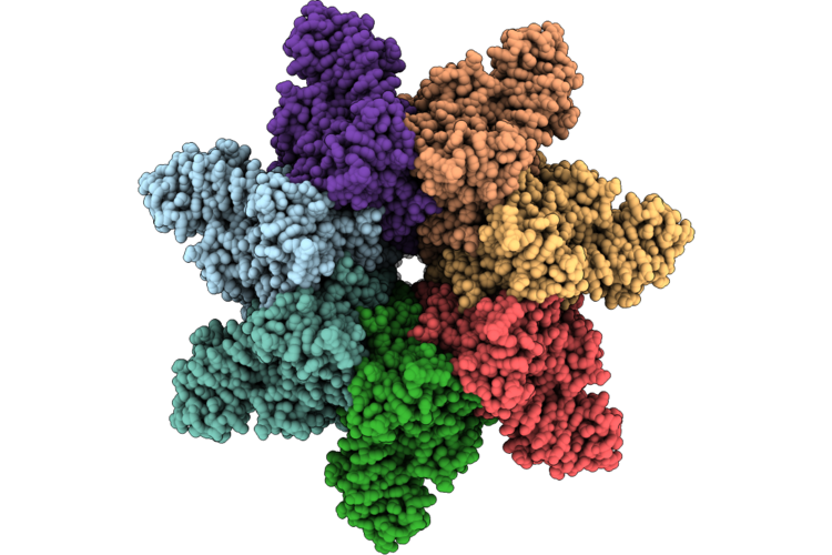 |
Vpb4Aa2 Pore Complex In C7 Symmetry
Organism: Bacillus thuringiensis
Method: ELECTRON MICROSCOPY Release Date: 2026-06-24 Classification: TOXIN Ligands: CA |
Organism: Bacillus thuringiensis
Method: ELECTRON MICROSCOPY
Release Date: 2026-06-24
Ligands: CA
 |
Native Structure Of Full-Length Pesticidal Protein Cry1Ca18 At Ph9, From Crystals Formed In Vivo
Organism: Bacillus thuringiensis
Method: X-RAY DIFFRACTION Resolution:1.65 Å Release Date: 2026-02-11 Classification: TOXIN Ligands: CA, ZN |
Organism: Bacillus thuringiensis
Method: X-RAY DIFFRACTION
Release Date: 2026-02-11
Ligands: CA, ZN
 |
Native Structure Of The Full-Length Pesticidal Protein Cry8Ba2, From Crystals Formed In Vivo (Form 1)
Organism: Bacillus thuringiensis
Method: X-RAY DIFFRACTION Resolution:2.27 Å Release Date: 2025-12-03 Classification: TOXIN |
Organism: Bacillus thuringiensis
Method: X-RAY DIFFRACTION
Release Date: 2025-12-03
 |
Native Structure Of The Full-Length Pesticidal Protein Cry8Ba2, From Crystals Formed In Vivo (Form 2)
Organism: Bacillus thuringiensis
Method: X-RAY DIFFRACTION Resolution:2.27 Å Release Date: 2025-12-03 Classification: TOXIN |
Organism: Bacillus thuringiensis
Method: X-RAY DIFFRACTION
Release Date: 2025-12-03
 |
Native Structure Of The Full-Length Pesticidal Protein Cry1Ca18 At Ph7, From Crystals Formed In Vivo
Organism: Bacillus thuringiensis
Method: X-RAY DIFFRACTION Resolution:1.80 Å Release Date: 2025-11-12 Classification: TOXIN Ligands: ZN, CA |
Organism: Bacillus thuringiensis
Method: X-RAY DIFFRACTION
Release Date: 2025-11-12
Ligands: ZN, CA
 |
Covalent Inhibitor Vvd-442 Bound To The Ras Binding Domain (Rbd) Of Pi3Ka
Organism: Homo sapiens
Method: X-RAY DIFFRACTION Resolution:2.83 Å Release Date: 2025-01-01 Classification: PROTEIN BINDING Ligands: A1BF9 |
Organism: Homo sapiens
Method: X-RAY DIFFRACTION
Release Date: 2025-01-01
Ligands: A1BF9
 |
Structure Of The Lysinibacillus Sphaericus Tpp49Aa1 Pesticidal Protein At Ph 3
Organism: Lysinibacillus sphaericus
Method: X-RAY DIFFRACTION Resolution:1.78 Å Release Date: 2023-11-01 Classification: TOXIN |
Organism: Lysinibacillus sphaericus
Method: X-RAY DIFFRACTION
Release Date: 2023-11-01
 |
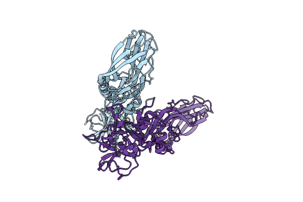
Structure Of The Lysinibacillus Sphaericus Tpp49Aa1 Pesticidal Protein At Ph 7
Organism: Lysinibacillus sphaericus
Method: X-RAY DIFFRACTION Resolution:1.62 Å Release Date: 2023-11-01 Classification: TOXIN |
Organism: Lysinibacillus sphaericus
Method: X-RAY DIFFRACTION
Release Date: 2023-11-01
 |
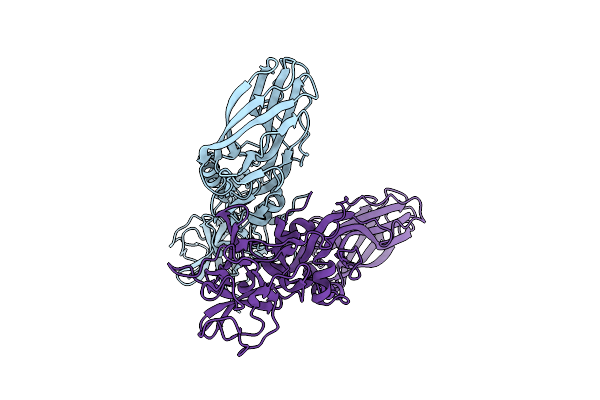
Structure Of The Lysinibacillus Sphaericus Tpp49Aa1 Pesticidal Protein At Ph 11
Organism: Lysinibacillus sphaericus
Method: X-RAY DIFFRACTION Resolution:1.75 Å Release Date: 2023-11-01 Classification: TOXIN |
Organism: Lysinibacillus sphaericus
Method: X-RAY DIFFRACTION
Release Date: 2023-11-01
 |
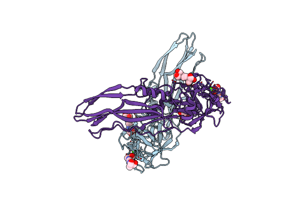
The Structure Of Natural Crystals Of The Lysinibacillus Sphaericus Tpp49Aa1 Pesticidal Protein Elucidated Using Serial Femtosecond Crystallography At An X-Ray Free Electron Laser
Organism: Lysinibacillus sphaericus
Method: X-RAY DIFFRACTION Resolution:2.20 Å Release Date: 2023-05-17 Classification: TOXIN |
Organism: Lysinibacillus sphaericus
Method: X-RAY DIFFRACTION
Release Date: 2023-05-17
 |
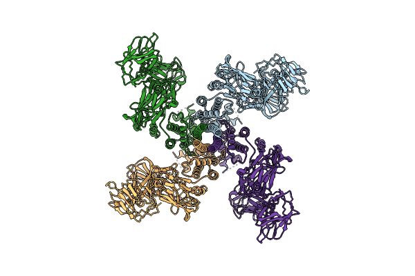
Tpp80Aa1
Organism: Bacillus thuringiensis
Method: X-RAY DIFFRACTION Resolution:1.81 Å Release Date: 2022-12-14 Classification: TOXIN Ligands: CA, B3P, CIT |
Organism: Bacillus thuringiensis
Method: X-RAY DIFFRACTION
Release Date: 2022-12-14
Ligands: CA, B3P, CIT
 |
Vip3Bc1 Tetramer In Processed, Activated State
Organism: Bacillus thuringiensis
Method: ELECTRON MICROSCOPY Release Date: 2021-04-14 Classification: TOXIN |
Organism: Bacillus thuringiensis
Method: ELECTRON MICROSCOPY
Release Date: 2021-04-14
 |
Bacillus Cereus Hbll1 Toxin Component
Organism: Bacillus cereus
Method: X-RAY DIFFRACTION Resolution:1.36 Å Release Date: 2021-04-07 Classification: TOXIN Ligands: PEG, EDO |
Organism: Bacillus cereus
Method: X-RAY DIFFRACTION
Release Date: 2021-04-07
Ligands: PEG, EDO
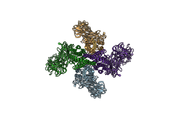 |
Vip3Bc1 Tetramer
Organism: Bacillus thuringiensis
Method: ELECTRON MICROSCOPY Release Date: 2021-03-17 Classification: TOXIN |
Organism: Bacillus thuringiensis
Method: ELECTRON MICROSCOPY
Release Date: 2021-03-17
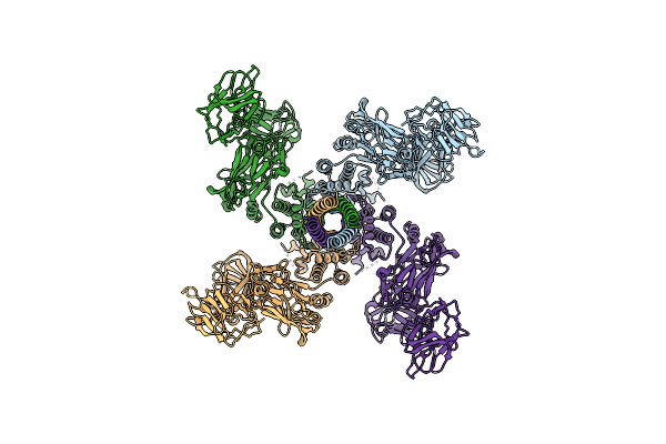 |
Vip3Bc1 Tetramer In Processed, Activated State
Organism: Bacillus thuringiensis
Method: ELECTRON MICROSCOPY Resolution:4.80 Å Release Date: 2021-03-17 Classification: TOXIN |
Organism: Bacillus thuringiensis
Method: ELECTRON MICROSCOPY
Release Date: 2021-03-17
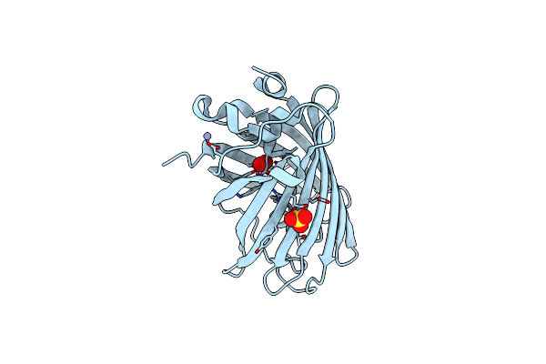 |
Venus 66 P-Azido-L-Phenylalanin (Azf) Variant, Dark Grown
Organism: Aequorea victoria
Method: X-RAY DIFFRACTION Resolution:1.91 Å Release Date: 2021-02-17 Classification: FLUORESCENT PROTEIN Ligands: OXY, ZN, SO4 |
Organism: Aequorea victoria
Method: X-RAY DIFFRACTION
Release Date: 2021-02-17
Ligands: OXY, ZN, SO4
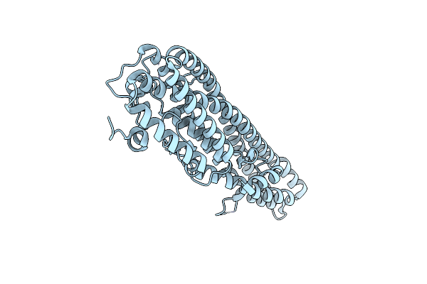 |
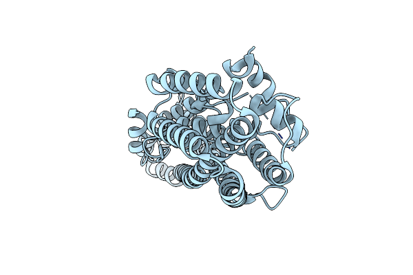
Crystal Structure Of Trypsin Activated Cry6Aa
Organism: Bacillus thuringiensis
Method: X-RAY DIFFRACTION Resolution:2.00 Å Release Date: 2016-08-03 Classification: TOXIN |
Organism: Bacillus thuringiensis
Method: X-RAY DIFFRACTION
Release Date: 2016-08-03
 |
Crystal Structure Of Full Length Cry6Aa
Organism: Bacillus thuringiensis
Method: X-RAY DIFFRACTION Resolution:2.70 Å Release Date: 2016-08-03 Classification: TOXIN |
Organism: Bacillus thuringiensis
Method: X-RAY DIFFRACTION
Release Date: 2016-08-03
 |
Crystal Structure At Ph 7.0 Of A Potato Sti-Kunitz Bi-Functional Inhibitor Of Serine And Aspartic Proteases In Space Group P4322 And Ph 9.0
Organism: Solanum tuberosum
Method: X-RAY DIFFRACTION Resolution:2.45 Å Release Date: 2016-07-06 Classification: HYDROLASE INHIBITOR |
Organism: Solanum tuberosum
Method: X-RAY DIFFRACTION
Release Date: 2016-07-06