

Search Count: 88
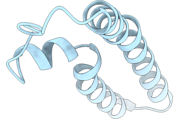 |
Cryoem Structure Of Phospholipid-Independent Cyclised Rp4 Pilus
Organism: Escherichia coli
Method: ELECTRON MICROSCOPY Resolution:2.74 Å Release Date: 2026-05-27 Classification: PROTEIN FIBRIL |
Organism: Escherichia coli
Method: ELECTRON MICROSCOPY
Release Date: 2026-05-27
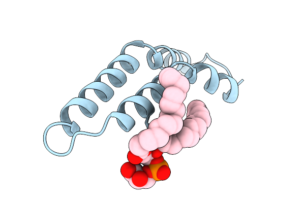 |
Cryoem Structure Of Cyclised H-Pilus (D69A)
Organism: salmonella enterica subsp. enterica serovar typhi
Method: ELECTRON MICROSCOPY Resolution:3.15 Å Release Date: 2025-12-24 Classification: PROTEIN FIBRIL Ligands: LHG |
Organism: salmonella enterica subsp. enterica serovar typhi
Method: ELECTRON MICROSCOPY
Release Date: 2025-12-24
Ligands: LHG
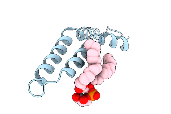 |
Cryoem Structure Of Cyclised H-Pilus (D69N)
Organism: Salmonella enterica subsp. enterica serovar typhi
Method: ELECTRON MICROSCOPY Resolution:2.88 Å Release Date: 2025-12-24 Classification: PROTEIN FIBRIL Ligands: LHG |
Organism: Salmonella enterica subsp. enterica serovar typhi
Method: ELECTRON MICROSCOPY
Release Date: 2025-12-24
Ligands: LHG
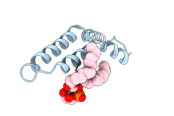 |
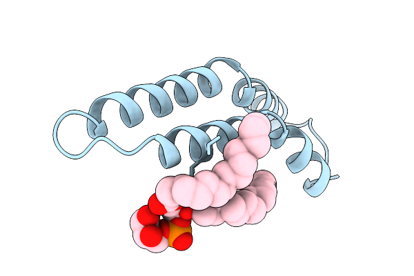
Cryoem Structure Of Cyclised H-Pilus (D69R)
Organism: Salmonella enterica subsp. enterica serovar typhi
Method: ELECTRON MICROSCOPY Resolution:2.89 Å Release Date: 2025-12-24 Classification: PROTEIN FIBRIL Ligands: LHG |
Organism: Salmonella enterica subsp. enterica serovar typhi
Method: ELECTRON MICROSCOPY
Release Date: 2025-12-24
Ligands: LHG
 |
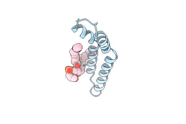
Cryoem Structure Of Cyclised H-Pilus (D69G)
Organism: Salmonella enterica subsp. enterica serovar typhi
Method: ELECTRON MICROSCOPY Resolution:3.11 Å Release Date: 2025-12-24 Classification: PROTEIN FIBRIL Ligands: LHG |
Organism: Salmonella enterica subsp. enterica serovar typhi
Method: ELECTRON MICROSCOPY
Release Date: 2025-12-24
Ligands: LHG
 |
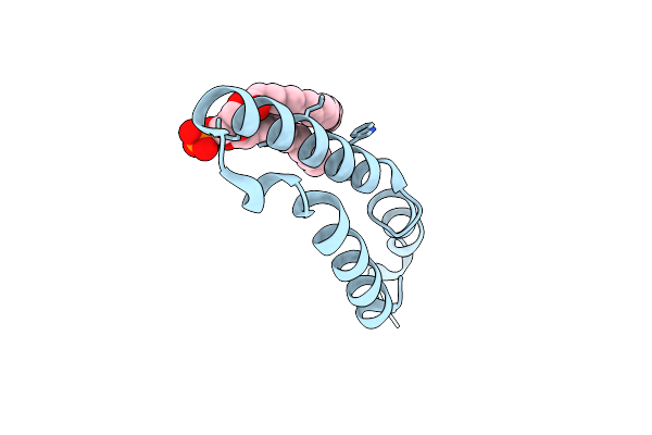
Cryoem Structure Of Cyclised H-Pilus
Organism: Salmonella
Method: ELECTRON MICROSCOPY Resolution:2.24 Å Release Date: 2025-03-26 Classification: PROTEIN FIBRIL Ligands: LHG |
Organism: Salmonella
Method: ELECTRON MICROSCOPY
Release Date: 2025-03-26
Ligands: LHG
 |
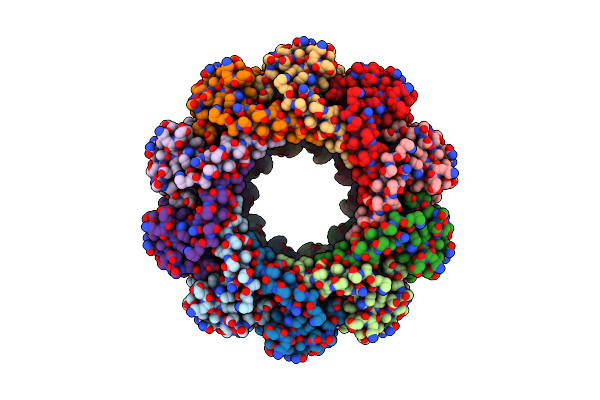
Cryo-Em Of F-Pilus
Organism: Escherichia coli
Method: ELECTRON MICROSCOPY Release Date: 2025-01-15 Classification: PROTEIN FIBRIL Ligands: X41 |
Organism: Escherichia coli
Method: ELECTRON MICROSCOPY
Release Date: 2025-01-15
Ligands: X41
 |
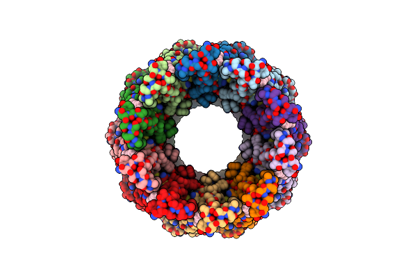
Cryo-Em Structure Of The Decameric Trat Surface Exclusion Lipoprotein From Escherichia Coli (F Plasmid)
Organism: Escherichia coli
Method: ELECTRON MICROSCOPY Release Date: 2024-12-11 Classification: MEMBRANE PROTEIN |
Organism: Escherichia coli
Method: ELECTRON MICROSCOPY
Release Date: 2024-12-11
 |
Cryo-Em Structure Of The Decameric Trat Surface Exclusion Lipoprotein From Klebsiella Pneumoniae (Pkpqil Plasmid)
Organism: Klebsiella pneumoniae
Method: ELECTRON MICROSCOPY Release Date: 2024-12-11 Classification: MEMBRANE PROTEIN Ligands: DGA |
Organism: Klebsiella pneumoniae
Method: ELECTRON MICROSCOPY
Release Date: 2024-12-11
Ligands: DGA
 |
Crystal Structure Of Chlorite Dismutase At 3000 Ev Based On Analytical Absorption Corrections
Organism: Cyanothece sp. pcc 7425
Method: X-RAY DIFFRACTION Resolution:2.70 Å Release Date: 2024-07-03 Classification: OXIDOREDUCTASE Ligands: HEM, SO4, GOL, CL |
Organism: Cyanothece sp. pcc 7425
Method: X-RAY DIFFRACTION
Release Date: 2024-07-03
Ligands: HEM, SO4, GOL, CL
 |
Crystal Structure Of Ompk36 Gd At 3500 Ev Based On Spherical Harmonics Absorption Corrections
Organism: Klebsiella pneumoniae
Method: X-RAY DIFFRACTION Resolution:2.34 Å Release Date: 2024-06-19 Classification: MEMBRANE PROTEIN Ligands: SO4 |
Organism: Klebsiella pneumoniae
Method: X-RAY DIFFRACTION
Release Date: 2024-06-19
Ligands: SO4
 |
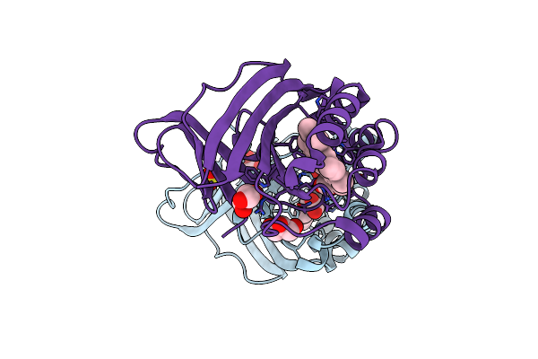
Crystal Structure Of Ompk36 Gd At 3500 Ev With No Absorption Corrections
Organism: Klebsiella pneumoniae
Method: X-RAY DIFFRACTION Resolution:2.34 Å Release Date: 2024-06-19 Classification: MEMBRANE PROTEIN Ligands: SO4 |
Organism: Klebsiella pneumoniae
Method: X-RAY DIFFRACTION
Release Date: 2024-06-19
Ligands: SO4
 |
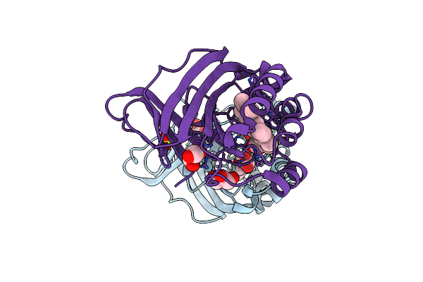
Crystal Structure Of Chlorite Dismutase At 3000 Ev Based On Spherical Harmonics Absorption Corrections
Organism: Cyanothece sp. pcc 7425
Method: X-RAY DIFFRACTION Resolution:2.70 Å Release Date: 2024-06-19 Classification: OXIDOREDUCTASE Ligands: HEM, SO4, GOL, CL |
Organism: Cyanothece sp. pcc 7425
Method: X-RAY DIFFRACTION
Release Date: 2024-06-19
Ligands: HEM, SO4, GOL, CL
 |
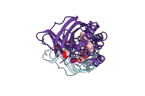
Crystal Structure Of Chlorite Dismutase At 3000 Ev With No Absorption Corrections
Organism: Cyanothece sp. pcc 7425
Method: X-RAY DIFFRACTION Resolution:2.70 Å Release Date: 2024-06-19 Classification: OXIDOREDUCTASE Ligands: HEM, SO4, GOL, CL |
Organism: Cyanothece sp. pcc 7425
Method: X-RAY DIFFRACTION
Release Date: 2024-06-19
Ligands: HEM, SO4, GOL, CL
 |
Crystal Structure Of Chlorite Dismutase At 3000 Ev Based On A Combination Of Spherical Harmonics And Analytical Absorption Corrections
Organism: Cyanothece sp. pcc 7425
Method: X-RAY DIFFRACTION Resolution:2.70 Å Release Date: 2024-06-19 Classification: OXIDOREDUCTASE Ligands: HEM, SO4, GOL, CL |
Organism: Cyanothece sp. pcc 7425
Method: X-RAY DIFFRACTION
Release Date: 2024-06-19
Ligands: HEM, SO4, GOL, CL
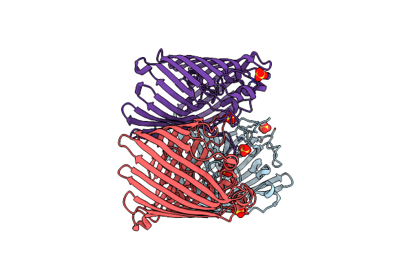 |
Crystal Structure Of Ompk36 Gd At 3500 Ev Based On A Combination Of Spherical Harmonics And Analytical Absorption Corrections
Organism: Klebsiella pneumoniae
Method: X-RAY DIFFRACTION Resolution:2.34 Å Release Date: 2024-06-19 Classification: MEMBRANE PROTEIN Ligands: SO4 |
Organism: Klebsiella pneumoniae
Method: X-RAY DIFFRACTION
Release Date: 2024-06-19
Ligands: SO4
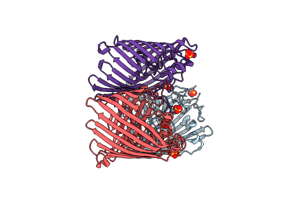 |
Crystal Structure Of Ompk36 Gd At 3500 Ev Based On Analytical Absorption Corrections
Organism: Klebsiella pneumoniae
Method: X-RAY DIFFRACTION Resolution:2.34 Å Release Date: 2024-06-19 Classification: MEMBRANE PROTEIN Ligands: SO4 |
Organism: Klebsiella pneumoniae
Method: X-RAY DIFFRACTION
Release Date: 2024-06-19
Ligands: SO4
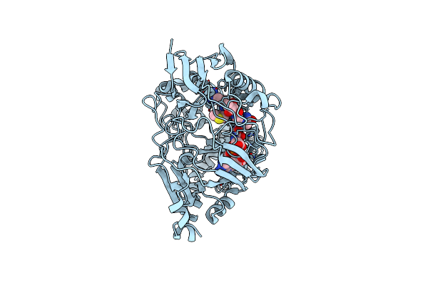 |
Complex Structure Of Yeja With Microcin C7
Organism: Escherichia coli
Method: X-RAY DIFFRACTION Resolution:1.76 Å Release Date: 2024-04-17 Classification: PEPTIDE BINDING PROTEIN/ANTIBIOTIC |
Organism: Escherichia coli
Method: X-RAY DIFFRACTION
Release Date: 2024-04-17
 |
Cryo-Em Structure Of The Rat Multidrug Resistance-Associated Protein 2 (Rmrp2) In An Autoinhibited State (Nucleotide-Free)
Organism: Rattus norvegicus
Method: ELECTRON MICROSCOPY Release Date: 2024-02-14 Classification: TRANSPORT PROTEIN |
Organism: Rattus norvegicus
Method: ELECTRON MICROSCOPY
Release Date: 2024-02-14
 |
Cryo-Em Structure Of The Rat Multidrug Resistance-Associated Protein 2 (Rmrp2) In Complex With Probenecid
Organism: Rattus norvegicus
Method: ELECTRON MICROSCOPY Release Date: 2024-02-14 Classification: TRANSPORT PROTEIN Ligands: Y01, RTO |
Organism: Rattus norvegicus
Method: ELECTRON MICROSCOPY
Release Date: 2024-02-14
Ligands: Y01, RTO