
Search Count: 74,342
All
Selected
 |
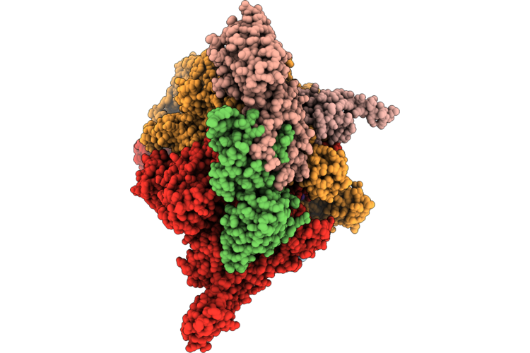
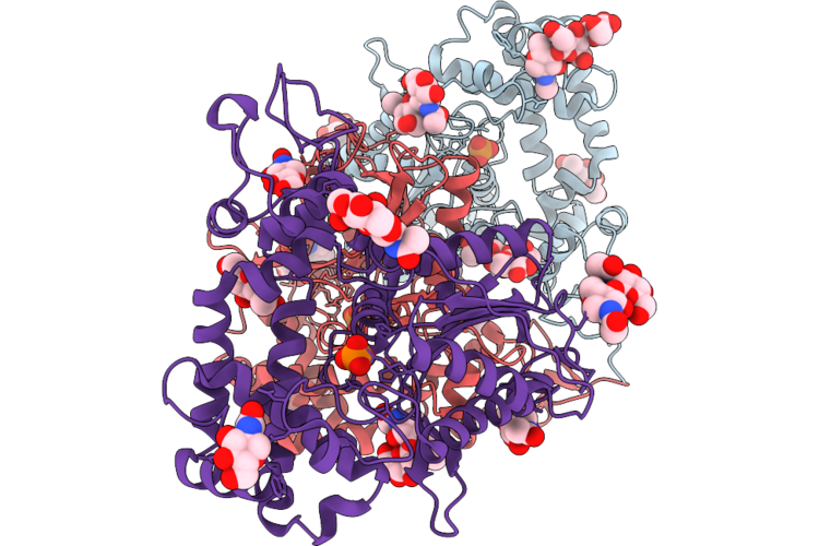
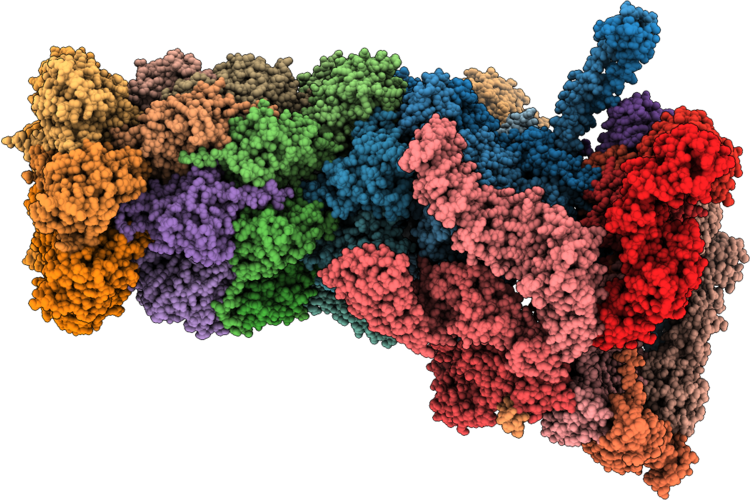
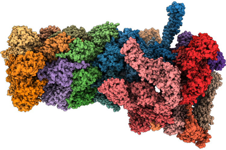
Closed Eco-Epec: Cryo-Em Structure Of Eco Rnap His-Elemental Paused Elongation Complex With A Closed Active Site (Closed Tl, Si3 And Rh-Fl)
Organism: Escherichia coli
Method: ELECTRON MICROSCOPY Resolution:2.90 Å Release Date: 2026-04-29 Classification: Transcription/DNA/RNA Ligands: 1N7, MG, ZN |
 |
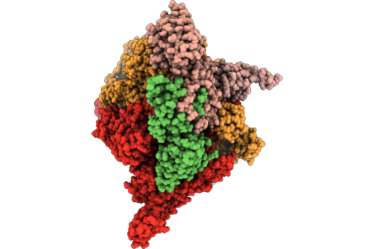
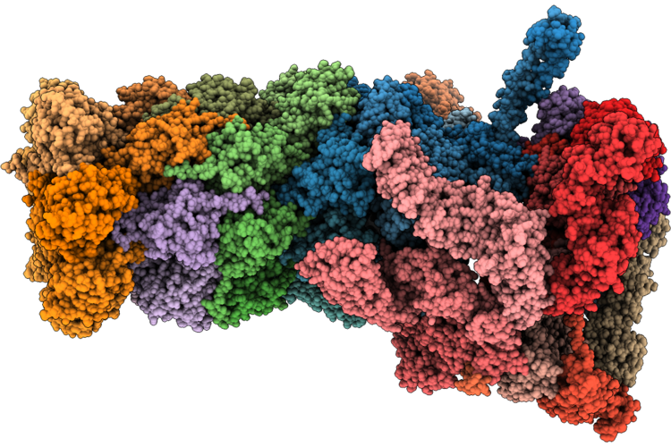
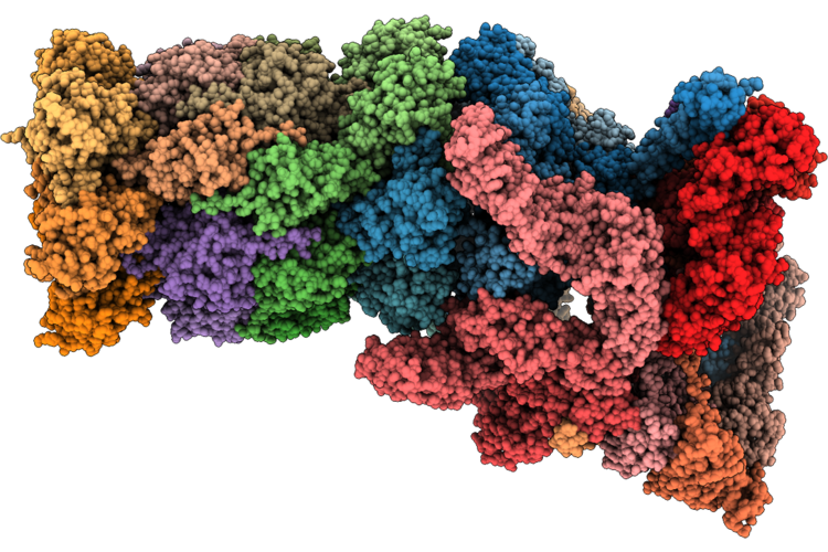
Open1 Eco-Epec: Cryo-Em Structure Of Eco Rnap His-Elemental Paused Elongation Complex With An Open Active Site (Open Tl, Si3 And Rh-Fl)
Organism: Escherichia coli
Method: ELECTRON MICROSCOPY Resolution:2.90 Å Release Date: 2026-04-29 Classification: TRANSCRIPTION/DNA/RNA Ligands: 1N7, MG, ZN |
 |
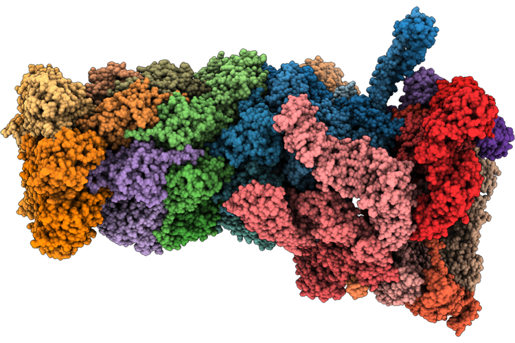
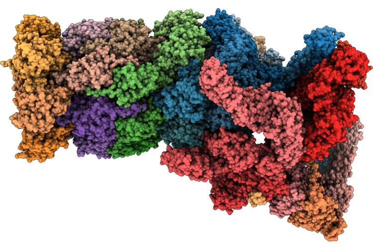
Open2 Eco-Epec: Cryo-Em Structure Of Eco Rnap His-Elemental Paused Elongation Complex With An Open Active Site (Open Tl, Si3 And Rh-Fl)
Organism: Escherichia coli
Method: ELECTRON MICROSCOPY Resolution:2.80 Å Release Date: 2026-04-29 Classification: Transcription/DNA/RNA Ligands: 1N7, MG, ZN |
 |
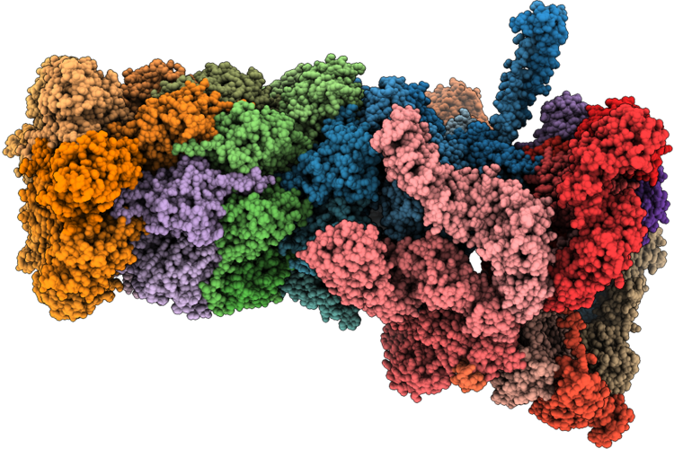
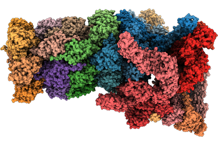
Open3 Eco-Epec: Cryo-Em Structure Of Eco Rnap His-Elemental Paused Elongation Complex With An Open Active Site (Open Tl, Si3 And Rh-Fl)
Organism: Escherichia coli
Method: ELECTRON MICROSCOPY Resolution:2.80 Å Release Date: 2026-04-29 Classification: TRANSCRIPTION Ligands: 1N7, MG, ZN |
 |
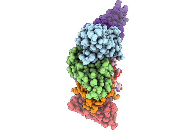
Epitope And Functional Classification Of Human Neutralizing Antibodies Against Sftsv Gn
Organism: Homo sapiens, Severe fever with thrombocytopenia syndrome virus
Method: ELECTRON MICROSCOPY Resolution:2.97 Å Release Date: 2026-04-29 Classification: VIRAL PROTEIN |
 |
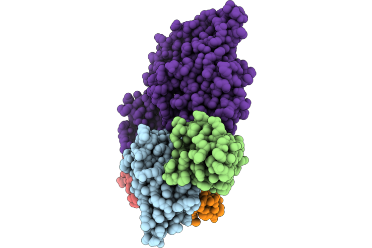
Epitope And Functional Classification Of Human Neutralizing Antibodies Against Sftsv Gn
Organism: Homo sapiens, Severe fever with thrombocytopenia syndrome virus
Method: ELECTRON MICROSCOPY Resolution:2.69 Å Release Date: 2026-04-29 Classification: VIRAL PROTEIN |
 |
Epitope And Functional Classification Of Human Neutralizing Antibodies Against Sftsv Gn
Organism: Homo sapiens, Severe fever with thrombocytopenia syndrome virus
Method: ELECTRON MICROSCOPY Resolution:2.69 Å Release Date: 2026-04-29 Classification: VIRAL PROTEIN |
 |
Organism: Leishmania mexicana
Method: ELECTRON MICROSCOPY Release Date: 2026-04-29 Classification: HYDROLASE Ligands: NAG, PO4 |
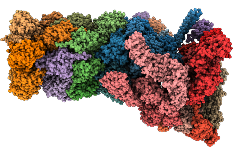 |
Structure Of Substrate-Engaged Human 26S Proteasome Rp-Cp Subcomplex In State Ea1.0
Organism: Homo sapiens
Method: ELECTRON MICROSCOPY Release Date: 2026-04-29 Classification: HYDROLASE Ligands: ATP, MG, ADP, ZN |
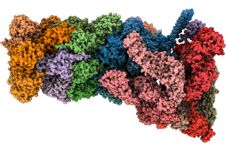 |
Structure Of Substrate-Engaged Human 26S Proteasome Rp-Cp Subcomplex In State Ea1.1
Organism: Homo sapiens
Method: ELECTRON MICROSCOPY Release Date: 2026-04-29 Classification: HYDROLASE Ligands: ATP, MG, ADP, ZN |
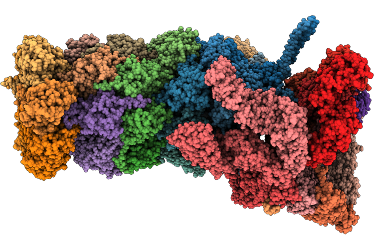 |
Structure Of Substrate-Engaged 26S Proteasome Rp-Cp Subcomplex In State Ea1.2
Organism: Homo sapiens
Method: ELECTRON MICROSCOPY Release Date: 2026-04-29 Classification: HYDROLASE Ligands: ATP, MG, ADP, ZN |
 |
Structure Of Substrate-Engaged Human 26S Proteasome Rp-Cp Subcomplex In State Ea2.1
Organism: Homo sapiens, Saccharomyces cerevisiae
Method: ELECTRON MICROSCOPY Release Date: 2026-04-29 Classification: HYDROLASE Ligands: ATP, MG, ADP, ZN |
 |
Structure Of Substrate-Engaged Human 26S Proteasome Rp-Cp Subcomplex In State Ea2.2
Organism: Homo sapiens, Saccharomyces cerevisiae
Method: ELECTRON MICROSCOPY Release Date: 2026-04-29 Classification: HYDROLASE Ligands: ATP, MG, ADP, ZN |
 |
Structure Of Substrate-Engaged Human 26S Proteasome Rp-Cp Subcomplex In State Ea2.3
Organism: Homo sapiens, Saccharomyces cerevisiae
Method: ELECTRON MICROSCOPY Release Date: 2026-04-29 Classification: HYDROLASE Ligands: ATP, MG, ADP, ZN |
 |
Structure Of Substrate-Engaged Human 26S Proteasome Rp-Cp Subcomplex In State Eb.1
Organism: Homo sapiens, Saccharomyces cerevisiae
Method: ELECTRON MICROSCOPY Release Date: 2026-04-29 Classification: HYDROLASE Ligands: ATP, MG, ADP, ZN |
 |
Structure Of Substrate-Engaged 26S Proteasome Rp-Cp Subcomplex In State Eb.2
Organism: Homo sapiens, Saccharomyces cerevisiae
Method: ELECTRON MICROSCOPY Release Date: 2026-04-29 Classification: HYDROLASE Ligands: ATP, MG, ADP, ZN |
 |
Structure Of Substrate-Engaged Human 26S Proteasome Rp-Cp Subcomplex In State Eb.3
Organism: Homo sapiens, Saccharomyces cerevisiae
Method: ELECTRON MICROSCOPY Release Date: 2026-04-29 Classification: HYDROLASE Ligands: ATP, MG, ADP, ZN |
 |
Structure Of Substrate-Engaged Human 26S Proteasome Rp-Cp Subcomplex In State Ec1
Organism: Homo sapiens, Saccharomyces cerevisiae
Method: ELECTRON MICROSCOPY Release Date: 2026-04-29 Classification: HYDROLASE Ligands: ADP, ATP, MG, ZN |
 |
Structure Of Substrate-Engaged Human 26S Proteasome Rp-Cp Subcomplex In State Ec2
Organism: Homo sapiens, Saccharomyces cerevisiae
Method: ELECTRON MICROSCOPY Release Date: 2026-04-29 Classification: HYDROLASE Ligands: ATP, MG, ADP, ZN |
 |
Structure Of Substrate-Engaged Human 26S Proteasome Rp-Cp Subcomplex In State Ed0.1
Organism: Homo sapiens, Saccharomyces cerevisiae
Method: ELECTRON MICROSCOPY Release Date: 2026-04-29 Classification: HYDROLASE Ligands: ATP, MG, ADP, ZN |