

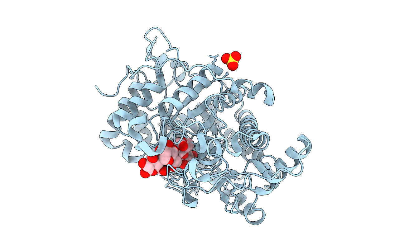
1V3I image
Deposition Date 2003-11-02
Release Date 2004-06-22
Last Version Date 2023-10-25
Entry Detail
PDB ID:
1V3I
Keywords:
Title:
The roles of Glu186 and Glu380 in the catalytic reaction of soybean beta-amylase
Biological Source:
Source Organism(s):
Glycine max (Taxon ID: 3847)
Expression System(s):
Method Details:
Experimental Method:
Resolution:
1.90 Å
R-Value Free:
0.19
R-Value Work:
0.15
R-Value Observed:
0.15
Space Group:
P 31 2 1

Macromolecular Entities
Polymer Type:polypeptide(L)
Molecule:Beta-amylase
Gene (Uniprot):BMY1
Mutagens:F76L, E380Q
Chain IDs:A
Chain Length:495
Number of Molecules:1
Biological Source:Glycine max
Ligand Molecules
Peptide-like Molecules
PRD_900001
PRD_900018
Primary Citation
The Roles of Glu186 and Glu380 in the Catalytic Reaction of Soybean beta-Amylase.
J. Mol. Biol. 339 1129 1140 (2004)
PMID: 15178253 DOI: 10.1016/j.jmb.2004.04.029

Abstact

It has previously been suggested that the glutamic acid residues Glu186 and Glu380 of soybean beta-amylase play critical roles as a general acid and a general base catalyst, respectively. In order to confirm the roles of Glu186 and Glu380, each residue was mutated to a glutamine residue and the crystal structures of the substrate (E186Q/maltopentaose) and product (E380Q/maltose) complexes were determined at resolutions of 1.6 Angstrom and 1.9 Angstrom, respectively. Both mutant enzymes exhibited 16,000- and 37,000-fold decreased activity relative to that of the wild-type enzyme. The crystal structure of the E186Q/maltopentaose complex revealed an unambiguous five-glucose unit at subsites -2 to +3. Two maltose molecules bind on subsites -2 to -1 and +2 to +3 in the E380Q/maltose complex, whereas they bind in tandem to -2 to -1 and +1 to +2 in the wild-type/maltose complex. The conformation of the glucose residue at subsite -1 was identified as a stable (4)C(1) alpha-anomer in the E380Q/maltose complex, whereas a distorted ring conformation was observed in the wild-type/maltose complex. The side-chain movement of Gln380 to the position of a putative attacking water molecule seen in the wild-type enzyme caused the inactivation of the E380Q mutant and an altered binding pattern of maltose molecules. These results confirm the critical roles played by Glu186 in the donation of a proton to the glycosidic oxygen of the substrate, and by Glu380 in the activation of an attacking water molecule. The observed difference between the backbones of E186Q/maltopentaose and E380Q/maltose in terms of Thr342 suggests that the side-chain of Thr342 may stabilize the deprotonated form of Glu186 after the cleavage of the glycosidic bond.

Legend

Protein

Chemical

Disease

Primary Citation of related structures
Feedback Form
Name
Email
Institute
Feedback