
Search Count: 34
All
Selected
 |
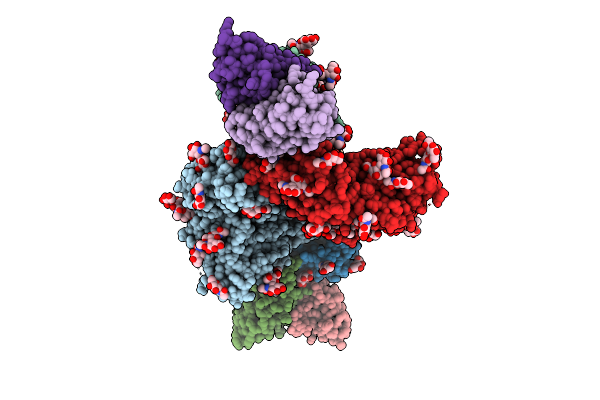
Organism: Homo sapiens, Human immunodeficiency virus 1
Method: ELECTRON MICROSCOPY Release Date: 2026-04-22 Classification: VIRAL PROTEIN/IMMUNE SYSTEM Ligands: NAG |
 |
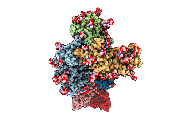
Organism: Human immunodeficiency virus 1, Homo sapiens
Method: ELECTRON MICROSCOPY Release Date: 2025-12-17 Classification: VIRAL PROTEIN/IMMUNE SYSTEM Ligands: NAG |
 |
Organism: Human immunodeficiency virus 1, Homo sapiens
Method: ELECTRON MICROSCOPY Release Date: 2025-12-17 Classification: VIRAL PROTEIN/IMMUNE SYSTEM Ligands: NAG |
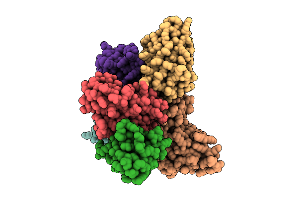 |
Organism: Homo sapiens, Plasmodium falciparum
Method: ELECTRON MICROSCOPY Release Date: 2025-12-10 Classification: IMMUNE SYSTEM/DE NOVO PROTEIN |
 |
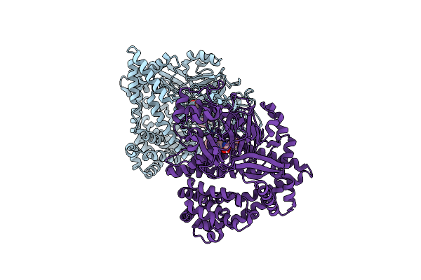
Organism: Homo sapiens
Method: ELECTRON MICROSCOPY Resolution:3.20 Å Release Date: 2025-08-20 Classification: CELL CYCLE |
 |
Organism: Homo sapiens
Method: ELECTRON MICROSCOPY Resolution:3.10 Å Release Date: 2025-08-20 Classification: CELL CYCLE |
 |
Organism: Homo sapiens
Method: ELECTRON MICROSCOPY Resolution:2.90 Å Release Date: 2025-08-20 Classification: CELL CYCLE |
 |
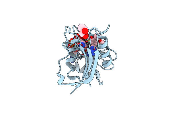
Crystal Structure Of High Affinity Tcr In Complex With Phla Harbouring Bacterial Peptide
Organism: Homo sapiens, Mycobacterium tuberculosis
Method: X-RAY DIFFRACTION Resolution:2.33 Å Release Date: 2024-05-15 Classification: IMMUNE SYSTEM Ligands: ACT |
 |
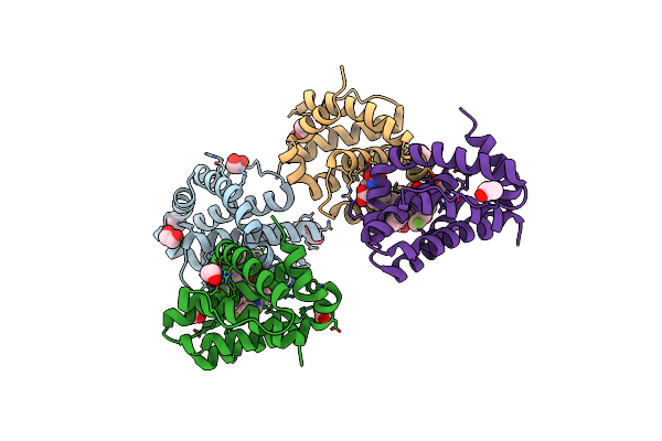
Small Dipeptide Analogues Developed By Co-Crystal Structure Of Stenotrophomonas Maltophilia Dipeptidyl Peptidase 7
Organism: Stenotrophomonas maltophilia (strain r551-3)
Method: X-RAY DIFFRACTION Resolution:2.59 Å Release Date: 2023-09-06 Classification: HYDROLASE Ligands: ALC, TYR |
 |
X-Ray Structure Of Escherichia Coli Dihydrofolate Reductase L28R Mutant In Complex With Trimethoprim
Organism: Escherichia coli
Method: X-RAY DIFFRACTION Resolution:2.10 Å Release Date: 2021-03-24 Classification: OXIDOREDUCTASE Ligands: NDP, GOL, TOP, CL |
 |
X-Ray Structure Of Escherichia Coli Dihydrofolate Reductase In Complex With Trimethoprim
Organism: Escherichia coli
Method: X-RAY DIFFRACTION Resolution:1.90 Å Release Date: 2021-03-24 Classification: OXIDOREDUCTASE Ligands: NDP, GOL, TOP, CL |
 |
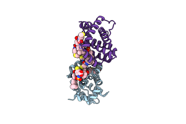
Organism: Homo sapiens
Method: X-RAY DIFFRACTION Resolution:2.75 Å Release Date: 2020-03-04 Classification: APOPTOSIS/INHIBITOR Ligands: Q0D |
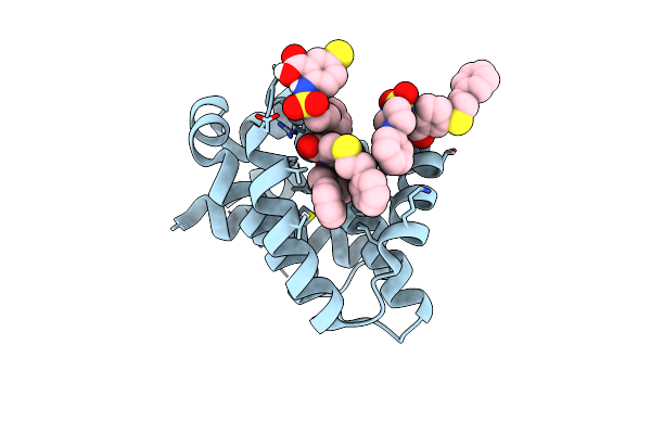 |
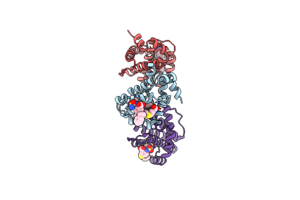
Organism: Homo sapiens
Method: X-RAY DIFFRACTION Resolution:2.55 Å Release Date: 2020-03-04 Classification: APOPTOSIS/INHIBITOR Ligands: Q0G |
 |
Organism: Homo sapiens
Method: X-RAY DIFFRACTION Resolution:2.09 Å Release Date: 2020-03-04 Classification: APOPTOSIS/INHIBITOR Ligands: Q0A, EDO, ACT, PEG |
 |
Organism: Homo sapiens
Method: X-RAY DIFFRACTION Resolution:1.84 Å Release Date: 2020-03-04 Classification: APOPTOSIS/INHIBITOR Ligands: Q01, BNL |
 |
The Crystal Structure Of Anti-Apoptotic Mcl-1 Protein In Complex With 2, 5-Substituted Benzoic Acid Inhibitor 21
Organism: Homo sapiens
Method: X-RAY DIFFRACTION Resolution:2.90 Å Release Date: 2020-03-04 Classification: APOPTOSIS/INHIBITOR Ligands: PZY |
 |
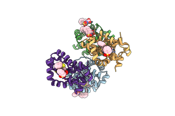
Organism: Saccharomyces cerevisiae (strain yjm789)
Method: X-RAY DIFFRACTION Resolution:2.60 Å Release Date: 2019-05-08 Classification: LYASE Ligands: ZN, SO4 |
 |
Organism: Saccharomyces cerevisiae (strain yjm789)
Method: X-RAY DIFFRACTION Resolution:2.90 Å Release Date: 2019-05-08 Classification: LYASE Ligands: ZN, CNN, SO4 |
 |
Organism: Saccharomyces cerevisiae (strain yjm789)
Method: X-RAY DIFFRACTION Resolution:2.90 Å Release Date: 2019-05-08 Classification: LYASE Ligands: ZN, SO4 |
 |
Organism: Saccharomyces cerevisiae (strain yjm789)
Method: X-RAY DIFFRACTION Resolution:3.00 Å Release Date: 2019-05-08 Classification: LYASE Ligands: ZN, SO4 |