

Search Count: 43
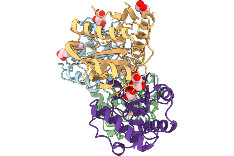 |
Crystal Structure Of The Complex Of Short Peptidoglycan Recognition Protein From Camelus Dromedarius With Nonanoic Acid At 1.83 A Resolution
Organism: Camelus dromedarius
Method: X-RAY DIFFRACTION Resolution:1.83 Å Release Date: 2026-06-17 Classification: IMMUNE SYSTEM Ligands: TLA, KNA, EDO, GOL |
Organism: Camelus dromedarius
Method: X-RAY DIFFRACTION
Release Date: 2026-06-17
Ligands: TLA, KNA, EDO, GOL
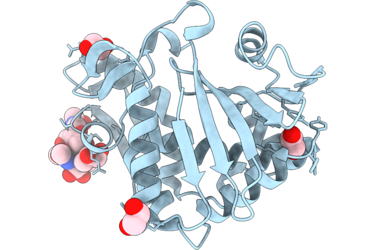 |
Crystal Structure Of A New Isoform Of Ribosome Inactivating Protein From Momordica Balsamina At 1.41 A Resolution
Organism: Momordica balsamina
Method: X-RAY DIFFRACTION Resolution:1.41 Å Release Date: 2026-05-20 Classification: HYDROLASE Ligands: NAG, EDO, GOL |
Organism: Momordica balsamina
Method: X-RAY DIFFRACTION
Release Date: 2026-05-20
Ligands: NAG, EDO, GOL
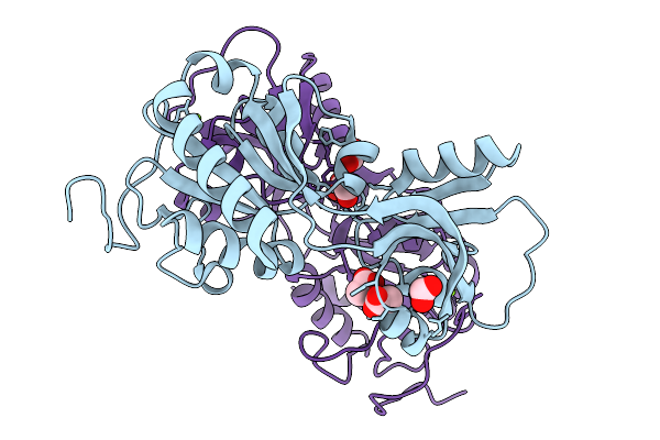 |
Crystal Structure Of Short-Form Adenosine Triphosphate Phosphoribosyltransferase From Acinetobacter Baumannii At 2.18 Angstrom Resolution.
Organism: Acinetobacter baumannii
Method: X-RAY DIFFRACTION Resolution:2.18 Å Release Date: 2026-02-18 Classification: TRANSFERASE Ligands: ACT, FMT, MG |
Organism: Acinetobacter baumannii
Method: X-RAY DIFFRACTION
Release Date: 2026-02-18
Ligands: ACT, FMT, MG
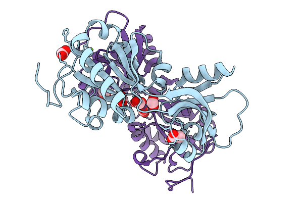 |
Crystal Structure Of Short-Form Adenosine Triphosphate Phosphoribosyltransferase From Acinetobacter Baumannii At 1.94 Angstrom Resolution
Organism: Acinetobacter baumannii
Method: X-RAY DIFFRACTION Resolution:1.94 Å Release Date: 2026-02-18 Classification: TRANSFERASE Ligands: FMT, ACT, MG |
Organism: Acinetobacter baumannii
Method: X-RAY DIFFRACTION
Release Date: 2026-02-18
Ligands: FMT, ACT, MG
 |
Crystal Structure Of The Chymotrypsin-Cleaved Iron-Free C-Lobe Of Bovine Lactoferrin At 2.82 Angstrom Resolution
Organism: Bos taurus
Method: X-RAY DIFFRACTION Resolution:2.82 Å Release Date: 2025-12-31 Classification: METAL BINDING PROTEIN Ligands: EDO, GOL, ACT, NAG, SO4 |
Organism: Bos taurus
Method: X-RAY DIFFRACTION
Release Date: 2025-12-31
Ligands: EDO, GOL, ACT, NAG, SO4
 |
Crystal Structure Of The Complex Of Camel Peptidoglycan Recognition Protein, Pgrp-S With Malic Acid And Oxalic Acid At 2.3 A Resolution
Organism: Camelus dromedarius
Method: X-RAY DIFFRACTION Resolution:2.31 Å Release Date: 2025-05-14 Classification: IMMUNE SYSTEM Ligands: MLT, OXD |
Organism: Camelus dromedarius
Method: X-RAY DIFFRACTION
Release Date: 2025-05-14
Ligands: MLT, OXD
 |
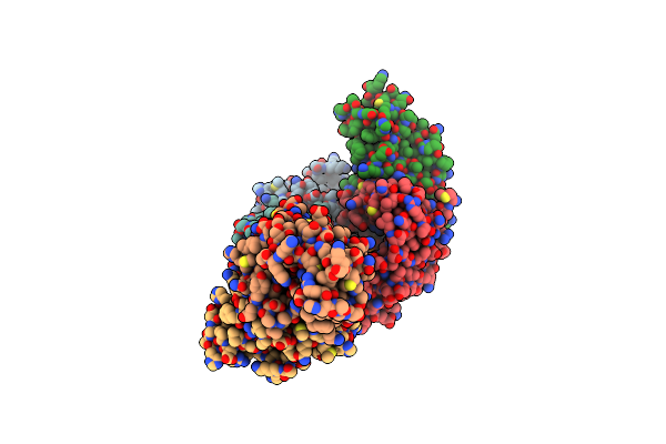
Structure Of Phosphopantetheine Adenylyltransferase (Ppat) From Enterobacter Spp. With The 17-Mer Expression Tag Bound In The Substrate Binding Site Of A Neighbouring Molecule At 2.10 A Resolution.
Organism: Enterobacter sp. 638
Method: X-RAY DIFFRACTION Resolution:2.10 Å Release Date: 2025-04-16 Classification: TRANSFERASE Ligands: CIT |
Organism: Enterobacter sp. 638
Method: X-RAY DIFFRACTION
Release Date: 2025-04-16
Ligands: CIT
 |
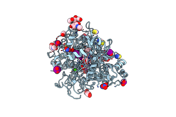
Structure Of Phosphopantetheine Adenylyltransferase (Ppat) From Enterobacter Spp. With The Expression Tag Bound In The Substrate Binding Site Of A Neighbouring Molecule At 2.20 A Resolution.
Organism: Enterobacter sp. 638
Method: X-RAY DIFFRACTION Resolution:2.20 Å Release Date: 2024-12-11 Classification: TRANSFERASE Ligands: PAE, GOL |
Organism: Enterobacter sp. 638
Method: X-RAY DIFFRACTION
Release Date: 2024-12-11
Ligands: PAE, GOL
 |
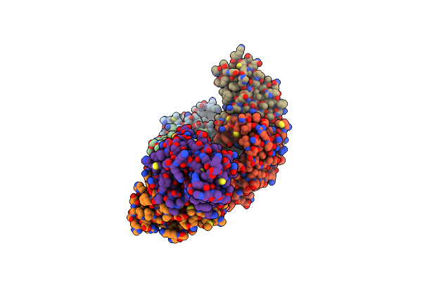
Crystal Structure Of The Ternary Complex Of Lactoperoxidase With Nitric Oxide And Nitrite Ion At 1.95 A Resolution
Organism: Bos taurus
Method: X-RAY DIFFRACTION Resolution:1.95 Å Release Date: 2024-09-11 Classification: OXIDOREDUCTASE Ligands: NAG, NO, NO2, NO3, SCN, IOD, HEM, EDO, CA, OSM |
Organism: Bos taurus
Method: X-RAY DIFFRACTION
Release Date: 2024-09-11
Ligands: NAG, NO, NO2, NO3, SCN, IOD, HEM, EDO, CA, OSM
 |
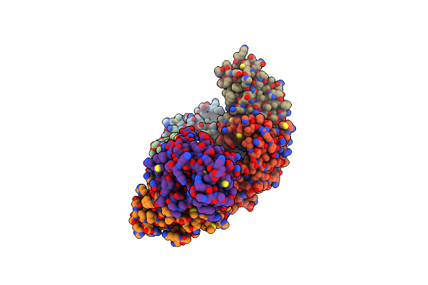
Structure Of Phosphopantetheine Adenylyltransferase (Ppat) From Enterobacter Spp. With The Expression Tag Bound In The Substrate Binding Site Of A Neighbouring Molecule At 2.39 A Resolution.
Organism: Enterobacter sp. 638
Method: X-RAY DIFFRACTION Resolution:2.39 Å Release Date: 2024-08-21 Classification: TRANSFERASE Ligands: PAE, GOL, EDO |
Organism: Enterobacter sp. 638
Method: X-RAY DIFFRACTION
Release Date: 2024-08-21
Ligands: PAE, GOL, EDO
 |
Structure Of Phosphopantetheine Adenylyltransferase (Ppat) From Enterobacter Spp. With The Expression Tag Bound In The Substrate Binding Site Of A Neighbouring Molecule At 2.37 A Resolution.
Organism: Enterobacter sp. 638
Method: X-RAY DIFFRACTION Resolution:2.37 Å Release Date: 2024-08-21 Classification: TRANSFERASE Ligands: PAE, GOL, EDO |
Organism: Enterobacter sp. 638
Method: X-RAY DIFFRACTION
Release Date: 2024-08-21
Ligands: PAE, GOL, EDO
 |
Structure Of Phosphopantetheine Adenylyltransferase (Ppat) From Enterobacter Spp. With The 17-Mer Expression Tag Bound In The Substrate Binding Site Of A Neighbouring Molecule At 2.60 A Resolution.
Organism: Enterobacter sp. 638
Method: X-RAY DIFFRACTION Resolution:2.60 Å Release Date: 2024-08-21 Classification: TRANSFERASE Ligands: CIT, GOL |
Organism: Enterobacter sp. 638
Method: X-RAY DIFFRACTION
Release Date: 2024-08-21
Ligands: CIT, GOL
 |
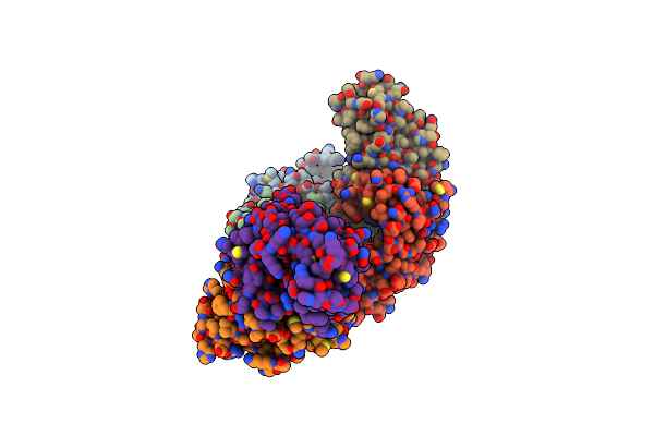
Structure Of Phosphopantetheine Adenylyltransferase (Ppat) From Enterobacter Spp. With The Expression Tag Bound In The Substrate Binding Site Of A Neighbouring Molecule At 2.25 A Resolution.
Organism: Enterobacter sp. 638
Method: X-RAY DIFFRACTION Resolution:2.25 Å Release Date: 2024-08-21 Classification: TRANSFERASE Ligands: PAE, GOL, EDO |
Organism: Enterobacter sp. 638
Method: X-RAY DIFFRACTION
Release Date: 2024-08-21
Ligands: PAE, GOL, EDO
 |
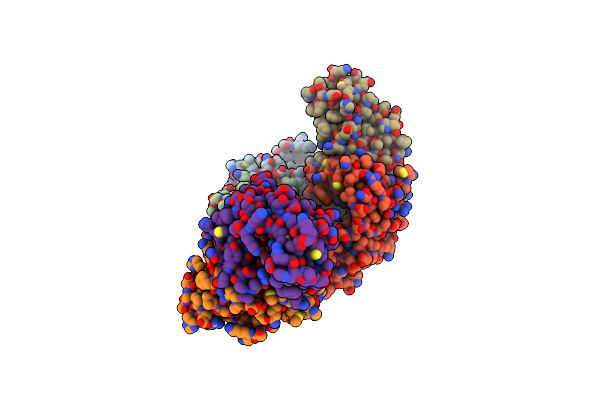
Structure Of Phosphopantetheine Adenylyltransferase (Ppat) From Enterobacter Sp. With The Expression Tag Bound In The Substrate Binding Site Of A Neighbouring Molecule At 2.65 A Resolution.
Organism: Enterobacter sp. 638
Method: X-RAY DIFFRACTION Resolution:2.65 Å Release Date: 2024-06-05 Classification: TRANSFERASE Ligands: PAE, GOL, EDO |
Organism: Enterobacter sp. 638
Method: X-RAY DIFFRACTION
Release Date: 2024-06-05
Ligands: PAE, GOL, EDO
 |
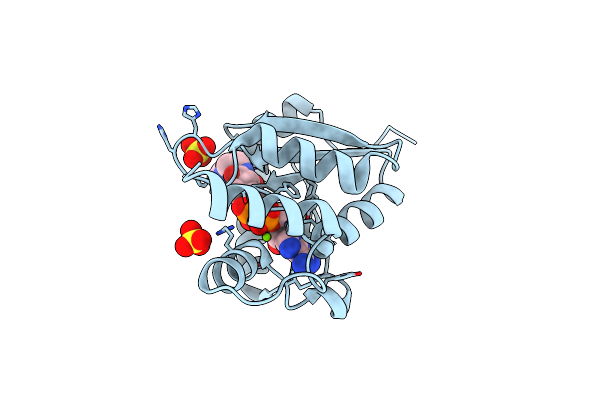
Structure Of Phosphopantetheine Adenylyltransferase (Ppat) From Enterobacter Sp. With The Expression Tag Bound In The Substrate Binding Site Of A Neighbouring Molecule At 2.41 A Resolution.
Organism: Enterobacter sp. 638
Method: X-RAY DIFFRACTION Resolution:2.41 Å Release Date: 2024-06-05 Classification: TRANSFERASE Ligands: PAE, GOL, EDO |
Organism: Enterobacter sp. 638
Method: X-RAY DIFFRACTION
Release Date: 2024-06-05
Ligands: PAE, GOL, EDO
 |
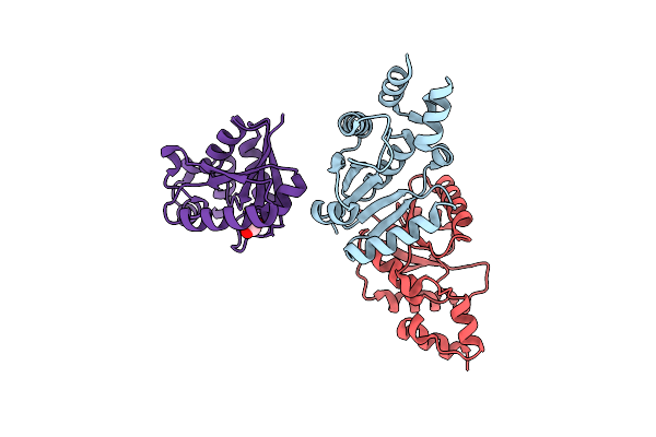
Crystal Structure Of The Complex Of Phosphopantetheine Adenylyltransferase From Acinetobacter Baumannii With Dephosphocoenzyme-A At 2.19 A Resolution.
Organism: Acinetobacter baumannii atcc 19606 = cip 70.34 = jcm 6841
Method: X-RAY DIFFRACTION Resolution:2.19 Å Release Date: 2023-04-12 Classification: TRANSFERASE Ligands: MG, COD, SO4, CL |
Organism: Acinetobacter baumannii atcc 19606 = cip 70.34 = jcm 6841
Method: X-RAY DIFFRACTION
Release Date: 2023-04-12
Ligands: MG, COD, SO4, CL
 |
Crystal Structure Of Phosphopantetheine Adenylyltransferase From Klebsiella Pneumoniae At 2.59 A Resolution
Organism: Klebsiella pneumoniae
Method: X-RAY DIFFRACTION Resolution:2.59 Å Release Date: 2023-02-22 Classification: TRANSFERASE Ligands: EDO |
Organism: Klebsiella pneumoniae
Method: X-RAY DIFFRACTION
Release Date: 2023-02-22
Ligands: EDO
 |
Structure Of The Complex Of Lactoperoxidase With Nitric Oxide Catalytic Product Nitrite At 1.89 A Resolution
Organism: Bos taurus
Method: X-RAY DIFFRACTION Resolution:1.89 Å Release Date: 2023-01-11 Classification: OXIDOREDUCTASE Ligands: CA, IOD, SCN, EDO, NO2, HEM, ZN, NAG |
Organism: Bos taurus
Method: X-RAY DIFFRACTION
Release Date: 2023-01-11
Ligands: CA, IOD, SCN, EDO, NO2, HEM, ZN, NAG
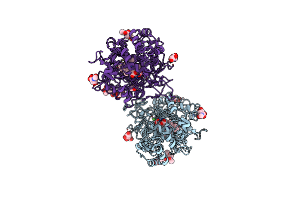 |
Crystal Structure Of The Complex Of Lactoperoxidase With Nitric Oxide At 2.50A Resolution
Organism: Bos grunniens
Method: X-RAY DIFFRACTION Resolution:2.50 Å Release Date: 2022-06-29 Classification: OXIDOREDUCTASE Ligands: MH0, CA, CL, NAG, NO, ACT |
Organism: Bos grunniens
Method: X-RAY DIFFRACTION
Release Date: 2022-06-29
Ligands: MH0, CA, CL, NAG, NO, ACT
 |
Structure Of The Complex Of Camel Peptidoglycan Recognition Protein-Short (Pgrp-S) With Heptanoic Acid At 2.15 A Resolution.
Organism: Camelus dromedarius
Method: X-RAY DIFFRACTION Resolution:2.15 Å Release Date: 2022-06-15 Classification: IMMUNE SYSTEM Ligands: EDO, PEO, CO3, NA, SHV, MPD |
Organism: Camelus dromedarius
Method: X-RAY DIFFRACTION
Release Date: 2022-06-15
Ligands: EDO, PEO, CO3, NA, SHV, MPD