
Search Count: 3,643
 |
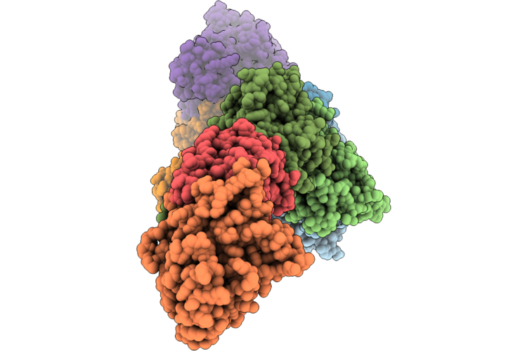
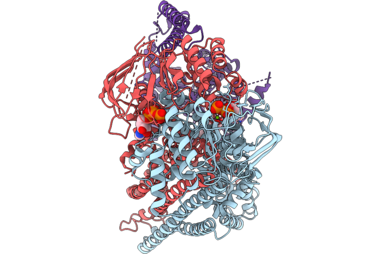
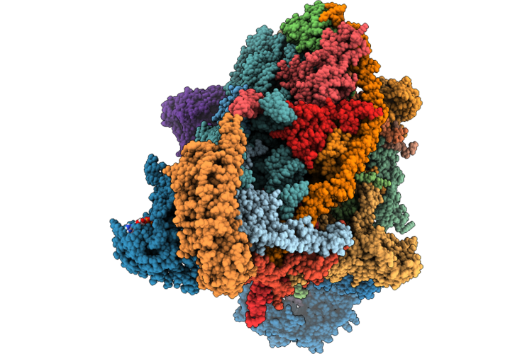
Crystal Structure Of Dihydroxyacetone Kinase From Komagataella Pastoris
Organism: Komagataella pastoris
Method: X-RAY DIFFRACTION Resolution:2.88 Å Release Date: 2026-07-22 Classification: BIOSYNTHETIC PROTEIN Ligands: MG, ATP |
Organism: Komagataella pastoris
Method: X-RAY DIFFRACTION
Release Date: 2026-07-22
Ligands: MG, ATP
 |
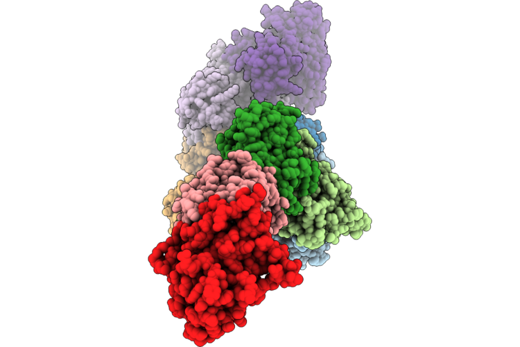
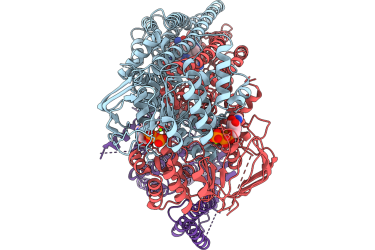
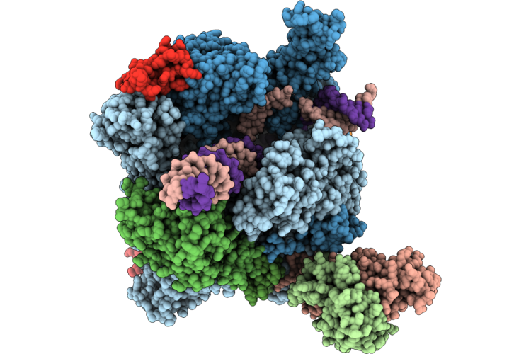
Rad55-Rad57-Shu Homologous Recombination Complex
Organism: Saccharomyces cerevisiae, Synthetic construct
Method: ELECTRON MICROSCOPY Release Date: 2026-07-22 Classification: DNA BINDING PROTEIN/DNA Ligands: ADP, MG, ATP, ZN |
Organism: Saccharomyces cerevisiae, Synthetic construct
Method: ELECTRON MICROSCOPY
Release Date: 2026-07-22
Ligands: ADP, MG, ATP, ZN
 |
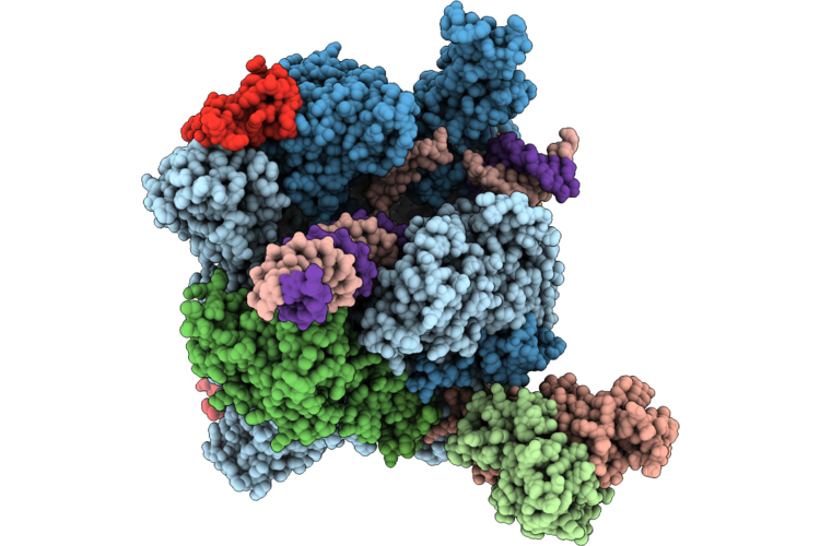
Rad55-Rad57(E161Q)-Shu-3Xrad51 Bound To Ssdna With Atp
Organism: Saccharomyces cerevisiae, Synthetic construct
Method: ELECTRON MICROSCOPY Release Date: 2026-07-22 Classification: DNA BINDING PROTEIN/DNA Ligands: ADP, MG, ATP, ZN |
Organism: Saccharomyces cerevisiae, Synthetic construct
Method: ELECTRON MICROSCOPY
Release Date: 2026-07-22
Ligands: ADP, MG, ATP, ZN
 |
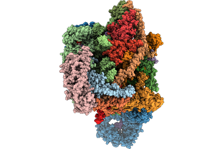
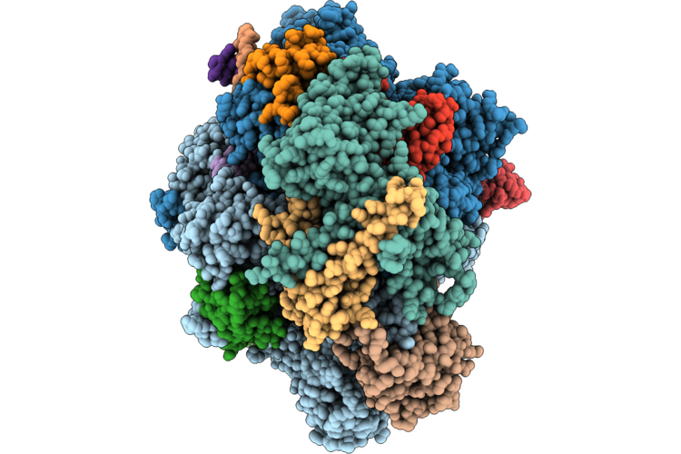
Human Mitoribosome In A Non-Rotated State, With Multiple Neomycin Molecules Bound, Featuring A/A-Site And P/P-Site Trnas
|
Organism: Homo sapiens
Method: ELECTRON MICROSCOPY
Release Date: 2026-07-22
Ligands: ZN, K, SPD, PUT, MG, NMY, VAL, FES, NAD, SPM, ATP, GDP
 |
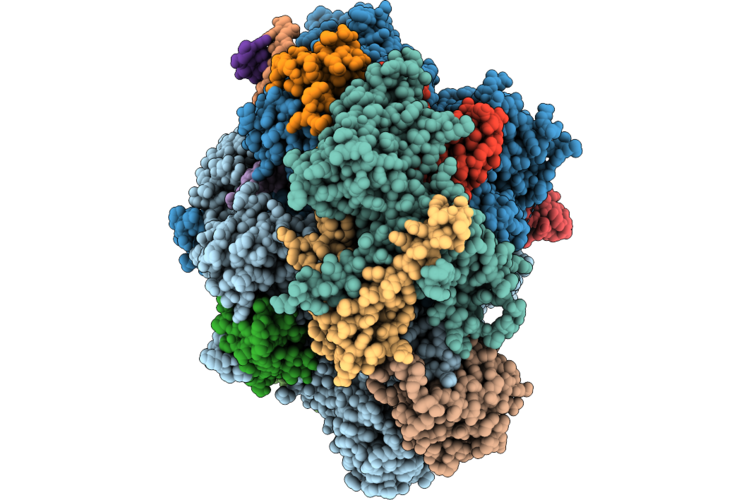
Human Mitoribosome Bound To Taco1 (Translational Activator Of Cox1), Mrna, A/A-, P/P- And (Partial) E/E-Trnas
Organism: Homo sapiens
Method: ELECTRON MICROSCOPY Resolution:2.64 Å Release Date: 2026-07-15 Classification: RIBOSOME Ligands: NAD, MG, K, 5F0, ZN, FES, ATP, GDP, SPD, VOL |
Organism: Homo sapiens
Method: ELECTRON MICROSCOPY
Release Date: 2026-07-15
Ligands: NAD, MG, K, 5F0, ZN, FES, ATP, GDP, SPD, VOL
 |
Assembly Intermediate Of Human Mitochondrial Ribosome Small Subunit In Complex With Noa1, Eral1, Mettl17, Mcat And Tfb1M (State N1)
Organism: Homo sapiens
Method: ELECTRON MICROSCOPY Resolution:3.90 Å Release Date: 2026-07-08 Classification: RIBOSOME Ligands: GDP, ZN, FES, MG, ATP, SF4 |
Organism: Homo sapiens
Method: ELECTRON MICROSCOPY
Release Date: 2026-07-08
Ligands: GDP, ZN, FES, MG, ATP, SF4
 |
Cryo-Et Structure Of Full-Length Membrane-Bound Ehd2 Complex
Organism: Mus musculus
Method: ELECTRON MICROSCOPY Release Date: 2026-07-08 Classification: STRUCTURAL PROTEIN Ligands: ATP, MG |
Organism: Mus musculus
Method: ELECTRON MICROSCOPY
Release Date: 2026-07-08
Ligands: ATP, MG
 |
Cryo-Et Structure Of N-Terminally Truncated Membrane-Bound Ehd2 Complex
Organism: Mus musculus
Method: ELECTRON MICROSCOPY Release Date: 2026-07-08 Classification: STRUCTURAL PROTEIN Ligands: ATP, MG |
Organism: Mus musculus
Method: ELECTRON MICROSCOPY
Release Date: 2026-07-08
Ligands: ATP, MG
 |
Cryo-Em Structure Of The Human Potassium Chloride Cotransporter T906A/T1007A Phospho-Knockout Mutants Kcc2B Bound Atp In Lmng (Outward-Facing State, Dimer)
Organism: Homo sapiens
Method: ELECTRON MICROSCOPY Resolution:3.86 Å Release Date: 2026-07-08 Classification: MEMBRANE PROTEIN Ligands: JUX, ATP, CL, NA |
Organism: Homo sapiens
Method: ELECTRON MICROSCOPY
Release Date: 2026-07-08
Ligands: JUX, ATP, CL, NA
 |
Structure Of Lumen-Open Abcd4-Lmbd1 Complex
Organism: Homo sapiens
Method: ELECTRON MICROSCOPY Resolution:2.80 Å Release Date: 2026-07-01 Classification: MEMBRANE PROTEIN Ligands: ATP, MG, CLR |
Organism: Homo sapiens
Method: ELECTRON MICROSCOPY
Release Date: 2026-07-01
Ligands: ATP, MG, CLR
 |
Structure Of Substrate-Bound Abcd4-Lmbd1 Complex
Organism: Homo sapiens
Method: ELECTRON MICROSCOPY Resolution:2.83 Å Release Date: 2026-07-01 Classification: MEMBRANE PROTEIN Ligands: ATP, B12, MG, CLR |
Organism: Homo sapiens
Method: ELECTRON MICROSCOPY
Release Date: 2026-07-01
Ligands: ATP, B12, MG, CLR
 |
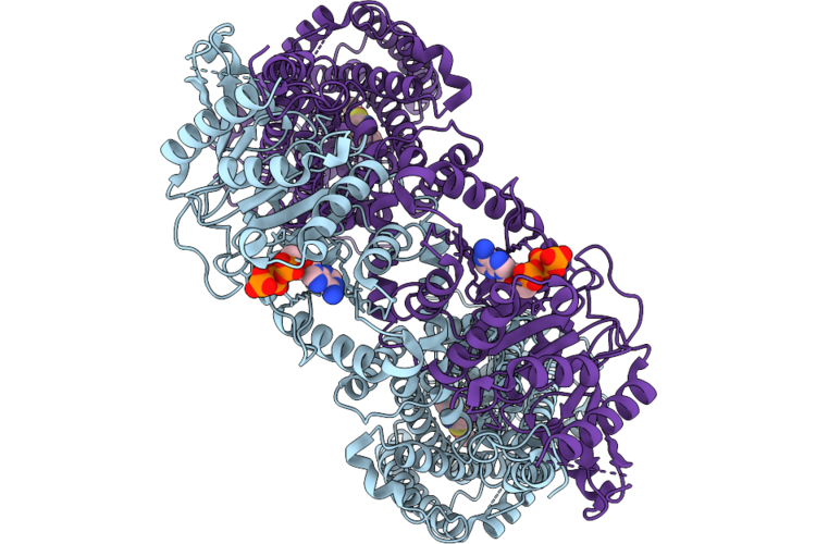
Cryo-Em Structure Of The Yeast Rna Polymerase Ii Elongation Complex With 19-Mer Rna In State Iii (Tl-Open), In The Presence Of Substrate Atp
Organism: Saccharomyces cerevisiae
Method: ELECTRON MICROSCOPY Resolution:2.55 Å Release Date: 2026-07-01 Classification: TRANSCRIPTION Ligands: MG, ZN, ATP |
Organism: Saccharomyces cerevisiae
Method: ELECTRON MICROSCOPY
Release Date: 2026-07-01
Ligands: MG, ZN, ATP
 |
Cryo-Em Structure Of The Yeast Rna Polymerase Ii Elongation Complex With 19-Mer Rna In State V (Tl-Closed), In The Presence Of Substrate Atp
Organism: Saccharomyces cerevisiae
Method: ELECTRON MICROSCOPY Resolution:2.54 Å Release Date: 2026-07-01 Classification: TRANSCRIPTION Ligands: ATP, MG, ZN |
Organism: Saccharomyces cerevisiae
Method: ELECTRON MICROSCOPY
Release Date: 2026-07-01
Ligands: ATP, MG, ZN
 |
Assembly Intermediate Of Human Mitochondrial Ribosome Small Subunit Bound To Mettl15 And Rbfa (Outward Conformation) (State M3)
Organism: Homo sapiens
Method: ELECTRON MICROSCOPY Release Date: 2026-07-01 Classification: RIBOSOME Ligands: FES, ATP, GDP, NAD, SPM, MG, K |
Organism: Homo sapiens
Method: ELECTRON MICROSCOPY
Release Date: 2026-07-01
Ligands: FES, ATP, GDP, NAD, SPM, MG, K
 |
Assembly Intermediate Of Human Mitochondrial Ribosome Small Subunit Bound To Mettl15, Rbfa, And Mtif2 (State M2.1)
Organism: Homo sapiens
Method: ELECTRON MICROSCOPY Release Date: 2026-07-01 Classification: RIBOSOME Ligands: ZN, FES, ATP, GDP, NAD, SPM, MG, K |
Organism: Homo sapiens
Method: ELECTRON MICROSCOPY
Release Date: 2026-07-01
Ligands: ZN, FES, ATP, GDP, NAD, SPM, MG, K
 |
Assembly Intermediate Of Human Mitochondrial Ribosome Small Subunit Bound To Mettl15 And Rbfa (Inward Conformation) (State M2)
Organism: Homo sapiens
Method: ELECTRON MICROSCOPY Release Date: 2026-07-01 Classification: RIBOSOME Ligands: K, ZN, FES, ATP, GDP, MG, NAD, SPM |
Organism: Homo sapiens
Method: ELECTRON MICROSCOPY
Release Date: 2026-07-01
Ligands: K, ZN, FES, ATP, GDP, MG, NAD, SPM
 |
Assembly Intermediate Of Human Mitochondrial Ribosome Small Subunit Bound To Mettl15 And Ms37 (State M4)
Organism: Homo sapiens
Method: ELECTRON MICROSCOPY Release Date: 2026-07-01 Classification: RIBOSOME Ligands: MG, ZN, FES, ATP, GDP, NAD, K |
Organism: Homo sapiens
Method: ELECTRON MICROSCOPY
Release Date: 2026-07-01
Ligands: MG, ZN, FES, ATP, GDP, NAD, K
 |
Structure Of Yeast Rna Polymerase Ii Elongation Complex With Atp Frame-1
Organism: Saccharomyces cerevisiae
Method: ELECTRON MICROSCOPY Resolution:4.28 Å Release Date: 2026-07-01 Classification: TRANSCRIPTION Ligands: MG, ZN, ATP |
Organism: Saccharomyces cerevisiae
Method: ELECTRON MICROSCOPY
Release Date: 2026-07-01
Ligands: MG, ZN, ATP
 |
Structure Of Yeast Rna Polymerase Ii Elongation Complex With Atp Frame-2
Organism: Saccharomyces cerevisiae
Method: ELECTRON MICROSCOPY Resolution:4.17 Å Release Date: 2026-07-01 Classification: TRANSCRIPTION Ligands: MG, ZN, ATP |
Organism: Saccharomyces cerevisiae
Method: ELECTRON MICROSCOPY
Release Date: 2026-07-01
Ligands: MG, ZN, ATP
 |
Structure Of Yeast Rna Polymerase Ii Elongation Complex With Atp Frame-3
Organism: Saccharomyces cerevisiae
Method: ELECTRON MICROSCOPY Resolution:3.95 Å Release Date: 2026-07-01 Classification: TRANSCRIPTION Ligands: MG, ZN, ATP |
Organism: Saccharomyces cerevisiae
Method: ELECTRON MICROSCOPY
Release Date: 2026-07-01
Ligands: MG, ZN, ATP