
Search Count: 4,742
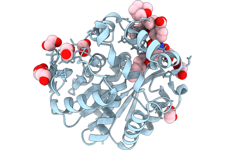 |
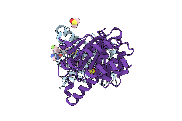
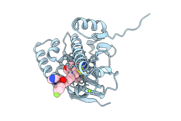
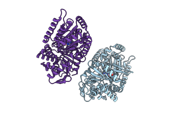
X-Ray Structure Of The Haloalkane Dehalogenase Halotag7 Labeled With Bd566-Htl Substrate
Organism: Escherichia
Method: X-RAY DIFFRACTION Resolution:1.48 Å Release Date: 2026-07-08 Classification: FLUORESCENT PROTEIN Ligands: CL, GLC, A1EQX |
Organism: Escherichia
Method: X-RAY DIFFRACTION
Release Date: 2026-07-08
Ligands: CL, GLC, A1EQX
 |
A Misfolded Structure Of An Fad-Binding Protein Ct375
Organism: Chlamydia trachomatis d/uw-3/cx
Method: X-RAY DIFFRACTION Resolution:2.30 Å Release Date: 2026-07-01 Classification: OXIDOREDUCTASE |
Organism: Chlamydia trachomatis d/uw-3/cx
Method: X-RAY DIFFRACTION
Release Date: 2026-07-01
 |
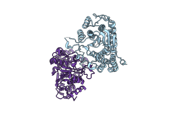
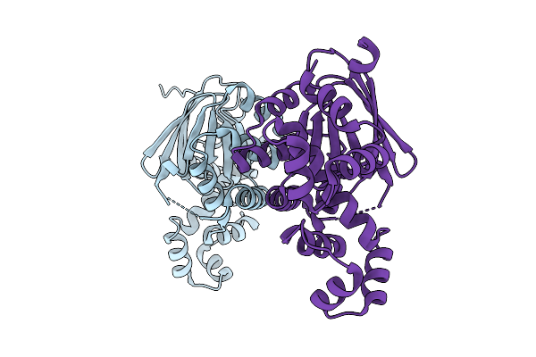
Structure Of Escherichia Vaps-Vapc Complex
Organism: Escherichia
Method: X-RAY DIFFRACTION Resolution:3.40 Å Release Date: 2026-06-03 Classification: TOXIN Ligands: MG |
Organism: Escherichia
Method: X-RAY DIFFRACTION
Release Date: 2026-06-03
Ligands: MG
 |
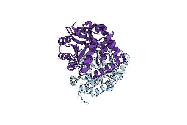
The Structure Of A Bacterial Cyanide Dihydratase From Bacillus Safensis Per-Urp-08
Organism: Bacillus safensis
Method: ELECTRON MICROSCOPY Release Date: 2026-02-25 Classification: HYDROLASE |
Organism: Bacillus safensis
Method: ELECTRON MICROSCOPY
Release Date: 2026-02-25
 |
Mutant S483T/G490N/V492M/S497T/A599G Of Hev-1 E2S Domain
Organism: Hepatitis e virus genotype 1 (isolate human/burma)
Method: X-RAY DIFFRACTION Resolution:1.35 Å Release Date: 2026-01-14 Classification: VIRAL PROTEIN Ligands: SO4 |
Organism: Hepatitis e virus genotype 1 (isolate human/burma)
Method: X-RAY DIFFRACTION
Release Date: 2026-01-14
Ligands: SO4
 |
Cryo-Em Structure Of Candida Albicans Fluoride Channel Fex In Complex With Fab Fragment
Organism: Homo sapiens, Hiv-1 m:b_mn, Candida albicans sc5314
Method: ELECTRON MICROSCOPY Resolution:4.05 Å Release Date: 2025-12-03 Classification: MEMBRANE PROTEIN |
Organism: Homo sapiens, Hiv-1 m:b_mn, Candida albicans sc5314
Method: ELECTRON MICROSCOPY
Release Date: 2025-12-03
 |
Crystal Structure Of Asfv Ep424R
Organism: African swine fever virus (isolate tick/south africa/pretoriuskop pr4/1996)
Method: X-RAY DIFFRACTION Resolution:2.79 Å Release Date: 2025-11-26 Classification: TRANSFERASE |
 |
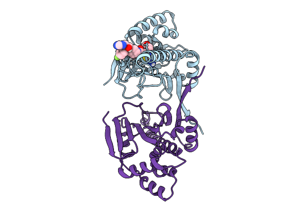
Candida Albicans Hsp90 Nucleotide Binding Domain In Complex With Bri2217
Organism: Candida albicans sc5314
Method: X-RAY DIFFRACTION Resolution:2.03 Å Release Date: 2025-09-17 Classification: CHAPERONE Ligands: A1A27, DMS, CL |
Organism: Candida albicans sc5314
Method: X-RAY DIFFRACTION
Release Date: 2025-09-17
Ligands: A1A27, DMS, CL
 |
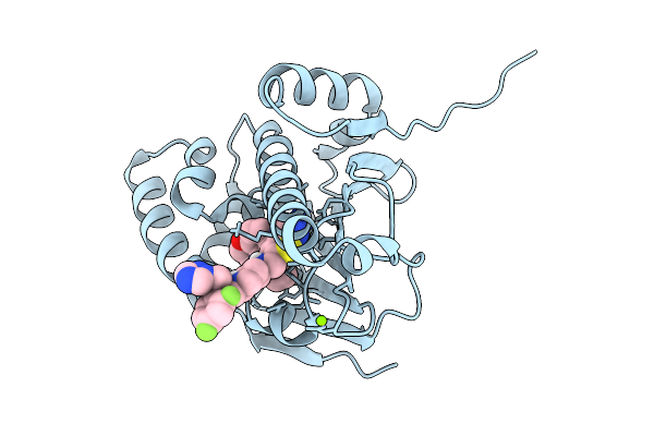
Candida Albicans Hsp90 Nucleotide Binding Domain In Complex With Bri2216
Organism: Candida albicans sc5314
Method: X-RAY DIFFRACTION Resolution:2.09 Å Release Date: 2025-09-10 Classification: CHAPERONE Ligands: A1A28 |
Organism: Candida albicans sc5314
Method: X-RAY DIFFRACTION
Release Date: 2025-09-10
Ligands: A1A28
 |
Candida Albicans Hsp90 Nucleotide Binding Domain In Complex With Bri2311
Organism: Candida albicans sc5314
Method: X-RAY DIFFRACTION Resolution:1.78 Å Release Date: 2025-09-10 Classification: CHAPERONE Ligands: A1A26, MG |
Organism: Candida albicans sc5314
Method: X-RAY DIFFRACTION
Release Date: 2025-09-10
Ligands: A1A26, MG
 |
Candida Albicans Hsp90 Nucleotide Binding Domain In Complex With Bri2312
Organism: Candida albicans sc5314
Method: X-RAY DIFFRACTION Resolution:1.68 Å Release Date: 2025-09-10 Classification: CHAPERONE Ligands: A1A24, MG |
Organism: Candida albicans sc5314
Method: X-RAY DIFFRACTION
Release Date: 2025-09-10
Ligands: A1A24, MG
 |
Crystal Structure Of Diferric Hrmi From Streptomyces Griseoflavus
Organism: Streptomyces griseoflavus
Method: X-RAY DIFFRACTION Resolution:2.13 Å Release Date: 2025-08-20 Classification: OXIDOREDUCTASE Ligands: FE, GOL |
Organism: Streptomyces griseoflavus
Method: X-RAY DIFFRACTION
Release Date: 2025-08-20
Ligands: FE, GOL
 |
Crystal Structure Of N-Oxygenase Hrmi With The Diferric Cofactor Partially Loaded
Organism: Streptomyces griseoflavus
Method: X-RAY DIFFRACTION Resolution:2.00 Å Release Date: 2025-08-20 Classification: OXIDOREDUCTASE Ligands: FE, SO4, GOL |
Organism: Streptomyces griseoflavus
Method: X-RAY DIFFRACTION
Release Date: 2025-08-20
Ligands: FE, SO4, GOL
 |
Crystal Structure Of N-Oxygenase Hrmi With The Diferrous Cofactor
Organism: Streptomyces griseoflavus
Method: X-RAY DIFFRACTION Resolution:2.14 Å Release Date: 2025-08-20 Classification: OXIDOREDUCTASE Ligands: FE2, SO4, GOL |
Organism: Streptomyces griseoflavus
Method: X-RAY DIFFRACTION
Release Date: 2025-08-20
Ligands: FE2, SO4, GOL
 |
Crystal Structure Of N-Oxygenase Hrmi With The Diferrous Cofactor And Substrate Bound
Organism: Streptomyces griseoflavus
Method: X-RAY DIFFRACTION Resolution:2.14 Å Release Date: 2025-08-20 Classification: OXIDOREDUCTASE Ligands: FE2, LYS, GOL |
Organism: Streptomyces griseoflavus
Method: X-RAY DIFFRACTION
Release Date: 2025-08-20
Ligands: FE2, LYS, GOL
 |
Crystal Structure Of N-Oxygenase Hrmi With The Diferric Cofactor And The N(6)-Hydroxy-L-Lysine Product Bound
Organism: Streptomyces griseoflavus
Method: X-RAY DIFFRACTION Resolution:1.96 Å Release Date: 2025-08-20 Classification: OXIDOREDUCTASE Ligands: FE, A1BX4, GOL |
Organism: Streptomyces griseoflavus
Method: X-RAY DIFFRACTION
Release Date: 2025-08-20
Ligands: FE, A1BX4, GOL
 |
Structure Of Candida Albicans Trehalose-6-Phosphate Synthase In Complex With 4456
Organism: Candida albicans sc5314
Method: X-RAY DIFFRACTION Resolution:3.35 Å Release Date: 2025-08-20 Classification: TRANSFERASE Ligands: A1BYI |
Organism: Candida albicans sc5314
Method: X-RAY DIFFRACTION
Release Date: 2025-08-20
Ligands: A1BYI
 |
Structure Of Candida Albicans Trehalose-6-Phosphate Synthase In Complex With Sj6675
Organism: Candida albicans sc5314
Method: X-RAY DIFFRACTION Resolution:3.50 Å Release Date: 2025-08-20 Classification: TRANSFERASE Ligands: A1BYV |
Organism: Candida albicans sc5314
Method: X-RAY DIFFRACTION
Release Date: 2025-08-20
Ligands: A1BYV
 |
Rns Pocket Mutant - R75A
Organism: Escherichia
Method: X-RAY DIFFRACTION Resolution:2.40 Å Release Date: 2025-07-23 Classification: DNA BINDING PROTEIN |
Organism: Escherichia
Method: X-RAY DIFFRACTION
Release Date: 2025-07-23
 |
High-Efficiency Kemp Eliminases By Complete Computational Design
Organism: Escherichia
Method: X-RAY DIFFRACTION Resolution:2.10 Å Release Date: 2025-06-11 Classification: LYASE |
Organism: Escherichia
Method: X-RAY DIFFRACTION
Release Date: 2025-06-11