

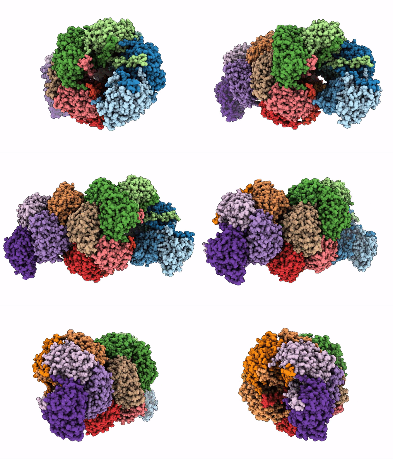
9ODT image
Deposition Date 2025-04-27
Release Date 2026-02-25
Last Version Date 2026-04-15
Entry Detail
PDB ID:
9ODT
Keywords:
Title:
The structure of a Bacterial Cyanide Dihydratase from Bacillus safensis PER-URP-08
Biological Source:
Source Organism(s):
Expression System(s):
Method Details:
Experimental Method:
Resolution:
2.04 Å
Aggregation State:
PARTICLE
Reconstruction Method:
SINGLE PARTICLE
Macromolecular Entities
Structures with similar UniProt ID
Protein Blast
Polymer Type:polypeptide(L)
Molecule:Cyanide dihydratase
Gene (Uniprot):cynD
Chain IDs:A (auth: B), B (auth: A), C (auth: R), D (auth: J), E (auth: I), F (auth: K), G (auth: L), H (auth: M), I (auth: N), J (auth: H), K (auth: G), L (auth: F), M (auth: O), N (auth: E), O (auth: P), P (auth: D), Q, R (auth: C)
Chain Length:330
Number of Molecules:18
Biological Source:Bacillus safensis
Ligand Molecules
Primary Citation
The single-particle cryo-EM structures of a bacterial cyanide dihydratase and a fungal cyanide hydratase.
Structure 34 599 610.e2 (2026)
PMID: 41709456 DOI: 10.1016/j.str.2026.01.009

Abstact

Cyanide is widely used in industries due to its affinity for metals, a property that also underlies its toxicity. Industries, therefore, must reduce cyanide concentration before the final disposal of wastewater. Physical, chemical, and biological methods have been developed for this; however, knowledge about the structure of enzymes involved in cyanide degradation remains limited. Structural characterization of these proteins could facilitate the development of enzymes with enhanced bioremediation potential. Here, we present the single-particle cryo-electron microscopy structures of a cyanide dihydratase from Bacillus safensis and a cyanide hydratase from Gloeocercospora sorghi at 2.2 A and 2.0 A resolution, respectively. We provide a comprehensive description and comparative analysis alongside all previously determined nitrilase structures. Importantly, our full-length structures reveal new features in the C-terminal as well as specific intermolecular interactions between protomer interfaces and within the helix lumen. Finally, our findings offer insights into the reaction mechanisms of these two enzymes.

Legend

Protein

Chemical

Disease

Primary Citation of related structures
Feedback Form
Name
Email
Institute
Feedback